Paper - A model of the left half of the human mandible at the 17 mm CRL stage
| Embryology - 27 Apr 2024 |
|---|
| Google Translate - select your language from the list shown below (this will open a new external page) |
|
العربية | català | 中文 | 中國傳統的 | français | Deutsche | עִברִית | हिंदी | bahasa Indonesia | italiano | 日本語 | 한국어 | မြန်မာ | Pilipino | Polskie | português | ਪੰਜਾਬੀ ਦੇ | Română | русский | Español | Swahili | Svensk | ไทย | Türkçe | اردو | ייִדיש | Tiếng Việt These external translations are automated and may not be accurate. (More? About Translations) |
Fawcett E. A model of the left half of the human mandible at the 17 mm CRL stage. (1930) J Anat Physiol. 47(2): 225-34. PMID:17104287 | PMC1250145
| Online Editor |
|---|
| This historic 1930 brief note by Fawcett is an early description of the development of the human mandible using a model reconstruction.
Prenatal Week 6 - Intramembranous ossification center develops lateral to Meckel's cartilage. Week 7 - Coronoid process begins differentiating. Week 8 - Coronoid process fuses with main mandibular mass.
Week 10 (approx) - Both condylar and coronoid processes are recognizable and anterior portion of Meckel's cartilage begins to ossify. Weeks 12-14 - Secondary cartilages for the condyle, coronoid, and symphysis appear. Weeks 14-16 - Deciduous tooth germs start to form.
|
| Historic Disclaimer - information about historic embryology pages |
|---|
| Pages where the terms "Historic" (textbooks, papers, people, recommendations) appear on this site, and sections within pages where this disclaimer appears, indicate that the content and scientific understanding are specific to the time of publication. This means that while some scientific descriptions are still accurate, the terminology and interpretation of the developmental mechanisms reflect the understanding at the time of original publication and those of the preceding periods, these terms, interpretations and recommendations may not reflect our current scientific understanding. (More? Embryology History | Historic Embryology Papers) |
A Model of the left half of the Human Mandible at the 17 mm CRL stage
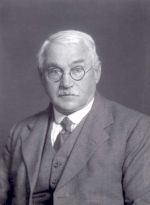
By Professor Fawcett, M.D., F.R.S.
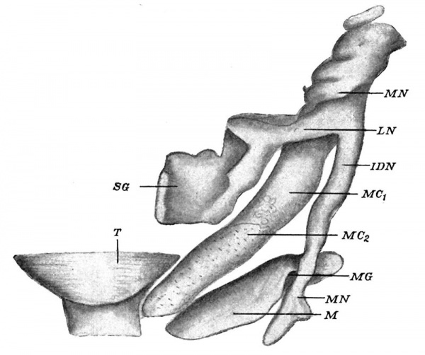
The sections from which this model was made were stained with Mallory’s stain, the most delicate known to me for bone. Fig. 1 shows the ossified mandible (M) as well as many other structures, e.g. Meckel’s cartilage, which is partially chondrified (M C1) and towards its anterior end unchondrified (M C2); the tongue (T) is also shown near its tip for topographical purposes: finally, the associated nerves, e.g. the mandibular (MN), the lingual (LN) with the submaxillary ganglion (SG), the inferior dental (I DN ), and the mental nerves (MN), are. introduced.
Fig. 1. Reconstruction of left mandible and annexes of a 17 millimetre human embryo viewed from above.
During the drawing for the production of the necessary wax plates a careful lookout was kept for tooth buds, but none were found. Furthermore, no sign of an incisive branch of the inferior dental nerve occurred, so the true termination of the inferior dental nerve would appear to be in the mental nerve and not a bifurcation into mental and incisive branches.
The mandible has ossified in the interval between the mental nerve and the unchondrified part of Meckel’s cartilage, and it reaches from not far from the middle line to just beyond the mental nerve, which is lodged in a groove on the upper margin of the bone. The groove is the mental groove (MG).
The bone is a simple plate of bone with no processes. Meckel’s cartilage is unchondrified from the front, back to the region of the mental groove.
The embryo from which this model was made I owe to the kindnessof my colleague, Prof. D. C. Rayner.
Cite this page: Hill, M.A. (2024, April 27) Embryology Paper - A model of the left half of the human mandible at the 17 mm CRL stage. Retrieved from https://embryology.med.unsw.edu.au/embryology/index.php/Paper_-_A_model_of_the_left_half_of_the_human_mandible_at_the_17_mm_CRL_stage
- © Dr Mark Hill 2024, UNSW Embryology ISBN: 978 0 7334 2609 4 - UNSW CRICOS Provider Code No. 00098G


