Paper - Factors leading to the development of a joint between the manubrium and the mesosternum
| Embryology - 27 Apr 2024 |
|---|
| Google Translate - select your language from the list shown below (this will open a new external page) |
|
العربية | català | 中文 | 中國傳統的 | français | Deutsche | עִברִית | हिंदी | bahasa Indonesia | italiano | 日本語 | 한국어 | မြန်မာ | Pilipino | Polskie | português | ਪੰਜਾਬੀ ਦੇ | Română | русский | Español | Swahili | Svensk | ไทย | Türkçe | اردو | ייִדיש | Tiếng Việt These external translations are automated and may not be accurate. (More? About Translations) |
Ashley GT. Factors leading to the development of a joint between the manubrium and the mesosternum. (1955) Thorax (1955), 10, 153-156.
| Historic Disclaimer - information about historic embryology pages |
|---|
| Pages where the terms "Historic" (textbooks, papers, people, recommendations) appear on this site, and sections within pages where this disclaimer appears, indicate that the content and scientific understanding are specific to the time of publication. This means that while some scientific descriptions are still accurate, the terminology and interpretation of the developmental mechanisms reflect the understanding at the time of original publication and those of the preceding periods, these terms, interpretations and recommendations may not reflect our current scientific understanding. (More? Embryology History | Historic Embryology Papers) |
Factors leading to the Development of a Joint between the Manubrium and the Mesosternum
By
G. T. Ashley
The sternum, though present in some form in the vast majority of vertebrates and always more or less constant in its position, is, nevertheless, a bone which varies greatly both in its form and in its function. For example, it may be broad and flat and devoid of joints, as in the tortoise; or it may be long and narrow and composed of many segments, as in the cat.
Fig. 1. Photomicrographs (natural size) of stained sections of foetal sterna showing (A, B, C, D) the presence of a fibrous band (arrowed) across the sternum at the level of the second costal, cartilages, and (E, F) the absence of such a band. Foetal age is indicated in each case.
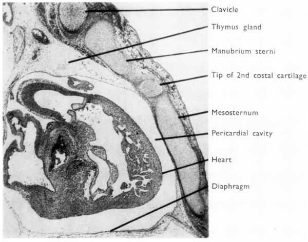
Fig. 2. Paramedian sagital section of a 28 mm human embryo to show the relation of the heart and pericardium to the overlying mesosternum.
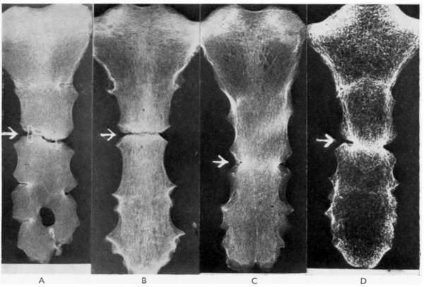
Fig.3. Examples of hylobatean-type human sterna showing the manubrio-sternal joint (free A,B: synostosed C,D) at the narrowest part of the sternum, in each case opposite the third costal cartilages.
Cite this page: Hill, M.A. (2024, April 27) Embryology Paper - Factors leading to the development of a joint between the manubrium and the mesosternum. Retrieved from https://embryology.med.unsw.edu.au/embryology/index.php/Paper_-_Factors_leading_to_the_development_of_a_joint_between_the_manubrium_and_the_mesosternum
- © Dr Mark Hill 2024, UNSW Embryology ISBN: 978 0 7334 2609 4 - UNSW CRICOS Provider Code No. 00098G


