Category:Musculoskeletal: Difference between revisions
From Embryology
mNo edit summary |
mNo edit summary |
||
| Line 3: | Line 3: | ||
{{Musculoskeletal Links}} | {{Musculoskeletal Links}} | ||
[[Category:Mesoderm]] | |||
Latest revision as of 10:35, 11 April 2018
This Embryology category covers content related to both muscle (skeletal muscle), cartilage, bone (skeleton) and joint development.
Subcategories
This category has the following 12 subcategories, out of 12 total.
Pages in category 'Musculoskeletal'
The following 159 pages are in this category, out of 159 total.
2
A
B
- Bone Development
- Bone Histology
- Template:Bone histology
- Template:Bone timeline
- Book - Handbook of Pathological Anatomy 2
- Book - Handbook of Pathological Anatomy 2.1
- Book - Handbook of Pathological Anatomy 2.10
- Book - Handbook of Pathological Anatomy 2.11
- Book - Handbook of Pathological Anatomy 2.12
- Book - Handbook of Pathological Anatomy 2.13
- Book - Handbook of Pathological Anatomy 2.14
- Book - Handbook of Pathological Anatomy 2.15
- Book - Handbook of Pathological Anatomy 2.2
- Book - Handbook of Pathological Anatomy 2.3
- Book - Handbook of Pathological Anatomy 2.4
- Book - Handbook of Pathological Anatomy 2.5
- Book - Handbook of Pathological Anatomy 2.6
- Book - Handbook of Pathological Anatomy 2.7
- Book - Handbook of Pathological Anatomy 2.8
F
L
M
- McMurrich1914 Chapter 7
- Template:Meckel1812-2 header
- File talk:Mesoderm cartoon 04.jpg
- Mouse Gene Expression Movie
- Template:Mouse limb links
- Template:Muscle timeline
- Template:Musculoskeletal
- Template:Musculoskeletal abnormalities
- Template:Musculoskeletal Links
- Musculoskeletal System - Abnormalities
- Musculoskeletal System - Appendicular Skeleton Development
- Musculoskeletal System - Axial Skeleton Development
- Musculoskeletal System - Bone Development
- Musculoskeletal System - Bone Development Timeline
- Musculoskeletal System - Cartilage Development
- Musculoskeletal System - Joint Development
- Musculoskeletal System - Limb Abnormalities
- Musculoskeletal System - Limb Development
- Musculoskeletal System - Muscle Development
- Musculoskeletal System - Muscle Development Timeline
- Musculoskeletal System - Pelvis Development
- Musculoskeletal System - Shoulder Development
- Musculoskeletal System - Skull Development
- Musculoskeletal System - Tendon Development
- Musculoskeletal System Development
P
- Paper - A contribution to the embryology of the fore-limb
- Paper - A model of the left half of the human mandible at the 17 mm CRL stage
- Paper - A study of the development of the mammalian pelvis
- Paper - Abnormal ribs and vertebrae in a human foetus (1919)
- Paper - An infrequent developmental abnormality of the foot (1928)
- Paper - Chondrification in the hands and feet of staged human embryos
- Paper - Congenital absence of ventrolateral abdominal musculature (1946)
- Paper - Congenital variation of the pectoral muscles, with report of a case (1915)
- Paper - Demonstration of the cartilaginous skeleton in mammalian fetuses
- Paper - Development and variation of the nerves and the musculature of the inferior extremity and of the neighboring regions of the trunk in man
- Paper - Development of the rectus abdominis and its sheath in the human fetus
- Paper - Development of the thoracic vertebrae in man (1905)
- Paper - Development of the ventral abdominal walls in man
- Paper - Developmental horizons in human embryos- A review of the histogenesis of cartilage and bone
- Paper - Difference in the ossification of the male and female skeleton (1928)
- Paper - Early development of the cervical vertebrae and the base of the occipital bone in man
- Paper - Extra digits and digital reductions
- Paper - Factors leading to the development of a joint between the manubrium and the mesosternum
- Paper - Fetal age assessment by centers of ossification
- Paper - Multiple malformations of the limbs (1928)
- Paper - Observations on the development of the human vertebral column
- Paper - Observations on the structure and development of bone
- Paper - On Ossification Centers in Human Embryos
- Paper - On the development and morphology of the human sphenoid bone
- Paper - On the mechanism controlling the growth in length of the long bones (1934)
- Paper - Pads on the palm and sole of the human foetus
- Paper - Principles of growth and repair in membrane bones (1946)
- Paper - Rare congenital malformation of hands and feet (1924)
- Paper - Roentgenographic observations of the times of appearance of epiphyses and their fusion with the diaphyses (1932)
- Paper - Some Gross Structural and Quantitative Aspects of the Developmental Anatomy of the Human Embryonic, Fetal and Circumnatal Skeleton
- Paper - Some rare muscular anomalies (1915)
- Paper - Studies of the development of the human skeleton (1905)
- Paper - The arrangement of the bursae in the superior extremities of the full-time foetus
- Paper - The development and ossification of the human clavicle
- Paper - The development of joints
- Paper - The development of muscle in the human foetus
- Paper - The development of nerve endings in the human foetus
- Paper - The development of synovial joints
- Paper - The development of the arm in man (1902)
- Paper - The development of the arteries of the human lower extremity
- Paper - The development of the human femoral artery, a correction
- Paper - The development of the limbs, body-wall and back
- Paper - The development of the limbs, body-wall and back (1901)
- Paper - The development of the patella
- Paper - The early development of the knee joint in staged human embryos
- Paper - The early development of the mammalian sternum
- Paper - The embryology of the human hip joint (1943)
- Paper - The epiphysis of the head of the femur (1915)
- Paper - The growth of the human foot
- Paper - The growth of the long bones in foetal life, as exemplified by a case of foetal syphilis (1929)
- Paper - The history of the earliest stages in the human clavicle
- Paper - The measurement of diaphysial growth in proximal and distal directions (1916)
- Paper - The ontogeny and phylogeny of the sternum (1919)
- Paper - The origin of the osteoblast and of the osteoclast (1913)
- Paper - The origin of the vertebrate limb (1912)
- Paper - The origin, growth, and fate of osteoclasts and their relation to bone resorption (1920)
- Paper - The proximo-distal sequence of origin of the parts of the chick wing and the role of the ectoderm
- Paper - The relation of the myotomes to the ventrolateral musculature and to the anterior limbs in amblystoma (1910)
- Paper - The relations of endogenous and exogenous factors in bone and tooth development (1937)
- Paper - The ribs in the second month of development (1912)
- Paper - The sexual differences of the fetal pelvis
- Paper - The sexual differences of the fetal pelvis (1899)
- Paper - The sternalis muscle in the anencephalous foetus (1936)
- Paper - The sternum - its early development and ossification in man and mammals
- Paper - Vertebral Regional Determination in Young Human Embryos
- Template:Pelvis
R
S
Media in category 'Musculoskeletal'
The following 62 files are in this category, out of 262 total.
(previous page) (next page) Mouse limb gene expression 01.mp4 ; 1.34 MB
- Mouse limb gene expression 02.mp4 ; 1.34 MB
 Mouse limb gene expression icon.jpg 446 × 550; 48 KB
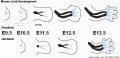 Mouse limb skeleton cartoon.jpg 1,000 × 487; 64 KB
 Mouse limb tissue development.jpg 1,280 × 767; 161 KB
 Mouse Lmx1b gene expression.jpg 1,624 × 550; 104 KB
 Mouse Prdm1.jpg 446 × 550; 35 KB
 Mouse Prox1.jpg 446 × 550; 36 KB
 Mouse Ptx1.jpg 446 × 550; 21 KB
 Mouse Sim2.jpg 446 × 550; 30 KB
 Mouse SmarcD2.jpg 446 × 550; 32 KB
 Mouse Sp6.jpg 446 × 550; 34 KB
 Mouse Tbx4.jpg 446 × 550; 37 KB
 Mouse Tbx5.jpg 446 × 550; 21 KB
 Mouse- axial skeleton intervertebral disc.jpg 600 × 300; 40 KB
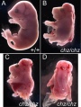 Mouse- chuzhoi mutant.jpg 754 × 1,000; 114 KB
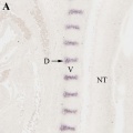 Mouse- intervertebral disc development 02.jpg 726 × 728; 36 KB
 Mouse- intervertebral disc development.jpg 1,454 × 728; 95 KB
 Mouse- postnatal muscle-extensor digitorum longus.jpg 600 × 800; 44 KB
 Muscle- centrosome protein localizes cytoplasmic site nuclear envelope.jpg 1,000 × 1,840; 293 KB
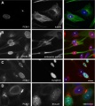 Muscle-reorganization microtububules and centrosome protein.jpg 1,000 × 1,125; 140 KB
 Musculoskeletal- adult hyoid.jpg 450 × 309; 45 KB
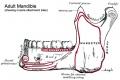 Musculoskeletal- adult mandible.jpg 600 × 402; 58 KB
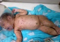 Neonatal varicella.jpg 795 × 561; 35 KB
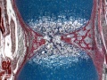
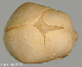
 Ossification centre.jpg 450 × 600; 101 KB
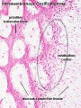
 Ossification endochondral 01.jpg 817 × 613; 198 KB
 Ossification endochondral 1.jpg 750 × 1,000; 147 KB
 Ossification endochondral 1a.jpg 600 × 800; 103 KB
 Ossification endochondral 1b.jpg 450 × 600; 64 KB
 Ossification endochondral 1c.jpg 300 × 400; 32 KB
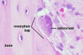 Osteoclast.jpg 500 × 333; 41 KB
 Oxycephaly.jpg 306 × 424; 21 KB
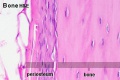 Periosteum.jpg 500 × 333; 34 KB
 Polydactylia.jpg 700 × 541; 37 KB
 Renal agenesis 01.jpg 600 × 662; 44 KB
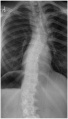 Scoliosis GH.jpg 600 × 1,053; 87 KB
 Shoulder cartoon.jpg 592 × 597; 36 KB
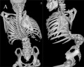
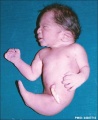 Sirenomelia 01.jpg 488 × 600; 23 KB
 Sirenomelia 02.jpg 498 × 598; 14 KB
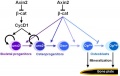 Skull - osteoblast lineage model.jpg 600 × 381; 20 KB
 Skull anterior.gif 200 × 205; 30 KB
 Skull lateral view.gif 200 × 147; 22 KB
 Skull superior.gif 200 × 163; 22 KB
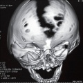 Skull vault defect and midface hypoplasia.jpg 800 × 798; 81 KB
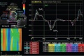 Speckle Tracking Echocardiograph of a dog affected with DMD.JPG 651 × 441; 67 KB
 Spina bifida.jpg 800 × 633; 77 KB
 Stage 13 image 096.jpg 1,000 × 469; 75 KB
 Stage 13 image 097.jpg 1,000 × 720; 162 KB
 Stage 13 image 098.jpg 1,000 × 623; 144 KB
 Stage 22 image 173.jpg 1,000 × 659; 125 KB
 Stage 22 image 196.jpg 1,000 × 671; 102 KB
 Stage 22 image 200.jpg 1,200 × 877; 563 KB
 Stage 22 image 203.jpg 1,200 × 754; 334 KB
 Stage20-23 limbs a.jpg 800 × 301; 19 KB
 Syndactyly.jpg 443 × 377; 16 KB
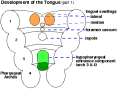 Tongue1.png 373 × 277; 9 KB
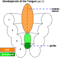 Tongue2.png 327 × 318; 6 KB
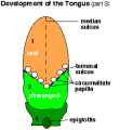 Tongue3.png 249 × 269; 6 KB
 Triphalangeal-thumb.jpg 450 × 273; 14 KB
 Utrophin effects compared to control.jpg 452 × 449; 114 KB
 Vertebra ossification placental sequence.jpg 500 × 440; 48 KB
 Vertebra ossification sequence.jpg 651 × 800; 97 KB


