Category:Gastrointestinal Tract: Difference between revisions
From Embryology
mNo edit summary |
|||
| Line 3: | Line 3: | ||
This category is a link to resource pages, images, podcasts and movies that relate to gastrointestinal tract development. | This category is a link to resource pages, images, podcasts and movies that relate to gastrointestinal tract development. | ||
{{ | {{Gastrointestinal Tract Links}} | ||
Revision as of 14:21, 6 May 2013
Introduction
This category is a link to resource pages, images, podcasts and movies that relate to gastrointestinal tract development.
Subcategories
This category has the following 16 subcategories, out of 16 total.
Pages in category 'Gastrointestinal Tract'
The following 134 pages are in this category, out of 334 total.
(previous page) (next page)P
- Paper - Retrogressive Changes in the Fetal Vessels and the Suspensory Ligament of the Liver
- Paper - Sequential innervation of the intestinal loop in the human embryo
- Paper - Some factors influencing the position of the small intestine (1915)
- Paper - Studies of the intestine and peritoneum in the human foetus - part 1
- Paper - Studies of the intestine and peritoneum in the human foetus - part 2
- Paper - Studies of the intestine and peritoneum in the human foetus - part 3
- Paper - Studies of the intestine and peritoneum in the human foetus - part 4
- Paper - Studies of the intestine and peritoneum in the human foetus - part 5
- Paper - Studies of the intestine and peritoneum in the human foetus - part 6
- Paper - The angiology, angiogenesis, and organogenesis of the submaxillary gland
- Paper - The bi-lobed form of the ventral pancreas in mammals
- Paper - The comparative anatomy of the lips and labial villi of vertebrates
- Paper - The critical period in the development of the intestines (1914)
- Paper - The development of the cloaca in human embryos
- Paper - The development of the form of the gastrointestinal canal in humans 1
- Paper - The development of the form of the gastrointestinal canal in humans 2
- Paper - The development of the great omentum and transverse mesocolon
- Paper - The development of the human pharynx
- Paper - The Development of the Infra-Umbilical Portion of the Abdominal Wall, with Remarks on the Aetiology of Ectopia Vesicae
- Paper - The development of the lobule of the pig's liver (1919)
- Paper - The development of the lobus quadratus of the liver with special reference to an unusual anomaly of this lobe in the adult (1914)
- Paper - The development of the mucous membrane oesophagus stomach and small intestine in human embryo
- Paper - The development of the mucous membrane of the large intestine and vermiform process in the human embryo
- Paper - The development of the rectum in the human embryo
- Paper - The development of the serous glands (von Ebner's) of the vallate papillae in man (1917)
- Paper - The development of the spiral coil in the large intestine of the pig
- Paper - The early looping of the alimentary canal in the mammalian and human foetus and the mechanisms assumed to be active in this process
- Paper - The early stages of the development of the ileo-colic sphincter (1924)
- Paper - The embryogenesis of human bile capillaries and ducts
- Paper - The form of the stomach in human embryos with notes upon the nomenclature of the stomach
- Paper - The formation of the duodenal curve
- Paper - The formation of the duodenal curve (1919)
- Paper - The formation of the umbilical cord and the umbilical region of the anterior abdominal wall
- Paper - The gall bladder and the extrahepatic biliary passages in late embryonic and early fetal life
- Paper - The genesis of Jackson's membrane (1914)
- Paper - The genesis of Jackson's membrane: notes on the genito-mesenteric fold of peritoneum and the supra-adhesion foramen
- Paper - The later development of the bursa pharyngea - Homo
- Paper - The morphology and development of intestinal folds and villi in vertebrates
- Paper - The nature of the malformations of the rectum and urogenital passages
- Paper - The regular occurrence of intestinal diverticula in embryos of the pig, rabbit and man
- Paper - The regular occurrence of intestinal diverticula in embryos of the pig, rabbit, and man
- Paper - The relations of endogenous and exogenous factors in bone and tooth development (1937)
- Paper - The relative frequency of the various positions of the vermiform appendix (1924)
- Paper - The role of the primitive mesothelium in the development of the mammalian spleen (1936)
- Paper - Transposition of Abdominal Viscera (1926)
- Paper - V. Meckel's diverticulum patent at the navel (1902)
- Template:Parotid gland
- Template:Phenylketonuria
R
- Template:Ref-Beattie1924
- Template:Ref-Bossy1981
- Template:Ref-BurnsOgryzlo1938
- Template:Ref-Carey1920a
- Template:Ref-Carey1920b
- Template:Ref-Cave1936
- Template:Ref-Crymble1915
- Template:Ref-deVries1974
- Template:Ref-Elze1909
- Template:Ref-Enbom1939
- Template:Ref-Flint1903
- Template:Ref-Franklin1948
- Template:Ref-Fraser1919b
- Template:Ref-Frazer1915
- Template:Ref-Frazer1919
- Template:Ref-Gladstone1913
- Template:Ref-Gladstone1924
- Template:Ref-Guthrie1945
- Template:Ref-Hilton1902
- Template:Ref-Hubbard1902
- Template:Ref-Hunter1927
- Template:Ref-Hunter1928
- Template:Ref-Jackson1909b
- Template:Ref-Johnson1910
- Template:Ref-Johnson1913
- Template:Ref-Johnson1914
- Template:Ref-Johnson1914b
- Template:Ref-Kirk1910
- Template:Ref-Latta1921
- Template:Ref-Lewis1912b
- Template:Ref-Lineback1916
- Template:Ref-Lineback1920
- Template:Ref-Lockwood1882
- Template:Ref-Lockwood1884
- Template:Ref-Mall1898a
- Template:Ref-Mall1898b
- Template:Ref-Mall1899
- Template:Ref-McGill1910
- Template:Ref-Minot1900a
- Template:Ref-Pernkoof1922
- Template:Ref-Pernkoof1925
- Template:Ref-Reid1908
- Template:Ref-Reid1911a
- Template:Ref-Reid1911b
- Template:Ref-Reid1912
- Template:Ref-Reid1913a
- Template:Ref-Reid1913b
- Template:Ref-Reid1913c
- Template:Ref-Reid1914a
- Template:Ref-Reid1914b
- Template:Ref-Snook1934b
- Template:Ref-Tandler1900
- Template:Ref-Wagstaffe1924
- Template:Ref-Wakeley1923
- Template:Ref-Wakeley1930
- Template:Ref-Woods-Jones1904
- Template:Reid DG.
S
- Template:Salivary gland
- Salivary Gland Development
- Site Map
- Talk:Site Map
- Template:Small intestine
- Template:Small Intestine Length table1
- Template:Splanchnic mesoderm
- Template:Stage 23 MRI movie 7
- Stage 23 MRI Movie 7
- Template:Stomach
- Template:Stomach Histology
- Template:Sublingual gland
- Template:Submandibular gland
- Template:Submaxillary gland
V
Media in category 'Gastrointestinal Tract'
The following 200 files are in this category, out of 523 total.
(previous page) (next page) Human week 10 fetus 26.jpg 1,200 × 900; 262 KB
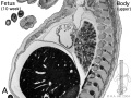 Human- fetal week 10 upper body A.jpg 600 × 450; 104 KB
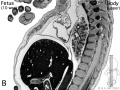 Human- fetal week 10 upper body B.jpg 600 × 450; 105 KB
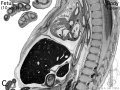 Human- fetal week 10 upper body C.jpg 600 × 450; 109 KB
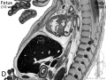 Human- fetal week 10 upper body D.jpg 600 × 450; 106 KB
 Hydrocolpos.jpg 375 × 361; 23 KB
 Interstitial Cells of Cajal 01.jpg 993 × 518; 142 KB
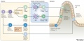
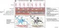
 Intestinal function and microbiota 01.jpg 1,200 × 845; 207 KB
 Intestinal lymphangiectasia 01.jpg 600 × 452; 138 KB
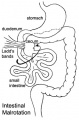 Intestinal malrotation.jpg 330 × 500; 32 KB
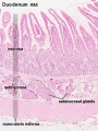
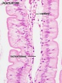 Intestine histology 001.jpg 450 × 600; 65 KB
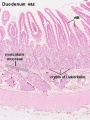
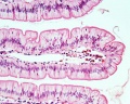 Intestine histology 002.jpg 800 × 640; 130 KB
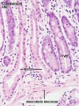
 Intestine histology 003.jpg 400 × 533; 64 KB
 Intestine histology 004.jpg 400 × 533; 81 KB
 Intestine histology 005.jpg 400 × 533; 78 KB
 Intestine histology 006.jpg 400 × 533; 77 KB
 Intestine histology 007.jpg 400 × 533; 82 KB
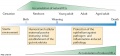
 Intraepithelial lymphocyte differentiation 01.jpg 1,200 × 585; 108 KB
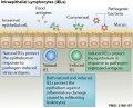
 Intraepithelial lymphocyte differentiation 02.jpg 1,200 × 557; 79 KB
 Intraepithelial lymphocyte differentiation 03.jpg 600 × 484; 68 KB
 Jejunal atresia 01.jpg 800 × 600; 75 KB
 Jejuno-ileal atresia 01.jpg 800 × 580; 95 KB
 Jejunum and ileum cartoon.jpg 500 × 704; 45 KB
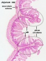
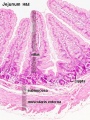
 Jejunum histology 01.jpg 480 × 600; 79 KB
 Johnson1914b fig01.jpg 700 × 525; 39 KB
 Johnson1917 plate02fig02.jpg 800 × 542; 55 KB
 Johnson1917 plate02fig03.jpg 800 × 447; 42 KB
 Johnson1917 plate02fig04.jpg 644 × 800; 67 KB
 Johnson1917 plate03fig03.jpg 599 × 800; 60 KB
 Johnson1917 plate03fig04.jpg 435 × 800; 45 KB
 Keibel Mall 2 223.jpg 1,000 × 719; 33 KB
 Keibel Mall 2 224.jpg 1,082 × 950; 197 KB
 Keibel Mall 2 225.jpg 976 × 979; 158 KB
 Keibel Mall 2 226.jpg 1,000 × 801; 90 KB
 Keibel Mall 2 227.jpg 572 × 1,171; 151 KB
 Keibel Mall 2 228.jpg 575 × 643; 68 KB
 Keibel Mall 2 229.jpg 771 × 1,200; 194 KB
 Keibel Mall 2 230.jpg 1,000 × 669; 180 KB
 Keibel Mall 2 231.jpg 1,019 × 1,000; 132 KB
 Keibel Mall 2 232.jpg 850 × 1,000; 205 KB
 Keibel Mall 2 233.jpg 1,000 × 498; 77 KB
 Keibel Mall 2 234.jpg 1,103 × 1,000; 187 KB
 Keibel Mall 2 235.jpg 1,200 × 469; 85 KB
 Keibel Mall 2 236.jpg 899 × 1,000; 108 KB
 Keibel Mall 2 237.jpg 1,105 × 800; 108 KB
 Keibel Mall 2 238.jpg 501 × 800; 28 KB
 Keibel Mall 2 239.jpg 1,007 × 1,000; 142 KB
 Keibel Mall 2 240.jpg 1,076 × 1,200; 176 KB
 Keibel Mall 2 241.jpg 1,039 × 1,000; 178 KB
 Keibel Mall 2 242.jpg 1,158 × 1,200; 191 KB
 Keibel Mall 2 243.jpg 944 × 1,200; 139 KB
 Keibel Mall 2 244.jpg 1,028 × 448; 116 KB
 Keibel Mall 2 245.jpg 920 × 800; 140 KB
 Keibel Mall 2 246.jpg 1,018 × 1,200; 156 KB
 Keibel Mall 2 247.jpg 1,200 × 619; 149 KB
 Keibel Mall 2 295.jpg 1,000 × 920; 83 KB
 Keibel Mall 325.jpg 301 × 577; 60 KB
 Keibel Mall 326.jpg 800 × 641; 147 KB
 Keibel Mall 327.jpg 735 × 900; 152 KB
 Keibel Mall 328.jpg 800 × 774; 125 KB
 Keibel Mall 329.jpg 800 × 464; 69 KB
 Keith1902 fig015b.jpg 1,000 × 719; 97 KB
 Keith1902 fig032.jpg 1,000 × 496; 81 KB
 Keith1902 fig212.jpg 1,123 × 750; 139 KB
 Keith1902 fig213a.jpg 854 × 800; 166 KB
 Keith1902 fig213b.jpg 800 × 564; 64 KB
 Keith1902 fig214.jpg 750 × 549; 58 KB
 Keith1902 fig215.jpg 704 × 600; 56 KB
 Keith1902 fig216.jpg 1,000 × 771; 170 KB
 Keith1902 fig217.jpg 1,000 × 726; 144 KB
 Keith1902 fig218.jpg 800 × 475; 70 KB
 Keith1902 fig219.jpg 1,000 × 768; 166 KB
 Keith1902 fig220.jpg 1,000 × 632; 116 KB
 Keith1902 fig221.jpg 1,000 × 652; 101 KB
 Keith1902 fig222.jpg 936 × 800; 118 KB
 Keith1902 fig223a.jpg 926 × 800; 95 KB
 Keith1902 fig223b.jpg 650 × 382; 49 KB
 Keith1902 fig224.jpg 861 × 750; 96 KB
 Keith1902 fig225a.jpg 774 × 520; 69 KB
 Keith1902 fig225b.jpg 783 × 580; 81 KB
 Keith1902 fig226.jpg 1,000 × 624; 112 KB
 Keith1921 fig038.jpg 1,200 × 636; 118 KB
 Keith1921 fig039.jpg 815 × 706; 128 KB
 Kollmann379.jpg 827 × 454; 45 KB
 Kollmann397.jpg 862 × 554; 64 KB
 Kollmann418.jpg 736 × 594; 82 KB
 Kollmann548.jpg 738 × 525; 72 KB
 Kyoto16834 stage17-umbilicus.jpg 1,536 × 1,316; 137 KB
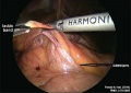 Ladd's band-01.jpg 600 × 423; 40 KB
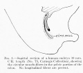 Lineback1920 fig01.jpg 600 × 530; 39 KB
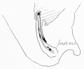 Lineback1920 fig03.jpg 800 × 660; 39 KB
 Lineback1920 fig04-5.jpg 1,200 × 756; 150 KB
 Lineback1920 fig06-7.jpg 1,200 × 808; 177 KB
 Lineback1920 fig08.jpg 244 × 800; 31 KB
 Liver animated cartoon.gif 300 × 200; 239 KB
 Liver development signaling.jpg 600 × 467; 45 KB
 Liver hepatocyte from stem cell.png 600 × 444; 96 KB
 Liver histology 001.jpg 400 × 533; 94 KB
 Liver histology 002.jpg 375 × 500; 54 KB
 Liver histology 003.jpg 375 × 500; 52 KB
 Liver histology 004.jpg 600 × 400; 70 KB
 Liver histology 005.jpg 800 × 664; 166 KB
 Liver histology 008.jpg 1,280 × 1,024; 214 KB
 Liver histology 009.jpg 1,280 × 1,024; 373 KB
 Liver histology 101.jpg 1,280 × 1,024; 410 KB
 Liver histology 102.jpg 1,280 × 1,024; 475 KB
 Liver histology 103.jpg 1,280 × 1,024; 330 KB
 Liver histology 104.jpg 800 × 664; 155 KB
 Liver structure cartoon.jpg 1,000 × 451; 78 KB
 Liver-sinusiod cartoon.jpg 600 × 523; 51 KB
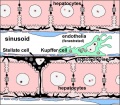 Liver-sinusoid colour cartoon.jpg 600 × 523; 64 KB
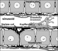 Liver-sinusoid-label cartoon.jpg 600 × 523; 58 KB
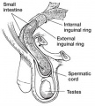 Male - inguinal hernia.jpg 361 × 400; 34 KB
 Mall1906-fig06.jpg 600 × 716; 111 KB
 Mall1906-fig07.jpg 503 × 753; 102 KB
 Mall1906-fig08.jpg 726 × 753; 75 KB
 Mall1906-fig09.jpg 452 × 744; 111 KB
 Mall1906-fig10.jpg 660 × 744; 71 KB
 Mall1906-fig11.jpg 681 × 735; 89 KB
 Mall1906-fig12.jpg 544 × 735; 68 KB
 Mall1906-fig13.jpg 592 × 706; 129 KB
 Mall1906-fig14.jpg 673 × 699; 50 KB
 Mall1906-fig15.jpg 887 × 1,000; 115 KB
 Mall1906-fig16.jpg 708 × 890; 155 KB
 Mall1906-fig17.jpg 705 × 881; 98 KB
 Mall1906-fig18.jpg 933 × 584; 66 KB
 Mall1906-fig19.jpg 596 × 610; 102 KB
 Mall1906-fig20.jpg 768 × 1,000; 115 KB
 Mall1906-fig21.jpg 1,000 × 763; 72 KB
 Mall1906-fig22.jpg 694 × 880; 195 KB
 Mall1906-fig23.jpg 650 × 850; 191 KB
 Mall1906-fig24.jpg 577 × 850; 176 KB
 Mall1906-fig25.jpg 1,165 × 887; 153 KB
 Mall1906-fig26.jpg 766 × 639; 78 KB
 Mall1906-fig27.jpg 890 × 800; 168 KB
 Mall1906-fig28.jpg 1,000 × 803; 146 KB
 Mall1906-fig29.jpg 780 × 700; 59 KB
 Mall1906-fig30.jpg 845 × 650; 67 KB
 Meckel's diverticulum 01.jpg 341 × 480; 74 KB
 Meckel's diverticulum 02.jpg 600 × 542; 58 KB
 Meckel's diverticulum 03.jpg 600 × 480; 69 KB
 Megacolon stoma.gif 200 × 202; 6 KB
 Megacolon stoma1.jpg 300 × 303; 8 KB
 Megacolon stoma2.jpg 300 × 302; 7 KB
 Megacolon surgery 01.jpg 300 × 416; 14 KB
 Megacolon surgery 02.jpg 300 × 420; 7 KB
 Megacolon surgery 03.jpg 300 × 416; 6 KB
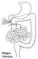 Midgut volvulus.jpg 323 × 500; 28 KB
 Minot1897 444.jpg 1,257 × 688; 107 KB
 MNGIE and altered ICC 01.jpg 1,946 × 925; 434 KB
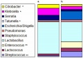 Mouse - analysis of colonic microbiota.jpg 600 × 422; 47 KB
 Mouse - stomach 01.png 599 × 600; 1.45 MB
 Mouse HOXA5 expression E12.5.jpg 1,000 × 577; 192 KB
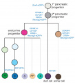 Mouse pancreas cell lineage.jpg 1,855 × 2,039; 291 KB
 Mouse pancreas development.jpg 600 × 939; 261 KB
 Mouse- pancreas differentiation model.jpg 1,200 × 435; 96 KB
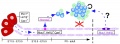
 Mouse-Gastrointestinal-tract-E10.5-01.jpg 1,000 × 306; 43 KB
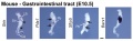
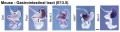 Mouse-Gastrointestinal-tract-E13.5-01.jpg 1,000 × 266; 48 KB
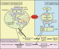 Nipbl heart and organ patterning.png 600 × 502; 164 KB
 Normal intestinal rotation cartoon.jpg 800 × 695; 86 KB
 Oesophageal atresia x-ray 01.jpg 894 × 588; 102 KB
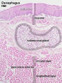
 Oesophagus histology 01.jpg 1,280 × 1,024; 290 KB
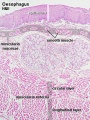
 Oesophagus histology 02.jpg 800 × 1,000; 196 KB
 Oesophagus histology 03.jpg 800 × 1,000; 209 KB
 Oesophagus histology 06.jpg 400 × 533; 100 KB
 Oesophagus histology 07.jpg 400 × 533; 100 KB
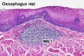 Oesophagus MALT.jpg 500 × 333; 73 KB
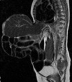 Omphalocoele MRI.jpg 553 × 631; 52 KB
 Pancreas acinar cell em01.jpg 1,280 × 928; 496 KB
 Pancreas adult.jpg 600 × 427; 50 KB
 Pancreas islet - structure human and rat.jpg 945 × 930; 259 KB
 Pancreas rotation.jpg 652 × 320; 23 KB
 Pancreatic duct developing.jpg 400 × 322; 15 KB
 Patten031.jpg 802 × 1,029; 178 KB
 Patten054.jpg 764 × 1,060; 179 KB
 Patten055.jpg 790 × 567; 124 KB
 Perineal fistula.jpg 800 × 596; 82 KB
 Persistent cloaca perineum.jpg 600 × 800; 57 KB
 Peyer's patch 01.jpg 450 × 600; 118 KB
 Peyer's patch 02.jpg 450 × 600; 69 KB
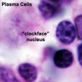 Plasma cell clockface nucleus 01.jpg 400 × 400; 27 KB
 PMID28514120-Chen et al-2017.pdf ; 2.87 MB
 Pyloric atresia 01.jpg 800 × 580; 35 KB
 Roux1911 fig01.jpg 1,108 × 1,200; 264 KB
 Roux1911 fig02.jpg 1,229 × 1,400; 175 KB
 Rugh 142.jpg 845 × 800; 108 KB
 Sabin1909 fig04.jpg 769 × 902; 97 KB
 Sabin1909 fig09.jpg 661 × 800; 34 KB
 Sabin1909 fig11.jpg 702 × 1,076; 109 KB
 Sabin1909 fig12.jpg 713 × 1,003; 89 KB
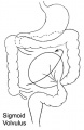 Sigmoid volvulus.jpg 325 × 500; 24 KB
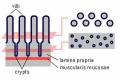 Small intestine villi and crypts.jpg 500 × 333; 26 KB
 Smooth muscle histology 001.jpg 600 × 750; 161 KB
 Smooth muscle histology 002.jpg 600 × 750; 112 KB
 Smooth muscle histology 003.jpg 1,280 × 1,024; 246 KB
 Smooth muscle histology 004.jpg 1,280 × 1,024; 310 KB
 Smooth muscle histology 005.jpg 1,280 × 1,024; 399 KB
 Smooth muscle histology 006.jpg 1,280 × 1,024; 481 KB
 Stage 11 historic-Atwell1930-1.jpg 538 × 1,000; 75 KB
 Stage 11 historic-Atwell1930-1a.jpg 430 × 800; 46 KB


