Lecture - Week 1 and 2 Development
2010 Lecture | Zygote | Blastocyst | Implantation
Objectives

- Understand the events during week 1 of development (Zygote, Blastomeres, Morula, Blastocyst)
- Understand the events during week 2 of development (Trophoblast, Syncytiotrophoblast, Cytotrophoblast, Embryoblast, Implantation)
- Brief understanding of early placentation
- Brief understanding of maternal changes
Week 1 and 2 Overview Animations
|
|
|
| Lectopia Lecture Audio
|
References
The Developing Human: Clinically oriented embryology

|
Citation: The Developing Human: clinically oriented embryology 9th ed. Keith L. Moore, T.V.N. Persaud, Mark G. Torchia. Philadelphia, PA: Saunders, 2011. |
Larsen's human embryology

|
Citation: Larsen's human embryology 4th ed. Schoenwolf, Gary C; Larsen, William J, (William James). Philadelphia, PA : Elsevier/Churchill Livingstone, c2009. |
UNSW Embryology

|
Hill, M.A. (2011) UNSW Embryology (11th ed.). Sydney:UNSW. |
Fertilization

|
|
Fertilization - Spermatozoa
- Sperm Binding - zona pellucida protein ZP3 acts as receptor for sperm
- Acrosome Reaction - exyocytosis of acrosome contents (Calcium mediated) MBoC - Figure 20-31. The acrosome reaction that occurs when a mammalian sperm fertilizes an egg
- enzymes to digest the zona pellucida
- exposes sperm surface proteins to bind ZP2
- Membrane Fusion - between sperm and egg, allows sperm nuclei passage into egg cytoplasm
Fertilization- Oocyte
- Membrane Depolarization - caused by sperm membrane fusion, primary block to polyspermy
- Cortical Reaction - IP3 pathway elevates intracellular Calcium, exocytosis of cortical granules MBoC - Figure 20-32. How the cortical reaction in a mouse egg is thought to prevent additional sperm from entering the egg
- enzyme alters ZP3 so it will no longer bind sperm plasma membrane
- Meiosis 2 - completion of 2nd meiotic division
- forms second polar body (a third polar body may be formed by meiotic division of the first polar body)
Zygote Formation
- zygote is the first diploid cell formed following fertilisation.
- male and female pronuclei, 2 nuclei approach each other and nuclear membranes break down.
- DNA replicates, first mitotic division
- sperm contributes centriole which organizes mitotic spindle

|

|
Movie - Pronuclear Fusion | Movie - Parental Genomes
- Conceptus - the term refers to all material derived from this fertilised zygote, includes both the embryo and the non-embryonic tissues (placenta, fetal membranes).
- Links: Carnegie stage 1
Cleavage of Zygote
- cleavage of zygote forms 2 blastomeres and is also cleavage with no cytoplasm synthesis.
- special "embryonic" cell cycle S phases and M phases alternate without any intervening G1 or G2 phases (MSMSMSMS, adult MG1SG2) therefore individual cell volume decreases.
- cell division is initially synchronous, then asynchronously
- slow- centre cells, larger fast- peripheral cells
- zona pellucid still intact (division occurs within the ZP)

Human Zygote to Blastocyst Development (day 1 to 6)
Morula

|

|
| Human Embryo (day 2) | Human Embryo (day 3) |
- about day 4 is a solid ball of 16-20 cells with peripheral cells flattened against zona pellucida
- compaction occurs forming a cavity and leading to the next blastocyst stage
Blastocyst

- about day 5 have 2 identifiable cell types and a fluid-filled cavity (blastoceol)
- outer cell layer - trophoblast, peripheral flattened cells, forms the placenta and placental membranes
- inner cell mass - embryoblast, mass of rounder cells located on one wall of the blastocoel, forms entire embryo
Blastula Cell Communication
Two forms of cellular junctions Junctions
- gap junctions, allow electrically couple cells of epithelium surrounding a fluid-filled cavity
- tight junctions, close to outer surface create a seal, isolates interior of embryo from external medium
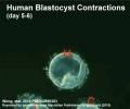
Blastocyst Hatching

Blastocyst Hatching - zona pellucida lost, ZP has sperm entry site, and entire ZP broken down by uterine secretions and possibly blastula secretions. Uterine Glands - secretions required for blastocyst motility and nutrition
|
||
| Day 3 to 6 | Contractions | Hatching |
| Quicktime | Flash | Quicktime | Flash | Quicktime | Flash |
Week 2 - Implantation
The second week of human development is concerned with the process of implantation and the differentiation of the blastocyst into early embryonic and placental forming structures.
Normal Implantation Sites - in uterine wall superior, posterior, lateral |
|
Endometrial Receptivity
- In humans, receptivity occurs 6 days after the post-ovulatory progesterone surge and lasts about 2 to 4 days.
- Similar "receptivity window" in other species (rat day 5 and mouse day 4.5).
- Many studies have looked into identifying markers for this receptivity period both to optimise and to block this process.
Abnormal Implantation

Abnormal implantation sites or Ectopic Pregnancy occurs if implantation is in uterine tube or outside the uterus.
- sites - external surface of uterus, ovary, bowel, gastrointestinal tract, mesentry, peritoneal wall
- If not spontaneous then, embryo has to be removed surgically
Tubal pregnancy - 94% of ectopic pregnancies
- if uterine epithelium is damaged (scarring, pelvic inflammatory disease)
- if zona pellucida is lost too early, allows premature tubal implantation
- embryo may develop through early stages, can erode through the uterine horn and reattach within the peritoneal cavity

|

|
Uterus

Uterus proliferative phase

Uterine gland proliferative phase

Uterus secretory phase

Uterine gland secretory phase


- Endometrium - 3 layers in secretory phase of menstrual cycle: compact, spongy, basal
- Myometrium - muscular layer outside endometrium, contracts in parturition
- Perimetrium - tunica serosa of the uterus continuous with the peritoneal wall
Endometrial Layers
- Compact - implantation occurs in this layer, dense stromal cells, uterine gland necks, capillaries of spiral arteries
- Spongy - swollen stromal cells, uterine gland bodies, spiral arteries
- Basal - not lost during menstruation or childbirth, own blood supply
Decidual Reaction
- transformation of endometrial stromal cells
- occurs initially at site of implantation and includes both cellular and matrix changes
- reaction spreads throughout entire uterus, not at cervix
- deposition of fibrinoid and glycogen and epithelial plaque formation (at anchoring villi)
- presence of decidual cells are indicative of pregnancy
Other Uterine Changes
- Cervix - at mouth of uterus, secretes mucus (CMP), forms a plug/barrier, mechanical and antibacterial
- Vascular - increased number of blood vessels
Decidua
The endometrium becomes the decidua and forms 3 distinct anatomical regions (at approx 3 weeks)
- Decidua Basalis at implantation site
- Decidua Capsularis enclosing the conceptus
- Decidua Parietalis the remainder of uterus
- Decidua Capsularis and Parietalis fuse eventually fuse and uterine cavity is lost by 12 weeks
Uterus Abnormalities
Endometriosis endometrial tissue located in other regions of the uterus or other tissues. This misplaced tissue develops into growths or lesions which respond to the menstrual cycle hormonal changes in the same way that the tissue of the uterine lining does; each month the tissue builds up, breaks down, and sheds.
Conceptus - Bilaminar Embryoblast
|
File:Chorion 001 icon.jpg</wikiflv> |
Conceptus - Bilaminar Trophoblast
Syncitiotrophoblasts
- secrete proteolytic enzymes, enzymes break down extracellular matrix around cells
- Allow passage of blastocyst into endometrial wall, totally surround the blastocyst
- generate spaces that fill with maternal blood- lacunae
- secrete Human Chorionic Gonadotropin (hCG), hormone, maintains decidua and Corpus Luteum, basis of pregnancy diagnostic test, present in urine is diagnostic of pregnancy
- levels peak at 8 to 10 weeks of pregnancy, then decline and are lower for rest of pregnancy
- 1-2 months: 5,000-200,000 mIU/ml; Non-pregnant females: <5.0 mIU/ml; Postmenopausal females: <9.5 mIU/ml)
- Later in development placenta will secrete hCG


Cytotrophoblasts
- form a continuous cellular layer that covers the developing placental villi.
Twinning
Twinning can be due to two separate fertilization events (dizygotic twins) or as an abnormality of a single fertilization (monozygotic twins) event during the early weeks of development.
Dizygotic Twinning
Dizygotic twins (fraternal, non-identical) arise from separate fertilization events involving two separate oocyte (egg, ova) and spermatozoa (sperm).
- In dizygotic twinning the genetic material is different and implantation and placentation is also different.
Monoygotic Twinning
- In monozygotic twinning the genetic material is initially identical and degree of twinning will depend upon the timing (early to late) from separate fetal membranes and placenta to conjoined twins.
- morula stage (diamniotic dichorionic), early blastocyst (diamniotic monochorionic), late blastocyst to bilaminar (monoamniotic monochorionic), bilaminar to trilaminar embryo (conjoined)
- Monozygotic twins are a unique research resource for comparing environmental effects on development and health.
- Congenital abnormality statistics for twins is generally increased in various conditions.
Monoygotic twins (identical) produced from a single fertilization event (one fertilised egg and a single spermatazoa, form a single zygote), these twins therefore share the same genetic makeup. Occurs in approximately 3-5 per 1000 pregnancies, more commonly with aged mothers. The later the twinning event, the less common are initially separate placental membranes and finally resulting in conjoined twins.
| Week | Week 1 | Week 2 | |||||||||||||
| Day | 0 | 1 | 2 | 3 | 4 | 5 | 6 | 7 | 8 | 9 | 10 | 11 | 12 | 13 | 14 |
| Cell Number | 1 | 1 | 2 | 16 | 32 | 128 | bilaminar | ||||||||
| Event | Ovulation | fertilization | First cell division | Morula | Early blastocyst | Late blastocyst
Hatching |
Implantation starts | X inactivation | |||||||
|
|
|

|
|||||||||||||
| Monoygotic
Twin Type |
Diamniotic
Dichorionic |
Diamniotic
Monochorionic |
Monoamniotic
Monochorionic |
Conjoined | |||||||||||
Table based upon recent Twinning Review.[1]
- ↑ <pubmed>12957099</pubmed>
- Links: Twinning | Australian Twin Registry
Now watch the Week 1 and 2 animation overview.
Glossary Links
- Glossary: A | B | C | D | E | F | G | H | I | J | K | L | M | N | O | P | Q | R | S | T | U | V | W | X | Y | Z | Numbers | Symbols | Term Link
Cite this page: Hill, M.A. (2024, June 25) Embryology Lecture - Week 1 and 2 Development. Retrieved from https://embryology.med.unsw.edu.au/embryology/index.php/Lecture_-_Week_1_and_2_Development
- © Dr Mark Hill 2024, UNSW Embryology ISBN: 978 0 7334 2609 4 - UNSW CRICOS Provider Code No. 00098G