Endocrine - Pancreas Development
Introduction

The pancreas is a two-headed organ, not only in origin but also in function. In origin, the pancreas develops from two separate primordia. In function, the organ has both endocrine function in relation to regulating blood glucose (and also other hormone secretions) and gastrointestinal function as an exocrine (digestive) organ, see Gastrointestinal Tract - Pancreas Development.
In recent years there has been much research due to the increasing incidence of diabetes in humans and the potential for stem cell therapeutics. Much is now known about the epithelial/mesenchymal and molecular regulation of pancres development.
At the foregut/midgut junction the septum transversum generates 2 pancreatic buds (dorsal and ventral endoderm) which will fuse to form the pancreas. The dorsal bud arises first and generates most of the pancreas. The ventral bud arises beside the bile duct and forms only part of the head and uncinate process of the pancreas.
In the fetal period islet cell clusters (icc) differentiate from pancratic bud endoderm. These cell clusters form acini and ducts (exocrine). On the edge of these cell clusters pancreatic islets (endocrine) also form. Pancreatic hormonal function is to secrete insulin and glucagon which together regulate blood glucose levels and also somaostatin.
The pancreas exocrine function begins after birth, while the endocrine function (hormone release) can be measured from 10 to 15 weeks onward. At this stage, it is not clear what the exact roles of these hormones are in regulating fetal growth.

| Lecture- Gastrointestinal Tract Development | Abnormal Development - Maternal Diabetes | Gastrointestinal Tract - Pancreas Development | original endocrine pancreas page
- Functions - exocrine (amylase, alpha-fetoprotein), 99% by volume; endocrine (pancreatic islets) 1% by volume
- Exocrine function - begins after birth
- Endocrine function - from 10 to 15 weeks onward hormone release
- exact roles of hormones in regulating fetal growth?
Some Recent Findings
|
- Links: Recent References | #References|References]]]]
Pancreas Development

- Pancreatic buds - duodenal level endoderm, splanchnic mesoderm forms dorsal and ventral mesentery, dorsal bud (larger, first), ventral bud (smaller, later)
- Pancreas Endoderm - pancreas may be opposite of liver
- Heart cells promote/notochord prevents liver formation
- Notochord may promote pancreas formation
- Heart may block pancreas formation
- Duodenum growth/rotation - brings ventral and dorsal buds together, fusion of buds
- Pancreatic duct - ventral bud duct and distal part of dorsal bud, exocrine function
- Islet cells - cords of endodermal cells form ducts, from which cells bud off to form islets
PMID 18508724
Human Pancreas Timeline

- Week 7 to 20 - pancreatic hormones secretion increases, small amount maternal insulin
- Week 10 - glucagon (alpha) differentiate first, somatostatin (delta), insulin (beta) cells differentiate, insulin secretion begins
- Week 15 - glucagon detectable in fetal plasma

Mouse pancreas duct development cartoon

|

|

|

|

Pig embryo (14 mm CRL) (ventral and dorsal)
Fetal Pancreas

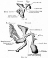
Fetal topographical anatomy of the pancreatic head and duodenum with special reference to courses of the pancreaticoduodenal arteries.[5]
A diagram showing joining processes between the dorsal and ventral primordia of the pancreas as well as the hypothetical rotation of the duodenum along a left-right axis. Viewed from the posterosuperior side of the body. A horizontal plane including most parts of the duodenum is shown to emphasize, in contrast to adults, the course of the second portion (D2) directing posteriorly rather than inferiorly.
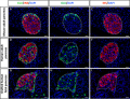
Developing Pancreatic Islets
| Model of endocrine cell and vessel organization in human islets[6]
|
A α-Cells (green) and β-cells (red) are organized into a thick folded plate lined at both sides with vessels (blue).
|

Adult Pancreatic Islets


The adult pancreatic islets (Islets of Langerhans) contain four distinct endocrine cell types.
Alpha Cells

- glucagon, mobilizes lipid
Beta Cells


- insulin, increase glucose uptake
- stimulate fetal growth, continue to proliferate to postnatal, in infancy most abundant
Delta Cells
- somatostatin, inhibits glucagon, insulin secretion
F-cells
- pancreatic polypeptide

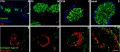
Rat - pancreatic islet development[7]
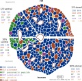
Islet size for Different Species
The following species comparison table has been slightly modified from Table 1 data in a recent paper by Kim etal., 2009.[8]
- Islet size is described as an effective diameter of a circle, which depicts the same area as a measured islet area.
- β-cell ratio is the area ratio of β-cells in an islet.
- Both data sets are expressed as the mean value with its standard deviation.
| Species | Age | Islet size (μm) | β-cell ratio |
| Human | 39 years (adult) | 50 ± 29 | 0.64 ± 0.21 |
| Monkey | 1 year | 67 ± 38* | 0.79 ± 0.14* |
| Pig | 6 month | 49 ± 15a | 0.89 ± 0.11* |
| Rabbit | 6 month | 64 ± 28* | 0.79 ± 0.17* |
| Bird | 40 day | 24 ± 6* | 0.46 ± 0.24* |
| Wild-type mouse | 6 month | 116 ± 80* | 0.85 ± 0.14* |
| Pregnant mouse | 3 month | 112 ± 94* | 0.84 ± 0.22* |
| ob/ob mouse | 15 week | 86 ± 76* | 0.92 ± 0.11* |
| db/db mouse | 15 week | 47 ± 24b | 0.53 ± 0.24c |
*p < 0.0001 ap = 0.65 bp = 0.42 cp = 0.0004 compared with human.
Hormones
Insulin
- Source - synthesized by the beta cells of the islets of Langerhans.
- Protein
- 2 dissimilar polypeptide chains, A and B, which are linked by 2 disulphide bonds.
- both chains are derived from a 1-chain precursor, proinsulin.
- proinsulin - converted to insulin by the enzymatic removal of a segment that connects the amino end of the A chain to the carboxyl end of the B chain.
Glucagon
Pancreas Histology


- Pancreas Histology Links: overview (label) | exocrine (label) | endocrine (label) | blood vessels (label) | insulin (label) | overview | exocrine | endocrine | blood vessels | insulin | Islet labeled for insulin and Glucagon | Insulin (Fl) | Glucagon (Fl) | GIT Histology
Diabetes

- Links: Maternal Diabetes
Abnormalities
Listed below are a number of pancreatic developmental abnormalities, see also the 2003 article "Lifetime consequences of abnormal fetal pancreatic development"[9].
Accessory Pancreatic Tissue - pancreatic tissue located in associated gastrointestinal tract tissues/organs such as the wall of the stomach, duodenum, jejunum or Meckel's diverticulum.
Annular Pancreas - (1 in 7,000 people) pancreas forms as a "ring" of tissue surrounding the duodenum which is subsequently narrowed.
Diabetes Mellitus - Maternal diabetes (and hyperglycaemia) have been shown to lead to increased fetal islet hyperplasia of the insulin producing beta cells and insulin secretion.
Intrauterine growth restriction - can lead to a delayed development of the insulin producing beta cells and low insulin secretion.
Tumours - Serous Cystadenoma (endocrine tumour), Somatostatinoma (tumour of delta cell origin), intraductal papillary-mucinous neoplasm
References
- ↑ <pubmed>18958289</pubmed>| PLoS ONE
- ↑ <pubmed>22084084</pubmed>| Proc Natl Acad Sci U S A.
- ↑ <pubmed>21909240</pubmed>| PLoS Biol.
- ↑ <pubmed>20377917</pubmed>
- ↑ <pubmed>20376893</pubmed>| Yonsei Med J.
- ↑ 6.0 6.1 <pubmed>20185817</pubmed>| PMC2857900 | Diabetes.
- ↑ <pubmed>19534767</pubmed>
- ↑ <pubmed>20606719</pubmed>
- ↑ <pubmed>12562919</pubmed>
Journals
- Pancreas The official journal of the American Pancreatic Association and the Japan Pancreas Society | PubMed
- Pancreatology Official Journal of the International Association of Pancreatology (IAP); European Pancreatic Club (EPC)and 16 other societies and study groups.
- Journal of the Pancreas electronic journal of pancreatology
- Diabetologia | PubMed
Online Textbooks
Endocrinology: An Integrated Approach Nussey, S.S. and Whitehead, S.A. Oxford, UK: BIOS Scientific Publishers, Ltd; 2001. table of Contents
NIH Genes & Disease Chapter 41 - Endocrine
Pathophysiology of the Endocrine System The Endocrine Pancreas
Developmental Biology (6th ed) Gilbert, Scott F. Sunderland (MA): Sinauer Associates, Inc.; c2000.
Molecular Biology of the Cell (4th Edn) Alberts, Bruce; Johnson, Alexander; Lewis, Julian; Raff, Martin; Roberts, Keith; Walter, Peter. New York: Garland Publishing; 2002. table 15-1. Some Hormone-induced Cell Responses Mediated by Cyclic AMP
Health Services/Technology Assessment Text (HSTAT) Bethesda (MD): National Library of Medicine (US), 2003 Oct.
Search NLM Online Textbooks- "pancreas development" : Endocrinology | Molecular Biology of the Cell | The Cell- A molecular Approach
Search Bookshelf Pancreas Development
Reviews
<pubmed>21084843</pubmed> <pubmed>19204986</pubmed> <pubmed>19013144</pubmed> <pubmed>18956314</pubmed> <pubmed>18676806</pubmed>
Articles
<pubmed>22110471</pubmed> <pubmed>22038519</pubmed> <pubmed>21926540</pubmed> <pubmed>21983268</pubmed> <pubmed>21537461</pubmed> <pubmed>20022941</pubmed> <pubmed>19365093</pubmed> <pubmed>19487809</pubmed> <pubmed>18810318</pubmed> <pubmed>18958289</pubmed> <pubmed>15072563</pubmed>| J Endocrinol.
Search Pubmed
Search April 2010
- Endocrine Development - All (14277) Review (4620) Free Full Text (3140)
Search Pubmed: pancreas development
Additional Images
Human - Pancreatic islet
Rat - pancreatic islet development
Pancreas_islet - structure human and rat

Historic Images
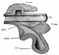
Fig. 273. From a model of the duodenum and the primary evaginations of the liver and pancreas in a 5 mm sheep embryo
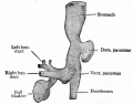
Fig. 274. From a reconstruction of the anlagen of the liver and pancreas and a part of the stomach and duodenum of a human embryo of 4 weeks
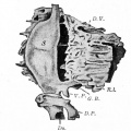
Fig. 275. From a reconstruction of the anlagen of the liver and pancreas and the stomach of a human embryo of 8 mm
Figs. 278 and 279. From models of the developing liver and pancreas of rabbit embryos
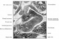
Fig. 280. From a transverse section through the region of the duodenum of a pig embryo of 14 mm
External Links
External Links Notice - The dynamic nature of the internet may mean that some of these listed links may no longer function. If the link no longer works search the web with the link text or name. Links to any external commercial sites are provided for information purposes only and should never be considered an endorsement. UNSW Embryology is provided as an educational resource with no clinical information or commercial affiliation.
- F1000 Reports - Recent advances in pancreas development
- Howard Hughes Medical Institute - Seung Kim Lab
Glossary Links
- Glossary: A | B | C | D | E | F | G | H | I | J | K | L | M | N | O | P | Q | R | S | T | U | V | W | X | Y | Z | Numbers | Symbols | Term Link
Cite this page: Hill, M.A. (2024, June 18) Embryology Endocrine - Pancreas Development. Retrieved from https://embryology.med.unsw.edu.au/embryology/index.php/Endocrine_-_Pancreas_Development
- © Dr Mark Hill 2024, UNSW Embryology ISBN: 978 0 7334 2609 4 - UNSW CRICOS Provider Code No. 00098G