Carnegie Stage 8 - "Dobbin" Embryo
| Embryology - 19 Jun 2024 |
|---|
| Google Translate - select your language from the list shown below (this will open a new external page) |
|
العربية | català | 中文 | 中國傳統的 | français | Deutsche | עִברִית | हिंदी | bahasa Indonesia | italiano | 日本語 | 한국어 | မြန်မာ | Pilipino | Polskie | português | ਪੰਜਾਬੀ ਦੇ | Română | русский | Español | Swahili | Svensk | ไทย | Türkçe | اردو | ייִדיש | Tiếng Việt These external translations are automated and may not be accurate. (More? About Translations) |
Introduction
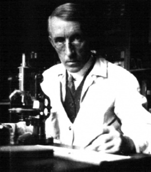
The historic "Dobbin" embryo was named after Dr Roy Dobbin (Cairo, Egypt) who originally provided the specimen to Dr James Hill.
This Carnegie stage 8 human embryo was 960 µm long and was subsequently serially sectioned and published in a series of papers by James Hill and Jan Florian and during the 1930's.[1][2][3]
The notes, photographs and sections are within the Hill Collection that form a part of the embryological collection relocated in 2004 to the Museum fur Naturkunde, Berlin and incorporated with the Hubrecht Collection.
- Links: Hill Embryo H91 | Carnegie stage 8 | Hill Collection | Dr James Hill | Hubrecht Collection | Museum fur Naturkunde - Embryological Collection
Description
The clinical history supplied by Dr. Dobbin is as follows: “ Coitus, 6.10.23 ; effort probably causing abortion, 21.10.23; first bleeding, 22.10.23; abortion (painless), 23.10.23." Although an abortion, we see no reason to regard the specimen as other than perfectly normal.
The chorionic vesicle (which was preserved in spirit) was, when received, somewhat flattened and shrunken. Except over a small area on one side (approximately 3 x 2 mm in diameter), which was almost bare, the vesicle possessed a fairly uniform covering of short, close~set, branched villi, to which at one point a small fragment of blood-clot adhered. Including the villi, its dimensions in alcohol were as follows : 11.5 mm (in long diameter) x 8.5 mm (in short diameter) x 45 mm (in thickness). After clearing in oil of cedar-wood, the corresponding internal diameters were 9 mm x 5.5 mm x 2.5 mm.
The vesicle, after being photographed and drawn, was dehydrated and cleared in oil of cedar-wood. A small portion of the chorion, including the bare area, was then carefully removed, and through the opening so made it was possible, fortunately enough, to locate the embryo under the binocular dissecting microscope. The embryo was then isolated along with the segment of the chorion to which it was attached, and stereo-photographs were successfully taken of it, in the cleared condition in oil of cedar-wood.
Hill catalogue number - (original) H159 | (new) H91
The original embryology catalogue numbers were consolidated and renumbered by Peter Hill's daughter, another embryologist.
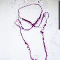
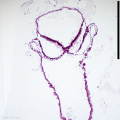
Embryo - Chorionic Vesicle
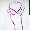
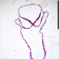
These are stereo photographs of the whole and opened vesicle before sectioning.
| Whole vesicle | |
|---|---|

|

|
| Opened vesicle | |

|

|
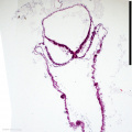
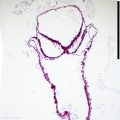
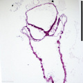
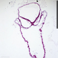
Histology
2-47
2-48
2-49
2-50
2-51
2-52
2-53
2-54
2-55
2-56
2-57


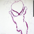
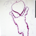
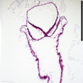
Transverse section, scale bar is 500 micron.
- Histology Slide: 2-47 | 2-48 | 2-49 | 2-50 | 2-51 | 2-52 | 2-53 | 2-54 | 2-55 | 2-56 | 2-57 | 2-70 | 2-75 | 2-80 | 2-89
References
- ↑ Hill JP. and Florian J. The development of head-process and prochordal plate in man (1931) J Anat. 65(2): 242-6. PMID 17104317
- ↑ Hill JP. and Florian J. A young human embryo (embryo dobbin) with head-process and prochordal plate. (1931) Phil. Tran. Roy. Soc. London B, 219: 443-486.
- ↑ Hill JP. and Florian J. Further note on the pro-chordal plate in man. (1931) J. Anat., 46: 46-47. PMID 17104356
Cite this page: Hill, M.A. (2024, June 19) Embryology Carnegie Stage 8 - "Dobbin" Embryo. Retrieved from https://embryology.med.unsw.edu.au/embryology/index.php/Carnegie_Stage_8_-_%22Dobbin%22_Embryo
- © Dr Mark Hill 2024, UNSW Embryology ISBN: 978 0 7334 2609 4 - UNSW CRICOS Provider Code No. 00098G