Category:Neural
From Embryology
This Embryology category shows pages and media related to Neural System Development. This includes related topics and undergraduate classes as well as pages and sub-categories describing specific components formed from the original ectoderm neural tube.
Subcategories
This category has the following 12 subcategories, out of 12 total.
Pages in category 'Neural'
The following 200 pages are in this category, out of 934 total.
(previous page) (next page)H
I
- Template:ICD-11 Anencephaly collapse table
- Template:ICD-11 Anencephaly table
- Template:ICD-11 Cerebral anomalies collapse table
- Template:ICD-11 Cerebral anomalies table
- Template:ICD-11 Cerebral palsy collapse table
- Template:ICD-11 Cerebral palsy table
- Template:ICD-11 Neural anomalies collapse table
- Template:ICD-11 Neural anomalies header table
- Template:ICD-11 Neural anomalies table
- Template:ICD-11 Rubella table
- Template:ICD-11 Spina bifida collapse table
- Template:ICD-11 Spina bifida table
- Interkinetic Nuclear Migration Movie
- Template:Iodine
- Template:Iodine deficiency
J
K
L
M
- McMurrich1914 Chapter 15
- Template:Medulla
- Template:Medulla oblongata
- Template:Medulloblastoma
- Template:Meninges
- Template:Mesencephalon
- Template:Metencephalon
- Template:Microcephaly
- Template:Microglia
- Template:Midbrain
- Template:Miller1913 table1
- Template:Miller1913 table2
- Template:Miller1913 table3
- Template:Miller1913 table4
- Template:Miller1913 table5
- Template:Miller1913 table6
- Template:Miller1913 table7
- Template:Miller1913 table8
- Template:Mouse neural tube 01 links
- Movie - Neural Tube Closure
- Movies
- Template:Multiple sclerosis
- Template:Myelencephalon
- Template:Myelination
N
- Template:Nanagas1925 figures
- Template:Neural
- Neural - Amygdala Development
- Neural - Basal Ganglia Development
- Neural - Cerebellum Development
- Neural - Cerebrum Development
- Talk:Neural - Cerebrum Development
- Neural - Cranial Nerve Development
- Neural - Diencephalon Development
- Neural - Epithalamus Development
- Neural - Hippocampus Development
- Neural - Medulla Oblongata Development
- Neural - Meninges Development
- Neural - Mesencephalon Development
- Neural - Metencephalon Development
- Neural - Myelencephalon Development
- Neural - Pons Development
- Neural - Prosencephalon Development
- Neural - Prosomere
- Neural - Rhinencephalon Development
- Neural - Rhombencephalon Development
- Neural - Spinal Cord Development
- Neural - Tectum Development
- Neural - Telencephalon Development
- Neural - Thalamus Development
- Neural - Vascular Development
- Neural - Ventricular System Development
- Neural 3D stage 13 Movie
- Neural Abnormal Exam - Newborn behaviour
- Neural Abnormal Exam - Newborn cranial nerve
- Neural Abnormal Exam - Newborn head circumference
- Neural Abnormal Exam - Newborn head shape and sutures
- Neural Abnormal Exam - Newborn positions - prone
- Neural Abnormal Exam - Newborn positions - ventral suspension
- Neural Abnormal Exam - Newborn positions - vertical suspension
- Neural Abnormal Exam - Newborn reflexes - deep tendon reflexes
- Neural Abnormal Exam - Newborn reflexes - Galant
- Neural Abnormal Exam - Newborn reflexes - grasp
- Neural Abnormal Exam - Newborn reflexes - Moro
- Neural Abnormal Exam - Newborn reflexes - plantar reflex
- Neural Abnormal Exam - Newborn reflexes - stepping
- Neural Abnormal Exam - Newborn reflexes - suck, root
- Neural Abnormal Exam - Newborn tone - arm recoil
- Neural Abnormal Exam - Newborn tone - arm traction
- Neural Abnormal Exam - Newborn tone - hand position
- Neural Abnormal Exam - Newborn tone - head control
- Neural Abnormal Exam - Newborn tone - head lag
- Neural Abnormal Exam - Newborn tone - heel to ear
- Neural Abnormal Exam - Newborn tone - leg recoil
- Neural Abnormal Exam - Newborn tone - leg traction
- Neural Abnormal Exam - Newborn tone - lower extremity
- Neural Abnormal Exam - Newborn tone - neck tone
- Neural Abnormal Exam - Newborn tone - popliteal angle
- Neural Abnormal Exam - Newborn tone - resting posture
- Neural Abnormal Exam - Newborn tone - scarf sign
- Neural Abnormal Exam - Newborn tone - upper extremity
- Template:Neural abnormalities
- Template:Neural cartoons
- Template talk:Neural cartoons
- Neural Crest - Enteric Nervous System
- Neural Crest - Peripheral Nervous System
- Neural Exam - 12 month Behaviour 1
- Neural Exam - 12 month Behaviour 2
- Neural Exam - 12 month Cranial Nerves
- Neural Exam - 12 month Head Circumference
- Neural Exam - 12 month Motor 1
- Neural Exam - 12 month Motor 2
- Neural Exam - 12 month Motor 3
- Neural Exam - 12 month Motor 4
- Neural Exam - 12 month Motor 5
- Neural Exam - 12 month Motor 6
- Neural Exam - 12 month Motor 7
- Neural Exam - 12 month Motor 8
- Neural Exam - 12 month Motor 9
- Neural Exam - 12 month reflexes - deep tendon reflexes
- Neural Exam - 12 month reflexes - plantar reflex
- Neural Exam - 12 month reflexes - postural reflex
- Neural Exam - 18 Behaviour 1
- Neural Exam - 18 Behaviour 2
- Neural Exam - 18 Behaviour 3
- Neural Exam - 18 Behaviour 4
- Neural Exam - 18 Cranial Nerves
- Neural Exam - 18 month Deep Tendon Reflex
- Neural Exam - 18 month Motor 1
- Neural Exam - 18 month Motor 2
- Neural Exam - 18 month Motor 3
- Neural Exam - 18 month Motor 4
- Neural Exam - 18 month Motor 5
- Neural Exam - 18 month Motor 6
- Neural Exam - 18 month Motor 7
- Neural Exam - 18 month Motor 8
- Neural Exam - 3 month - hand movements
- Neural Exam - 3 month - head and trunk control
- Neural Exam - 3 month - prone position
- Neural Exam - 3 month - supine position
- Neural Exam - 3 month behaviour
- Neural Exam - 3 month cranial nerves
- Neural Exam - 3 month positions - ventral suspension
- Neural Exam - 3 month positions - vertical suspension
- Neural Exam - 3 month reflexes - asymmetric tonic neck
- Neural Exam - 3 month reflexes - deep tendon reflexes
- Neural Exam - 3 month reflexes - Galant
- Neural Exam - 3 month reflexes - grasp
- Neural Exam - 3 month reflexes - Moro
- Neural Exam - 3 month reflexes - plantar reflex
- Neural Exam - 3 month reflexes - root
- Neural Exam - 3 month tone - lower extremity
- Neural Exam - 3 month tone - upper extremity
- Neural Exam - 30 month Behaviour 1
- Neural Exam - 30 month Behaviour 2
- Neural Exam - 30 month Behaviour 3
- Neural Exam - 30 month Behaviour 4
- Neural Exam - 30 month Behaviour 5
- Neural Exam - 30 month Behaviour 6
- Neural Exam - 30 month Deep Tendon Reflex
- Neural Exam - 30 month Motor 1
- Neural Exam - 30 month Motor 2
- Neural Exam - 30 month Motor 3
- Neural Exam - 30 month Motor 4
- Neural Exam - 30 month Motor 5
- Neural Exam - 30 month Motor 6
- Neural Exam - 30 month Motor 7
- Neural Exam - 6 month - hand movements
- Neural Exam - 6 month - head and trunk control
- Neural Exam - 6 month - prone position
- Neural Exam - 6 month - supine position
- Neural Exam - 6 month behaviour
- Neural Exam - 6 month cranial nerves
- Neural Exam - 6 month positions - ventral suspension
Media in category 'Neural'
The following 200 files are in this category, out of 1,070 total.
(previous page) (next page) PediNeuroLogic Intro 01.jpg 320 × 240; 9 KB
 PediNeuroLogic Intro 02.flv ; 147 KB
 PediNeuroLogic Intro 02.jpg 320 × 240; 14 KB
- PediNeuroLogic Intro 03.flv ; 468 KB
 PediNeuroLogic Intro 03.jpg 320 × 240; 11 KB
- PediNeuroLogic Intro 04.flv ; 407 KB
 PediNeuroLogic Intro 04.jpg 320 × 240; 21 KB
- PediNeuroLogic Intro 05.flv ; 923 KB
 PediNeuroLogic Intro 05.jpg 320 × 240; 6 KB
- PediNeuroLogic Intro 06.flv ; 872 KB
 PediNeuroLogic Intro 06.jpg 320 × 240; 16 KB
- PediNeuroLogic Intro 07.flv ; 294 KB
 PediNeuroLogic Intro 07.jpg 320 × 240; 11 KB
- PediNeuroLogic Intro 08.flv ; 781 KB
 PediNeuroLogic Intro 08.jpg 320 × 240; 13 KB
- PediNeuroLogic Larsen welcome.flv ; 5.3 MB
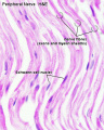
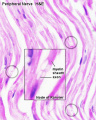
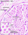 Peripheral nerve histology 01.jpg 640 × 800; 56 KB
 Peripheral nerve histology 02.jpg 640 × 800; 53 KB
 Peripheral nerve histology 03.jpg 640 × 800; 51 KB
 Peripheral nerve histology 04.jpg 640 × 800; 79 KB
 Peripheral nerve histology 05.jpg 640 × 800; 78 KB
 Postnatal cortex development trajectory.jpg 563 × 290; 37 KB
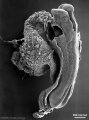
 Proboscis histology.jpg 600 × 1,041; 166 KB
 Rabbithhdrocephalus.jpg 795 × 485; 73 KB
 Rat neural cadherin 01.jpg 1,154 × 470; 62 KB
 Rat neural cadherin 02.jpg 991 × 502; 68 KB
 Rat neural cadherin 03.jpg 1,182 × 680; 58 KB
 Rat neural cadherin 04.jpg 792 × 749; 64 KB
 Rat neural cadherin 05.jpg 985 × 745; 67 KB
 Rat neural cadherin 06.jpg 546 × 479; 34 KB
 Rat neural cadherin 07.jpg 1,229 × 475; 62 KB
 Rat neural cadherin 08.jpg 597 × 678; 31 KB
 Rat thyroid system and neural development.jpg 1,032 × 740; 127 KB
 Rat-hindbrain E11.5.jpg 600 × 931; 164 KB
 Retinoid acid model in olfactory development.jpg 800 × 616; 58 KB
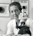 Rita Levi-Montalcini.jpg 500 × 545; 29 KB
 Rugh 021.jpg 1,000 × 626; 117 KB
 Rugh 092.jpg 846 × 1,000; 183 KB
 Rugh 117.jpg 1,000 × 805; 159 KB
 Rugh 118.jpg 1,000 × 692; 161 KB
 Rugh 132.jpg 735 × 1,000; 195 KB
 Rugh 133.jpg 1,000 × 707; 141 KB
 Rugh 134.jpg 1,200 × 764; 121 KB
 Schmidt-Lanterman cleft cartoon.jpg 703 × 600; 91 KB
 Schwann and axon interactions.jpg 1,280 × 807; 196 KB
 Screen Shot 2017-10-16 at 1.43.55 pm.png 1,183 × 535; 704 KB
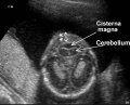 Second Trimester Cerebellum.jpeg 475 × 385; 26 KB
 Secondary neurulation 01.mov ; 445 KB
 Sensenig1951 plate01.jpg 1,979 × 2,591; 1.35 MB
 Sensenig1951 plate02.jpg 2,078 × 2,619; 1.52 MB
 Sensenig1951 plate03.jpg 1,963 × 2,664; 1.33 MB
 Sensenig1951 plate04.jpg 1,990 × 2,627; 1.29 MB
 Shanklin1940 fig01.jpg 1,000 × 657; 136 KB
 Shanklin1940 fig02.jpg 1,000 × 682; 131 KB
 Shanklin1940 fig04.jpg 884 × 1,018; 112 KB
 Shanklin1940 fig06.jpg 1,280 × 837; 92 KB
 Shanklin1940 fig07.jpg 1,280 × 1,080; 125 KB
 Shanklin1940 plate01.jpg 1,522 × 2,138; 362 KB
 Shanklin1940 plate02.jpg 1,411 × 2,131; 210 KB
 Sonic hedgehog expression.jpg 150 × 150; 7 KB
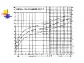
 Special educational need by gestational age.jpg 600 × 443; 19 KB
 Spina Bifida 1.jpg 393 × 599; 49 KB
 Spina bifida front.JPG 477 × 550; 52 KB
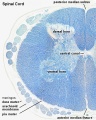
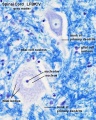
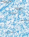
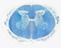
 Spinal cord histology 01.jpg 480 × 600; 116 KB
 Spinal cord histology 02.jpg 480 × 600; 121 KB
 Spinal cord histology 03.jpg 480 × 600; 103 KB
 Spinal cord histology 04.jpg 480 × 600; 119 KB
 Spinal cord histology 05.jpg 1,280 × 1,024; 463 KB
 Spinal cord histology 06.jpg 1,280 × 1,024; 318 KB
 Spinal cord histology 07.jpg 1,280 × 1,024; 365 KB
 Spinal cord histology 08.jpg 1,280 × 1,024; 418 KB
 Spinal cord histology 09.jpg 1,280 × 1,024; 227 KB
 Spinal cord histology 10.gif 480 × 600; 456 KB
 Spinal cord histology 11.jpg 481 × 600; 117 KB
 Spinal cord histology 12.jpg 480 × 600; 130 KB
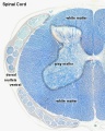
 Spinal cord tracts.png 1,000 × 430; 204 KB
 Stage 10 historic-Corner1929-1.jpg 654 × 1,000; 145 KB
 Stage 10 historic-Corner1929-1a.jpg 523 × 800; 87 KB
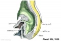
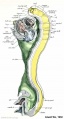 Stage 11 historic-Atwell1930-1.jpg 538 × 1,000; 75 KB
 Stage 11 historic-Atwell1930-1a.jpg 430 × 800; 46 KB
 Stage 11 historic-Atwell1930-1b.jpg 323 × 600; 26 KB
 Stage 11 historic-Atwell1930-1c.jpg 215 × 400; 14 KB
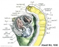
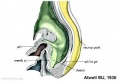
 Stage 11 historic-Atwell1930-2.jpg 800 × 639; 87 KB
 Stage 11 historic-Atwell1930-2a.jpg 800 × 639; 87 KB
 Stage 11 historic-Atwell1930-2b.jpg 600 × 479; 50 KB
 Stage 11 historic-Atwell1930-2c.jpg 400 × 319; 23 KB
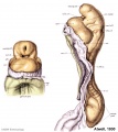
 Stage 11 historic-Atwell1930-3.jpg 1,000 × 679; 87 KB
 Stage 11 historic-Atwell1930-3a.jpg 800 × 543; 57 KB
 Stage 11 historic-Atwell1930-3b.jpg 600 × 407; 32 KB
 Stage 11 historic-Atwell1930-3c.jpg 400 × 271; 15 KB
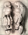
 Stage 11 historic-Atwell1930-4.jpg 1,000 × 1,121; 114 KB
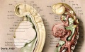
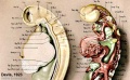
 Stage 11 historic-Davis1923-1.jpg 804 × 1,000; 87 KB
 Stage 11 historic-Davis1923-1a.jpg 643 × 800; 61 KB
 Stage 11 historic-Davis1923-1b.jpg 482 × 600; 41 KB
 Stage 11 historic-Davis1923-1c.jpg 321 × 400; 20 KB
 Stage 11 historic-Davis1923-2.jpg 768 × 1,000; 128 KB
 Stage 11 historic-Davis1923-2a.jpg 614 × 800; 83 KB
 Stage 11 historic-Davis1923-2b.jpg 461 × 600; 48 KB
 Stage 11 historic-Davis1923-2c.jpg 307 × 400; 23 KB
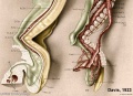
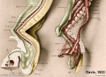
 Stage 11 historic-Davis1923-3.jpg 1,000 × 615; 84 KB
 Stage 11 historic-Davis1923-3a.jpg 800 × 492; 59 KB
 Stage 11 historic-Davis1923-3b.jpg 600 × 369; 41 KB
 Stage 11 historic-Davis1923-3c.jpg 400 × 246; 22 KB
 Stage 11 historic-Davis1923-4.jpg 1,000 × 722; 90 KB
 Stage 11 historic-Davis1923-4a.jpg 800 × 578; 63 KB
 Stage 11 historic-Davis1923-4b.jpg 600 × 434; 39 KB
 Stage 11 historic-Davis1923-4c.jpg 400 × 289; 21 KB
 Stage 11 historic-Heuser1930-1.jpg 521 × 1,000; 94 KB
 Stage 11 historic-Heuser1930-1a.jpg 417 × 800; 57 KB
 Stage 11 historic-Heuser1930-1b.jpg 313 × 600; 30 KB
 Stage 11 historic-Heuser1930-1c.jpg 209 × 400; 15 KB
 Stage 13 image 001.jpg 1,000 × 357; 44 KB
 Stage 13 image 002.jpg 1,000 × 359; 52 KB
 Stage 13 image 003.jpg 1,000 × 436; 71 KB
 Stage 13 image 004.jpg 1,000 × 386; 65 KB
 Stage 13 image 005.jpg 1,000 × 451; 81 KB
 Stage 13 image 006.jpg 1,000 × 439; 83 KB
 Stage 13 image 007.jpg 1,000 × 514; 93 KB
 Stage 13 image 022.jpg 1,000 × 473; 101 KB
 Stage 13 image 023.jpg 1,000 × 544; 110 KB
 Stage 13 image 050.jpg 1,000 × 370; 52 KB
 Stage 13 image 051.jpg 1,000 × 382; 55 KB
 Stage 13 image 052.jpg 1,000 × 476; 81 KB
 Stage 13 image 053.jpg 1,000 × 423; 76 KB
 Stage 13 image 056.jpg 1,000 × 516; 102 KB
 Stage 13 image 057.jpg 1,000 × 511; 99 KB
 Stage 13 image 058.jpg 1,000 × 481; 94 KB
 Stage 13 image 059.jpg 1,000 × 513; 92 KB
 Stage 13 image 060.jpg 1,000 × 486; 96 KB
 Stage 13 image 061.jpg 1,000 × 600; 101 KB
 Stage 13 image 098.jpg 1,000 × 623; 144 KB
 Stage 22 image 050.jpg 1,000 × 650; 128 KB
 Stage 22 image 051.jpg 1,000 × 653; 113 KB
 Stage 22 image 052.jpg 1,000 × 665; 177 KB
 Stage 22 image 053.jpg 1,000 × 687; 132 KB
 Stage 22 image 054.jpg 1,000 × 636; 119 KB
 Stage 22 image 055.jpg 1,000 × 632; 145 KB
 Stage 22 image 056.jpg 1,000 × 635; 138 KB
 Stage 22 image 065.jpg 1,000 × 637; 100 KB
 Stage 22 image 152.jpg 1,000 × 670; 127 KB
 Stage 22 image 153.jpg 1,000 × 662; 191 KB
 Stage 22 image 155.jpg 1,000 × 672; 239 KB
 Stage 22 image 157.jpg 1,000 × 655; 148 KB
 Stage 22 image 162.jpg 1,000 × 657; 176 KB
 Stage 22 image 176.jpg 1,000 × 659; 223 KB
 Stage 22 image 205.jpg 1,112 × 909; 261 KB
 Stage 22 image 206.jpg 1,200 × 754; 245 KB
 Stage 22 image 207.jpg 1,200 × 753; 208 KB
 Stage 22 image 208.jpg 1,200 × 903; 368 KB
 Stage 22 image 209.jpg 1,200 × 808; 305 KB
 Stage 22 image 211.jpg 1,200 × 760; 242 KB
 Stage 22 image 212.jpg 1,200 × 753; 269 KB
 Stage 22 image 217.jpg 1,200 × 820; 323 KB
 Stage 22 image 219.jpg 1,250 × 892; 295 KB
 Stage 22 image 222.jpg 1,180 × 999; 225 KB
 Stage 22 image 322.jpg 1,180 × 999; 254 KB
 Stage 22 vomeronasal organ.jpg 600 × 554; 125 KB
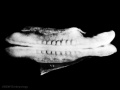 Stage10 bf5.jpg 1,000 × 750; 54 KB
 Stage10 bf6.jpg 1,000 × 750; 91 KB
 Stage10 bf6a.jpg 800 × 600; 63 KB
 Stage10 bf6b.jpg 600 × 450; 41 KB
 Stage10 bf6c.jpg 400 × 300; 22 KB
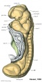
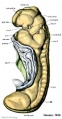
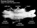
 Stage10 neural sm.jpg 665 × 499; 22 KB
 Stage10 sem10.jpg 1,000 × 740; 68 KB
 Stage10 sem10a.jpg 800 × 592; 48 KB
 Stage10 sem10b.jpg 600 × 444; 32 KB
 Stage10 sem10c.jpg 400 × 296; 16 KB
 Stage10 sem2.jpg 1,000 × 590; 60 KB
 Stage10 sem5.jpg 845 × 1,000; 83 KB
 Stage10 sem6 annotated.jpg 720 × 960; 122 KB
 Stage10 sem6.jpg 614 × 1,000; 57 KB
 Stage10 sem7.jpg 1,243 × 1,000; 116 KB
 Stage10 sem9.jpg 740 × 1,000; 72 KB
 Stage10 sem9a.jpg 592 × 800; 51 KB
 Stage10 sem9b.jpg 444 × 600; 32 KB
 Stage10 sem9c.jpg 296 × 400; 16 KB
 Stage11 histology-neural tube roof plate 1.jpg 800 × 594; 134 KB
 Stage11 histology-neural tube roof plate 2.jpg 800 × 552; 145 KB
 Stage11 histology-optic vesicle-hindbrain.jpg 800 × 536; 158 KB
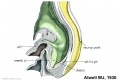
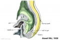
 Stage11 historic-Atwell1930-2.jpg 1,000 × 799; 131 KB
 Stage11 sem100.jpg 1,000 × 898; 109 KB
 Stage11 sem100c.jpg 400 × 359; 30 KB
 Stage11 sem101.jpg 1,000 × 898; 175 KB
 Stage11 sem11c.jpg 311 × 400; 32 KB
 Stage11 sem21.jpg 600 × 447; 79 KB
 Stage11 sem7a.jpg 729 × 1,000; 141 KB
 Stage11 sem9.jpg 1,203 × 1,653; 301 KB
 Stage11 sem9a.jpg 728 × 1,000; 145 KB
 Stage12 sem1.jpg 472 × 1,000; 69 KB
 Stage12 sem1a.jpg 378 × 800; 24 KB
 Stage12 sem1b.jpg 284 × 600; 15 KB
 Stage12 sem1c.jpg 189 × 400; 8 KB
 Stage12 sem2.jpg 1,787 × 2,303; 313 KB
 Stage12 sem2a.jpg 776 × 1,000; 100 KB
 Stage12 sem2b.jpg 621 × 800; 73 KB
 Stage12 sem2c.jpg 466 × 600; 47 KB
 Stage12 SEM3.jpg 507 × 600; 68 KB
 Stage12 sem3.jpg 1,462 × 2,253; 297 KB
 Stage12 sem3a.jpg 649 × 1,000; 95 KB
 Stage12 sem3b.jpg 519 × 800; 67 KB


