Early Circulation
Three components of early circulation
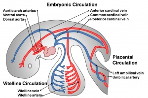
The vascular system of the embryo is formed from blood islands that appear in the extraembryonic mesoderm of the yolk sac and the embryonic mesoderm (primarily splanchnic mesoderm). Both of these clusters fuse together and extend, forming a vast network.
The early circulation has 3 components: Vitelline, Embryonic, Placental (each of these has its own system of arteries and veins).
- Vitelline - the vitelline arteries branch off the dorsal aortas and enter the yolk sac, covering its entire surface. The vitelline veins return red blood cells from the capillary beds to the sinus venosus, posterior to the heart. The vitelline vessels eventually contribute to the portal system of the liver in the adult.
- Embryonic - blood from the dorsal aorta enters intersegmental arteries, including the arteries of the pharyngeal arches. The blood returns to the heart via the anterior and posterior cardinal veins.
- Placental - the umbilical arteries receive blood from the aorta. This is carried to the chorionic villi, where exchange occurs with the mother. Waste products are disposed of, nutrients and oxygen are collected, and then the umbilical veins convey the blood to the sinus venosus.
Heart
The heart develops from cardiogenic mesoderm, a region of splanchnic mesoderm lying above the buccopharyngeal membrane. Development begins in week 3 with the formation of a pair of heart tubes. These fuse and form a single tube in week 4, as a result of the embryonic folding processes that occur. As the heart grows, septation events occur, transforming it into a 4-chambered pump. Initially, the ventricles develop above the atria; however simultaneous growth and bending of the tube bring the structures into correct position. In humans, the heart begins to beat on day 22-23. (This will be covered in detail in a later Lab)
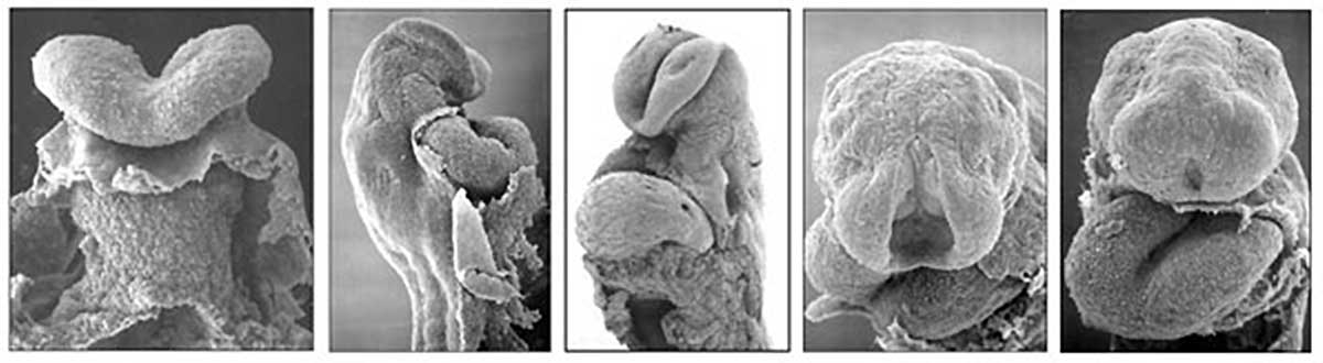
Scanning electron micrograph images of the early human (day 21 to 25) embryonic heart tube. Note the anterior body wall has been removed exposing the pericardial cavity in which the heart tube lies.
Cardiovascular Movies
We will not be covering later heart development, including septation, in today's practical.
Systemic Circulation
Compare the mid-embryonic cardiovascular system with that existing at the end of embryonic development. Then follow blood flow through the embryo using the Stage 13 embryo cross-sections.
About Stage 13 Embryo Sections - This image is from a serial section of a 6mm CRL pig embryo with some features of the Stage 14 embryo. This embryo is approximately equal to the day 42 human embryo. Use these serial images to identify internal features and relationships that exist within the embryo at this stage. Then compare these images with the later features of the Carnegie stage 22 human embryo.
- Links: Carnegie stage 13 - serial sections | Carnegie stage 13 | Embryo Serial Sections


