2010 Lecture | placentation | gastrulation | Notochord
Introduction
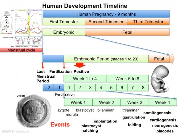
In this lecture I will talk about week 3 of embryonic development. I will cover early placentation, gastrulation and embryonic folding. The PDF for this lecture can be found here: File:ANAT2341 Lecture 3 - Beverdam - Week 3.pdf
Objectives
- Understand broadly the events of week 3 of human development
- Understand the process early placentation, villi formation
- Understand the process of gastrulation
- Understand the process of axis formation
- Brief understanding of folding
Textbooks
UNSW Embryology

|
Hill, M.A. (2011) UNSW Embryology (11th ed.). Sydney:UNSW.
|
The Developing Human: Clinically Oriented Embryology
|
Citation: The Developing Human: clinically oriented embryology 9th ed. Keith L. Moore, T.V.N. Persaud, Mark G. Torchia. Philadelphia, PA: Saunders, 2011. (links available to UNSW students)
|
Larsen's Human Embryology
|
Citation: Larsen's human embryology 4th ed. Schoenwolf, Gary C; Larsen, William J, (William James). Philadelphia, PA : Elsevier/Churchill Livingstone, c2009. (links available to UNSW students)
|
Movies
Early Placentation
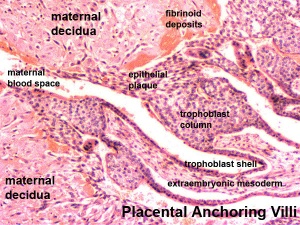
Early placenta anchoring villi
The trophoblast layer has now differentiated into two morphologically distinct cellular layers.
- Syncitiotrophoblasts - form a multinucleated cytoplasmic mass by cytotrophoblast cell fusion and both invade the decidua and secrete hCG
- Cytotrophoblasts - form a cellular layer around the blastocyst, proliferates and extends behind syncitiotrophoblasts
Early Utero-Placental exchange
- transfer of nutrition from maternal lacunae filled with secretions from uterine glands and maternal blood from blood vessels.
- development of trophoblast villi extending into the uterine decidua.
- ongoing process with placental development (covered in future lecture)
There are three stages of villi development:
- Primary Villi - cytotrophoblast
- Secondary Villi - cytotrophoblast + extraembryonic mesoderm
- Tertiary Villi - cytotrophoblast + extraembryonic mesoderm+ blood vessels
There are two main types of early villi:
- Anchoring villi - attached to decidua
- Floating villi - not attached to decidua, floating in maternal lacunae.
- Links: Development Animation - Implantation
Gastrulation
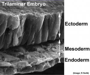
Embryonic Disc
- Gastrulation, (Greek = belly) means the formation of gut.
- used in a more looser sense to to describe the formation of the trilaminar embryo.
- Epiblast layer - consists of totipotential cells forming all 3 embryo layers (germ layers) that form the entire embryo.
- Historically, gastrulation was one of the earliest observable morphological event occurring in the frog embryo.
- Currently, the molecular and physical mechanisms that regulate patterning and migration during this key event are being investigated in several different animal models.
- In humans, it is proposed that similar mechanisms regulate gastrulation to those found in other vertebrates.
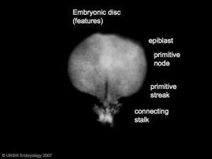
Embryonic Disc showing primitive streak
|
|
|
- Ectoderm (epithelium) - forms the central and peripheral nervous system, parts of the sensory systems, and the epithelium of the skin.
- Mesoderm (connective tissue) - forms the body connective tissues: blood, bone, muscle, connective tissue skin, gastrointestinal and respiratory tracts.
- Endoderm (epithelium) - forms gastrointestinal tract organs and the epithelium of the gastrointestinal and respiratory tracts.
- This is a very simplified explanation of the 3 layer contributions, many body tissues have contributions from all 3 origins.
|
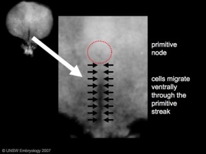
Primitive Streak
The primitive streak is the visible feature which represents the site of cell migration to form the additional layers.
- Midline of embryonic disc running between the primitive node to near the edge of the disc at the caudal end (near the connecting stalk).
- Region of cell migration from the epiblast layer forming sequentially the two germ cell layers (endoderm and mesoderm).
- Cells that do not migrate form the ectoderm.
|
|
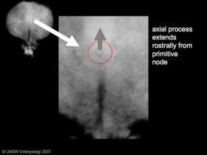
Primitive node
- (Hensen's node) region in the middle of the early embryonic disc epiblast from which the primitive streak extends caudally (tail).
- nodal cilia establish the embryo left/right axis
- axial process extends from the nodal epiblast
|
|
|
- Links: Development Animation - Mesoderm | Gastrulation
Epithelial to Mesenchymal Transition
- Epithelial cells (organised cellular layer) which loose their organisation and migrate/proliferate as a mesenchymal cells (disorganised cellular layers) are said to have undergone an Epithelial Mesenchymal Transition (EMT).
- Mesenchymal cells have an embryonic connective tissue-like cellular arrangement
- cells that have undergone this process may at a later time and under specific signaling conditions undergo the opposite process, mesenchyme to epithelia.
- In development, this process can be repeated several times during tissue differentiation.
This process occurs at the primitive streak where epiblast cells undergo an epithelial to mesenchymal transition in order to delaminate and migrate.
MH - there are a number of common cellular changes that occur during embryonic development at different times and in different tissues, which we can classify into "developmental mechanisms".
Notochord
- The notochord is a structure which has an early mechanical role in embryonic disc folding and a major signaling role in patterning surrounding embryonic tissue development.
- A developmental feature not present in the adult anatomy.
- This signaling role patterns many different tissues (neural plate, neural tube, somites, endodermal organs).
- Has own sequence of development from a primitive axial process
- axial process an initial epiblast hollow epithelial tube which extends in the midline from the primitive pit, cranially in the embryonic disc (toward the oral membrane).
- neuroenteric canal is a transient communication between the amnionic cavity and the yolk sac cavity formed by the axial process.
- notochordal plate forms from the axial process merging with the endoderm layer.
- notochord forms from the notochordal plate which then separates back into the mesoderm layer as a solid column of cells lying in the midline of the embryonic disc and running rostro-caudally (head to tail).
- An alternate name for the notochord is "axial mesoderm".
MH - Much of our knowledge of this structure comes from the study of animal models of development.
- Links: Development Animation - Notochord | Mesoderm
Embryo Folding
- embryonic disc now has 3 germ layers (ectoderm, mesoderm, endoderm)
- this flat 3 layer disc then begins to fold ventrally at the edges
- eventually forming a "hollow tube" of these 3 germ layers
|
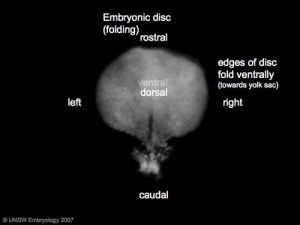
Embryonic Disc Folding - all edges of the embryonic disc will fold ventrally, forming a rostro-caudal "C" shaped tube.
|
|
Timeline
- 2012 Course: Week 1 Lecture 1 Lecture 2 Lab 1 | Week 2 Lecture 3 Lecture 4 Lab 2 | Week 3 Lecture 5 Lecture 6 Lab 3 | Week 4 Lecture 7 Lecture 8 Lab 4 | Week 5 Lecture 9 Lecture 10 Lab 5 | Week 6 Lecture 11 Lecture 12 Lab 6 | Week 7 Lecture 13 Lecture 14 | Lab 7 | Week 8 Lecture 15 Lecture 16 Lab 8 | Week 9 Lecture 17 Lecture 18 Lab 9 | Week 10 Lecture 19 Lecture 20 Lab 10 | Week 11 Lecture 21 Lecture 22 Lab 11 | Week 12 Lecture 23 Lecture 24 Lab 12
Glossary Links
- Glossary: A | B | C | D | E | F | G | H | I | J | K | L | M | N | O | P | Q | R | S | T | U | V | W | X | Y | Z | Numbers | Symbols | Term Link
Cite this page: Hill, M.A. (2024, June 14) Embryology Lecture - Week 3 Development. Retrieved from https://embryology.med.unsw.edu.au/embryology/index.php/Lecture_-_Week_3_Development
- What Links Here?
- © Dr Mark Hill 2024, UNSW Embryology ISBN: 978 0 7334 2609 4 - UNSW CRICOS Provider Code No. 00098G


