Gastrointestinal Tract - Intestine Development: Difference between revisions
| Line 78: | Line 78: | ||
{| | {| | ||
|-bgcolor="CEDFF2" | |-bgcolor="CEDFF2" | ||
| width 200px|'''Age''' (weeks gestational age) | | width=200px|'''Age''' (weeks gestational age) | ||
| width 200px|'''Average Length''' (cm) | | width=200px|'''Average Length''' (cm) | ||
|- | |- | ||
| 20 | | 20 | ||
| Line 103: | Line 103: | ||
|} | |} | ||
Table data <ref><pubmed>1752463</pubmed>| [http://www.ncbi.nlm.nih.gov/pmc/articles/PMC1379160 PMC1379160] | [http://gut.bmj.com/content/32/11/1321.long Gut.]</ref> | Table data <ref><pubmed>1752463</pubmed>| [http://www.ncbi.nlm.nih.gov/pmc/articles/PMC1379160 PMC1379160] | [http://gut.bmj.com/content/32/11/1321.long Gut.]</ref> based upon 8 published reports of necropsy measurement of 1010 guts. | ||
==Abnormalities== | ==Abnormalities== | ||
Revision as of 17:19, 2 May 2011
Introduction

The part of the gastrointestinal tract (GIT) lying between the stomach and anus, is described as the intestines or bowel. This region is further divided anatomically and functionally into the small intestine or bowel (duodenum, jejunum and ileum) and large intestine or bowel (cecum and colon). Initially development concerns the midgut region, connected to the yolk sac, and the hindgut region, ending at the cloacal membrane. This is followed by two mechanical processes of elongation and rotation. Elongation, growth in length, leaves the midgut "herniated" at the umbilicus and external to the abdomen. Rotation, around a mesentery axis, establishes the anatomical position of the large intestine within the peritoneal space.
Migration of neural crest cells into the wall establishes the enteric nervous system, which has a role in peristalsis and secretion. Prenatally, secretions also accumulate in this region and are the first postnatal bowel movement, the meconium.
Like most of the gut, this region is not "functional" until after birth, when development continues by populating the large intestine with commensal bacteria and the establishment of the immune structure in the wall.
Some Recent Findings
|
Adult Intestine

Intestinal Regions
Small intestine or bowel
- Duodenum (adult 25 cm length)
- Jejunum (adult 1.4 m length)
- Ileum (adult 3.5 m length)
Large intestine or bowel
- Cecum (caecum)
- Vermiform appendix ("appendix", adult 2 to 20 cm length)
- Colon
- Ascending colon (adult 25 cm length)
- Transverse colon
- Descending colon
- Sigmoid colon
Intestinal Functions
Small Intestine
- absorption of nutrients and minerals found in food
- Duodenum -principal site for iron absorption
Cecum
- connects the ileum with the ascending colon
- separated by the ileocecal valve (ICV, Bauhin's valve)
- connected to the vermiform appendix ("appendix")
Colon
- absorbs fluid, water and salts, from solid wastes
- site of commensal bacteria (flora) fermentation of unabsorbed material
Embryonic Development
Week 4
| Quicktime | Flash |
Week 8
| Quicktime | Flash |


Late embryonic small intestine commencing at the duodenum, continuing as ventrally herniated and returning to join the colon.
- Links: Carnegie stage 22 | Week 8
Small Intestine Length
| Age (weeks gestational age) | Average Length (cm) |
| 20 | 125 |
| 30 | 200 |
| term | 275 |
| 1 year postnatal | 380 |
| 5 years | 450 |
| 10 years | 500 |
| 20 years | 575 |
Table data [2] based upon 8 published reports of necropsy measurement of 1010 guts.
Abnormalities
- Abnormality Links: Gastrointestinal Tract - Abnormalities | Intestine Development | Gastrointestinal Tract
- Lumen Abnormalities: Image - Duplication sites | Pyloric atresia | Jejunal atresia
- Rotation: Image - Midgut volvulus | Image - Intestinal malrotation | Image - Cecal volvulus | Image - Sigmoid volvulus | Ladd's band
- Meckel's Diverticulum: Meckel's Image 1 | Meckel's Image 2 | Meckel's Image 3 |
- Intestinal Aganglionosis: Image - Ostomy | Image - Stoma | Surgery 1 | Surgery 2 | Surgery 3
Cite this page: Hill, M.A. (2024, June 14) Embryology Gastrointestinal Tract - Intestine Development. Retrieved from https://embryology.med.unsw.edu.au/embryology/index.php/Gastrointestinal_Tract_-_Intestine_Development
- © Dr Mark Hill 2024, UNSW Embryology ISBN: 978 0 7334 2609 4 - UNSW CRICOS Provider Code No. 00098G
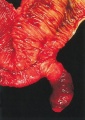
Meckel's diverticulum
Meckel's diverticulum
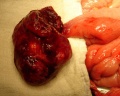
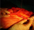
Meckel's diverticulum and tumor
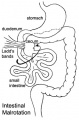
Intestinal malrotation
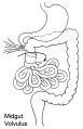
midgut volvulus
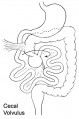
cecal volvulus
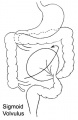
sigmoid volvulus
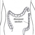
Megacolon
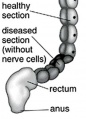
Megacolon


Appendix Duplication
Appendix duplication is an extremely rare congenital anomaly (0.004% to 0.009% of appendectomy specimens) first classified according to their anatomic location by Cave in 1936[3] and a later modified by Wallbridge in 1963[4], subsequently two more types of appendix abnormalities have been identified.[5][6]
Modified Cave-Wallbridge Classification (table from[7])
| Classification of types of appendix duplication |
Features |
| A | Single cecum with various degrees of incomplete duplication |
| B1 (bird type) | Two appendixes symmetrically placed on either side of the ileocecal valve |
| B2 (tenia coli type) | ne appendix arises from the cecum at the usual site, and the second
appendix branches from the cecum along the lines of the tenia at various distances from the first |
| B3 | One appendix arises from the usual site, and the second appendix arises from
the hepatic flexura |
| B4 | One appendix arises from the usual site, and the second appendix arises from
the splenic flexura |
| C | Double cecum, each with an appendix |
| Horseshoe appendix | One appendix has two openings into a common cecum |
| Triple appendix | One appendix arises from the cecum at the usual site, and two additional appendixes arise from the colon |
Molecular Factors
- Cdx (Caudal-type homeobox) group of ParaHox genes (mouse Cdx1, Cdx2 and Cdx4)[8]
- FGF9
References
Reviews
Articles
Search Pubmed
Search Bookshelf Intestine Development
Search Pubmed Now: Intestine Embryology | Intestine Development
Glossary Links
- Glossary: A | B | C | D | E | F | G | H | I | J | K | L | M | N | O | P | Q | R | S | T | U | V | W | X | Y | Z | Numbers | Symbols | Term Link
Cite this page: Hill, M.A. (2024, June 14) Embryology Gastrointestinal Tract - Intestine Development. Retrieved from https://embryology.med.unsw.edu.au/embryology/index.php/Gastrointestinal_Tract_-_Intestine_Development
- © Dr Mark Hill 2024, UNSW Embryology ISBN: 978 0 7334 2609 4 - UNSW CRICOS Provider Code No. 00098G