Second Trimester: Difference between revisions
| Line 7: | Line 7: | ||
:{{Template:Fetal Links}}| [[:Category:Second Trimester|Category:Second Trimester]] | :{{Template:Fetal Links}} | [[:Category:Second Trimester|Category:Second Trimester]] | ||
==Neural Development== | ==Neural Development== | ||
Revision as of 23:33, 22 February 2011
Introduction


Ultrasound Image of an early Fetus (12 week)
| Fetal Links: fetal | Week 10 | Week 12 | second trimester | third trimester | fetal neural | Fetal Blood Sampling | fetal growth restriction | birth | birth weight | preterm birth | Developmental Origins of Health and Disease | macrosomia | BGD Practical | Medicine Lecture | Science Lecture | Lecture Movie | Category:Human Fetus | Category:Fetal | |||
|
Neural Development
The images below are from a recent MRI study of fixed fetal brains at different weeks of development during the second trimester.[1]
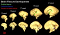
Three-dimensional reconstruction of the lateral (top row) and medial (bottom row) surface of 13–21 week brains to reveal the development of the Sylvian fissure or lateral sulcus (green arrow), the calcarine fissure (blue arrow), and the parieto-occipital sulcus (red arrow), respectively.
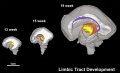
3D depiction of developmental white matter fibers. A lateral view of limbic tracts where pink fibers in 13, 15, and 19 week brains are the fornix and stria terminalis and purple fibers in the 19 week brains indicate the cingulum bundle.
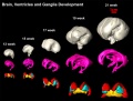
Three-dimensional reconstruction of the basal ganglia and ganglionic eminence. Different colors represent different brain structures: whole brain (gray), ventricle (pink), ganglionic eminence (red), putamen and globus pallidus together (cyan), thalamus (yellow), and caudate nucleus (green).
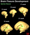
Three-dimensional reconstruction of the lateral (top row) and medial (bottom row) surface of 13–21 week brains to reveal the development of the Sylvian or lateral fissure (green arrow).


Second Trimester Timeline
(Clinical Week 14)
| Event | ||
| Clinical second trimester |  Week 12 - CRL 85 mm, femur length 15 mm, biparietal diameter 25 mm Week 12 - CRL 85 mm, femur length 15 mm, biparietal diameter 25 mm
Hearing Week 12-16 - Capsule adjacent to membranous labrynth undegoes vacuolization to form a cavity (perilymphatic space) around membranous labrynth and fills with perilymph Genital male and female external genital differences observable Respire Month 3-6 - lungs appear glandular, end month 6 alveolar cells type 2 appear and begin to secrete surfactant Tongue Week 12 - first differentiated epithelial cells (Type II and III) female genital canal (80 days) formed with absorption of the median septum | |
| Tongue Week 12 to 13 - maximum synapses between cells and afferent nerve fibers
| ||
| Tongue Week 14 to 15 - taste pores develop, mucous | ||
| Pancreas glucagon detectable in fetal plasma | ||
| 14 cm | Hearing Week 16-24 - Centres of ossification appear in remaining cartilage of otic capsule form petrous portion of temporal bone. Continues to ossify to form mastoid process of temporal bone.
Pituitary adenohypophysis fully differentiated Respire Week 16 to 25 lung histology - canalicular Skin 4 months - basal cell- proliferation generates folds in basement membrane; neural crest cells- (melanocytes) migrate into epithelium; embryonic connective tissue- differentiates into dermis, a loose ct layer over a dense ct layer. Beneath the dense ct layer is another loose ct layer that will form the subcutaneous layer. Ectoderm contributes to nails, hair follictles and glands. Nails form as thickening of ectoderm epidermis at the tips of fingers and toes. These form germinative cells of nail field. Cords of these cells extend into mesoderm forming epithelial columns. These form hair follocles, sebaceous and sweat glands. primary follicles begin to form in the ovary and are characterized by an oocyte glandular urethra forms and skin folds present | |
| Tongue Week 18 - substance P detected in dermal papillae, not in taste bud primordia
Skin vernix caseosa covers skin Spleen SMA-positive reticulum cells increase in number and begin to form a reticular framework. PMID: 1925578 | ||
| Pituitary week 20 to 24 growth hormone levels peak, then decline
Skin lanugo, skin hair Skin 5 months - Hair growth initiated at base of cord, lateral outgrowths form associated sebaceous glands; Other cords elongate and coil to form sweat glands; Cords in mammary region branch as they elongate to form mammary glands. Uterus Development uterine horn fimbrial development begins and continues after birth | ||
| Neural brain cortical sulcation - sylvian fissure, interhemispheric fissure, callosal sulcus, parietooccipital fissure, and hippocampic fissures present(PMID:11158907
Spleen antigenic reticular framework diversity, T and B lymphocytes segregated in the framework PMID: 1925578 | ||
| Respire Week 24 to 40 lung histology - terminal sac
Earliest potential survival expected if born ovarian follicles can consist of growing oocytes surrounded by several layers of granulosa cells | ||
| Respire end month 6 alveolar cells type 2 appear and begin to secrete surfactant |
References
- ↑ <pubmed>19339620</pubmed>
Search Pubmed: Second Trimester
Next: Third Trimester
Glossary Links
- Glossary: A | B | C | D | E | F | G | H | I | J | K | L | M | N | O | P | Q | R | S | T | U | V | W | X | Y | Z | Numbers | Symbols | Term Link
Cite this page: Hill, M.A. (2024, June 21) Embryology Second Trimester. Retrieved from https://embryology.med.unsw.edu.au/embryology/index.php/Second_Trimester
- © Dr Mark Hill 2024, UNSW Embryology ISBN: 978 0 7334 2609 4 - UNSW CRICOS Provider Code No. 00098G