Second Trimester
| Embryology - 26 Jul 2026 |
|---|
| Google Translate - select your language from the list shown below (this will open a new external page) |
|
العربية | català | 中文 | 中國傳統的 | français | Deutsche | עִברִית | हिंदी | bahasa Indonesia | italiano | 日本語 | 한국어 | မြန်မာ | Pilipino | Polskie | português | ਪੰਜਾਬੀ ਦੇ | Română | русский | Español | Swahili | Svensk | ไทย | Türkçe | اردو | ייִדיש | Tiếng Việt These external translations are automated and may not be accurate. (More? About Translations) |
Introduction


Ultrasound Image of an early Fetus (12 week)
| Fetal Links: fetal | Week 10 | Week 12 | second trimester | third trimester | fetal neural | Fetal Blood Sampling | fetal growth restriction | birth | birth weight | preterm birth | Developmental Origins of Health and Disease | macrosomia | BGD Practical | Medicine Lecture | Science Lecture | Lecture Movie | Category:Human Fetus | Category:Fetal | |||
|
Some Recent Findings
|
| More recent papers |
|---|
 This table allows an automated computer search of the external PubMed database using the listed "Search term" text link.
More? References | Discussion Page | Journal Searches | 2019 References | 2020 References Search term: Second Trimester |
| Older papers |
|---|
| These papers originally appeared in the Some Recent Findings table, but as that list grew in length have now been shuffled down to this collapsible table.
See also the Discussion Page for other references listed by year and References on this current page.
In this study, we aimed to compare serum 25(OH)D levels in women with and without gestational diabetes mellitus (GDM), and to identify the serum 25(OH)D levels associated with GDM. We recruited 40 women with GDM and 40 healthy pregnant women, aged 20-40 years and in the second trimester, at Gulhane Education and Research Hospital. We excluded women with chronic diseases, preeclampsia, pre-GDM, multiple pregnancies, and those taking medications related to calcium or vitamin D metabolism. We took anthropometric measurements and blood samples during the second trimester. Of the 80 pregnant women, pre-pregnancy body mass index was significantly higher among the GDM group than the healthy group (26.4 ± 5.73 kg/m2 vs. 22.6 ± 3.56 kg/m2, p = .001). Serum 25(OH)D levels in women with GDM were significantly lower than those in healthy women (16.8 ± 9.90 ng/mL vs. 20.9 ± 8.16 ng/mL, p = .016). The prevalence of severe vitamin D deficiency was as high as 72.5% among women in the GDM group, with a 1.74-fold increased risk of deficient status. Levels of 25(OH)D lower than a cutoff value of 14.0 ng/mL were determined to be related to GDM. These study results suggest that maternal vitamin D deficiency in mid-pregnancy is significantly associated with development of GDM."
|
Neural Development

Three-dimensional reconstruction of the lateral (top row) and medial (bottom row) surface of second trimester (13 to 21 week) brains to reveal the development of the Sylvian fissure or lateral sulcus (green arrow), the calcarine fissure (blue arrow), and the parieto-occipital sulcus (red arrow), respectively.

The images below are from a recent MRI study of fixed fetal brains at different weeks of development during the second trimester.[9]
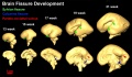
Three-dimensional reconstruction of the lateral (top row) and medial (bottom row) surface of 13–21 week brains to reveal the development of the Sylvian fissure or lateral sulcus (green arrow), the calcarine fissure (blue arrow), and the parieto-occipital sulcus (red arrow), respectively.
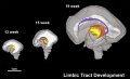
3D depiction of developmental white matter fibers. A lateral view of limbic tracts where pink fibers in 13, 15, and 19 week brains are the fornix and stria terminalis and purple fibers in the 19 week brains indicate the cingulum bundle.
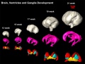
Three-dimensional reconstruction of the basal ganglia and ganglionic eminence. Different colors represent different brain structures: whole brain (gray), ventricle (pink), ganglionic eminence (red), putamen and globus pallidus together (cyan), thalamus (yellow), and caudate nucleus (green).
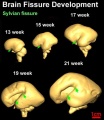
Three-dimensional reconstruction of the lateral (top row) and medial (bottom row) surface of 13–21 week brains to reveal the development of the Sylvian or lateral fissure (green arrow).


- Links: Neural System - Fetal
Second Trimester Timeline
| Event | ||
| Fertilisation Age FA | Gestational Age GA | |
| 14 |  Week 12 - CRL 85 mm, femur length 15 mm, biparietal diameter 25 mm Week 12 - CRL 85 mm, femur length 15 mm, biparietal diameter 25 mm
Hearing Week 12-16 - Capsule adjacent to membranous labrynth undegoes vacuolization to form a cavity (perilymphatic space) around membranous labrynth and fills with perilymph Genital male and female external genital differences observable Respiratory Month 3-6 - lungs appear glandular, end month 6 alveolar cells type 2 appear and begin to secrete surfactant Tongue Week 12 - first differentiated epithelial cells (Type II and III) Genital female genital canal (80 days) formed with absorption of the median septum | |
| 15 | Tongue Week 12 to 13 - maximum synapses between cells and afferent nerve fibers
Hearing - Outer Ear Development Week 13 - Meatal plug disc-like, innermost surface in contact with the primordial malleus, contributes to the formation of the tympanic membrane. | |
| 16 | Tongue Week 14 to 15 - taste pores develop, mucous
Ovary Development 100 days - primary follicles present Nail Development toenails appear Head Development facial skeleton remodelling begins | |
| 17 | Pancreas glucagon detectable in fetal plasma.
Spleen Week 15 -alpha-smooth muscle actin (alpha-SMA)-positive reticulum cells scattered around the arterioles. [10] | |
| 18 | 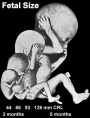 Hearing Week 16-24 - Centres of ossification appear in remaining cartilage of otic capsule form petrous portion of temporal bone. Continues to ossify to form mastoid process of temporal bone. Hearing Week 16-24 - Centres of ossification appear in remaining cartilage of otic capsule form petrous portion of temporal bone. Continues to ossify to form mastoid process of temporal bone.
Pituitary adenohypophysis fully differentiated Respiratory Week 16 to 25 lung histology - canalicular Hearing - Outer Ear Development Week 16.5 - External auditory meatus is fully patent throughout its length, lumen is still narrow and curved. Skin 4 months - basal cell- proliferation generates folds in basement membrane; neural crest cells- (melanocytes) migrate into epithelium; embryonic connective tissue- differentiates into dermis, a loose ct layer over a dense ct layer. Beneath the dense ct layer is another loose ct layer that will form the subcutaneous layer. Ectoderm contributes to nails, hair follictles and glands. Nails form as thickening of ectoderm epidermis at the tips of fingers and toes. These form germinative cells of nail field. Cords of these cells extend into mesoderm forming epithelial columns. These form hair follocles, sebaceous and sweat glands. primary follicles begin to form in the ovary and are characterized by an oocyte glandular urethra forms and skin folds present | |
| 19 |  Neural - Brain development histology week 17 Neural - Brain development histology week 17
| |
| 20 |  Tongue Week 18 - substance P detected in dermal papillae, not in taste bud primordia Tongue Week 18 - substance P detected in dermal papillae, not in taste bud primordia
Skin vernix caseosa covers skin Spleen Week 18 - alpha-SMA-positive reticulum cells increase in number and began to form a reticular framework. An accumulation of T and B lymphocytes occurred within the framework, and a primitive white pulp was observed around the arterioles. [10] Hearing - Outer Ear Development week 18 - External auditory meatus is already fully expanded to its complete form. | |
| 21 | ||
| 22 | Pituitary week 20 to 24 growth hormone levels peak, then decline
Skin lanugo, skin hair Skin 5 months - Hair growth initiated at base of cord, lateral outgrowths form associated sebaceous glands; Other cords elongate and coil to form sweat glands; Cords in mammary region branch as they elongate to form mammary glands. | |
| 23 | ||
| 24 |  Neural brain cortical sulcation - sylvian fissure, interhemispheric fissure, callosal sulcus, parietooccipital fissure, and hippocampic fissures present[11] Neural brain cortical sulcation - sylvian fissure, interhemispheric fissure, callosal sulcus, parietooccipital fissure, and hippocampic fissures present[11]
Spleen - Week 22 - antigenic diversity of the reticular framework was observed, and T and B lymphocytes were segregated in the framework. T lymphocytes were sorted into the alpha-smooth muscle actin-positive reticular framework, and the periarteriolar lymphoid sheath (PALS) was formed around the arteriole. B lymphocytes aggregated in eccentric portions to the PALS and formed the lymph follicle (LF). The reticular framework of the LF was alpha-SMA-negative. [10] | |
| 25 | ||
| 26 | Respiratory Week 24 to 40 lung histology - terminal sac
Spleen Week 24 - marginal zone appeared in the alpha-smooth muscle actin-positive reticular framework around the white pulp.[10] Earliest potential survival expected if born ovarian follicles can consist of growing oocytes surrounded by several layers of granulosa cells | |
| 27 | Respiratory end month 6 alveolar cells type 2 appear and begin to secrete surfactant | |
References
- ↑ Xu F, Ge X, Shi Y, Zhang Z, Tang Y, Lin X, Teng G, Zang F, Gao N, Liu H, Toga AW & Liu S. (2020). Morphometric development of the human fetal cerebellum during the early second trimester. Neuroimage , 207, 116372. PMID: 31751665 DOI.
- ↑ Turk E, van den Heuvel MI, Benders MJ, de Heus R, Franx A, Manning JH, Hect JL, Hernandez-Andrade E, Hassan SS, Romero R, Kahn RS, Thomason ME & van den Heuvel MP. (2019). Functional connectome of the fetal brain. J. Neurosci. , , . PMID: 31685648 DOI.
- ↑ Zhang Z, Lin X, Yu Q, Teng G, Zang F, Wang X, Liu S & Hou Z. (2019). Fetal ocular development in the second trimester of pregnancy documented by 7.0 T postmortem Magnetic Resonance Imaging. PLoS ONE , 14, e0214939. PMID: 30947240 DOI.
- ↑ Nunthapiwat S, Sekararithi R, Wanapirak C, Sirichotiyakul S, Tongprasert F, Srisupundit K, Luewan S & Tongsong T. (2019). Second Trimester Serum Biomarker Screen for Fetal Aneuploidies as a Predictor of Preterm Delivery: A Population-Based Study. Gynecol. Obstet. Invest. , , 1-8. PMID: 30602167 DOI.
- ↑ Ede G, Keskin U, Cemal Yenen M & Samur G. (2019). Lower vitamin D levels during the second trimester are associated with developing gestational diabetes mellitus: an observational cross-sectional study. Gynecol. Endocrinol. , , 1-4. PMID: 30599810 DOI.
- ↑ Akhter N & Chakraborty RK. (2014). Second trimester sonographic features of fetal chromosomal defects. Mymensingh Med J , 23, 412-6. PMID: 24858178
- ↑ Savasan ZA, Goncalves LF & Bahado-Singh RO. (2014). Second- and third-trimester biochemical and ultrasound markers predictive of ischemic placental disease. Semin. Perinatol. , 38, 167-76. PMID: 24836829 DOI.
- ↑ Report of the Workshop on Acute Perinatal Asphyxia in Term Infants, U.S. Department of Health and Human Services, Public Health Service, National Institutes of Health, National Institute of Child Health and Human Development, NIH Publication No. 96-3823, March 1996.
- ↑ Huang H, Xue R, Zhang J, Ren T, Richards LJ, Yarowsky P, Miller MI & Mori S. (2009). Anatomical characterization of human fetal brain development with diffusion tensor magnetic resonance imaging. J. Neurosci. , 29, 4263-73. PMID: 19339620 DOI.
- ↑ 10.0 10.1 10.2 10.3 <pubmed>19255788</pubmed>
- ↑ <pubmed>11158907</pubmed>
Reviews
Raines DA & Cooper DB. (2020). Braxton Hicks Contractions. , , . PMID: 29262073
Articles
Zhang Z, Hou Z, Lin X, Teng G, Meng H, Zang F, Fang F & Liu S. (2013). Development of the fetal cerebral cortex in the second trimester: assessment with 7T postmortem MR imaging. AJNR Am J Neuroradiol , 34, 1462-7. PMID: 23413246 DOI.
Toi A, Lister WS & Fong KW. (2004). How early are fetal cerebral sulci visible at prenatal ultrasound and what is the normal pattern of early fetal sulcal development?. Ultrasound Obstet Gynecol , 24, 706-15. PMID: 15586358 DOI.
Search Pubmed: Second Trimester
Next: Third Trimester
Glossary Links
- Glossary: A | B | C | D | E | F | G | H | I | J | K | L | M | N | O | P | Q | R | S | T | U | V | W | X | Y | Z | Numbers | Symbols | Term Link
Cite this page: Hill, M.A. (2026, July 26) Embryology Second Trimester. Retrieved from https://embryology.med.unsw.edu.au/embryology/index.php/Second_Trimester
- © Dr Mark Hill 2026, UNSW Embryology ISBN: 978 0 7334 2609 4 - UNSW CRICOS Provider Code No. 00098G