Lecture - Renal Development: Difference between revisions
m (→Mesonephros) |
m (→Metanephros) |
||
| Line 143: | Line 143: | ||
===Metanephros=== | ===Metanephros=== | ||
* Human E35-37, Mouse E11 epithelia bud at end of mesonephric duct uteric bud and associated metanephric mesenchyme | * Human E35-37, Mouse E11 epithelia bud at end of mesonephric duct uteric bud and associated metanephric mesenchyme | ||
{{Urogenital stage 22 movie}} | |||
===Uteric Bud=== | ===Uteric Bud=== | ||
Revision as of 12:00, 19 September 2016
| Embryology - 26 Jun 2024 |
|---|
| Google Translate - select your language from the list shown below (this will open a new external page) |
|
العربية | català | 中文 | 中國傳統的 | français | Deutsche | עִברִית | हिंदी | bahasa Indonesia | italiano | 日本語 | 한국어 | မြန်မာ | Pilipino | Polskie | português | ਪੰਜਾਬੀ ਦੇ | Română | русский | Español | Swahili | Svensk | ไทย | Türkçe | اردو | ייִדיש | Tiếng Việt These external translations are automated and may not be accurate. (More? About Translations) |
Introduction

| <html5media height="520" width="360">File:Urogenital_sinus_001.mp4</html5media> |
Urogenital Sinus and Renal Development
This animation gives an overview of both early renal and genital (urogenital) development associated with the urogenital sinus. The paired adult kidneys filter blood, excrete waste, reabsorb water and have endocrine functions. In the embryo, there are several stages in their development closely linked to genital development. The nephron, the functional unit of the kidney, is also a classical epithelial/mesenchyme type of interaction. The urinary system is developmentally and anatomically associated with genital development, often described as the urogenital system.
|
Lecture 2016 - Renal Development PDF
Objectives

- Understand the 3 main stages of kidney development.
- Understand development of the nephron and renal papilla.
- Brief understanding of the mechanisms of nephron development.
- Understand the development of the cloaca, ureter and bladder.
- Brief understanding of abnormalities of the urinary system.
Lecture Resources
| Movies | ||||||||||||||||||||||||||||
|---|---|---|---|---|---|---|---|---|---|---|---|---|---|---|---|---|---|---|---|---|---|---|---|---|---|---|---|---|
|
| References | |
|---|---|
 |
2016 | 2016 PDF | 2015 | 2015 PDF | |
 |
The following chapter links only work with a UNSW connection. |
 Schoenwolf, G.C., Bleyl, S.B., Brauer, P.R., Francis-West, P.H. & Philippa H. (2015). Larsen's human embryology (5th ed.). New York; Edinburgh: Churchill Livingstone. Schoenwolf, G.C., Bleyl, S.B., Brauer, P.R., Francis-West, P.H. & Philippa H. (2015). Larsen's human embryology (5th ed.). New York; Edinburgh: Churchill Livingstone.
|
The following chapter links only work with UNSW Library subscription (with student Zpass log-in). |

Nussey, S. and Whitehead, S. (2001). Endocrinology - An Integrated Approach. UK Oxford: BIOS Scientific Publishers. ISBN-10: 1-85996-252-1 |
Detailed Table of Contents | Bookshelf Link |
Background
- Mesoderm then intermediate mesoderm
- Vascular Development
- Gastrointestional
- Cloacal development
- Endocrine - covered in future lecture/lab
Renal Anatomy

Each adult human kidney typically contains about 750,000 nephrons, though the total number can vary significantly from as few as 250,000 to as many as 2,000,000.
Kidney
|
Ureter
Urinary Bladder
Urethra
|
Germ layers
|
Intermediate Mesoderm

- development occurs laterally symmetrical (left right)
- intermediate mesoderm lying beside the dorsal aorta
- initially form mesonephric tubules (epithelial)
- these tubules connect to a common duct, mesonephric duct
- the mesonephric duct then extends within the mesoderm, rostro-caudally
- eventually making contact with the cloaca
Mesonephric Duct
Later in development, both the mesonephric duct and the cloaca both continue to differentiate and undergo extensive remodelling (and renaming)
Uteric Bud
- arise near the cloacal connection of the mesonephric duct
- branch from the mesonephric duct laterally into the intermediate mesoderm
- induce the surrounding mesoderm to differentiate - metanephric blastema
- this mesoderm will in turn signal back to differentiate the uteric bud
Epithelial - mesenchymal interaction
Uteric Bud forms - ureter, pelvis, calyces, collecting ducts
Metanephric Blastema
- forms glomeruli, capsule, nephron tubules
- this development continues through fetal period
Nephros Development
The 3 main stages and pairs during development:
- pronephros
- mesonephros
- metanephros
Pronephros
- week 3-4 few cells in cervical region (fish type kidney)
- Human E18, Mouse E7.5 - pronephric duct forms first with associated nephrogenic mesenchyme
- grows rostro caudally cervical -> cloaca
- E22 nephrogenic mesenchyme differentiates to form pronephroi not functional in mammals degenerates rapidly
Mesonephros


- Human E24, Mouse E9.5 caudal to pronephros
- forms by induction from pronephros
- pronephric duct now becomes mesonephric duct (also called Wolffian Duct)
Metanephros
- Human E35-37, Mouse E11 epithelia bud at end of mesonephric duct uteric bud and associated metanephric mesenchyme
| Urogenital |
| Page | Play |
Uteric Bud
- induced by metanephric mesenchyme to differentiate
- forms collecting tubules, renal pelvis, ureter
- metanephric mesenchyme induced by uteric to differentiate forms nephron
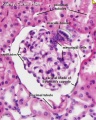
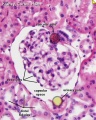
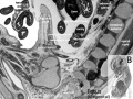
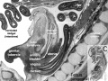
Week 5 and Week 8

|

|
| Embryo Stage 13 mesonephros (week 5) | Embryo Stage 22 metanephros (week 8) |

Fetal


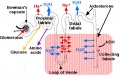
Nephron

| Nephron |
| Page | Play |
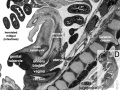
Early Renal Development
Legend
- Uteric Bud - developing ureter, pelvis, calyces, collecting ducts
- Metanephric Blastema (intermediate mesoderm) - developing glomeruli, capsule, nephron tubules
Development has four developmental stages:
- vesicle (V) stage (13-19 weeks)
- S-shaped body (S) stage ( 20-24 weeks)
- capillary loop (C) stage (25-29 weeks)
- maturation (M) stage (infants aged 1-6 months)
Links: Quicktime version | Animation - Urogenital Sinus | Renal System Development
Nephron Development
- disorganised mesenchymal cells become a highly organised epithelial tubule
- Condensation - groups of about 100 cells condense tightly together to form a distinct mass
- Epithelialisation - condensed cells lose their mesenchymal character and gain epithelial
- At end of this period formed a small epithelial cyst complete with a basement membrane, cell-cell junctions and a defined cellular apico-basal polarity.
Early morphogenesis

- cyst invaginates twice to form a comma
- then a S-shaped body one invagination site later becomes the glomerular cleft
- At about this time blood vessel progenitors invade cleft to begin construction of vascular component of glomerulus
- Tubule maturation specialised transporting segments of nephron differentiate complex of convoluted tubules is created

Adult nephron structure
- mean glomerular number shown to level at 36 weeks
- about 15,000 at 15 weeks
- about 740,000 at 40 weeks.
- key structure of the adult nephron is the glomerulus (renal corpuscle), which represents the vascular/renal interface.
Glomerulus structure
Vascular and renal poles


Related Images: Nephron histology overview | glomerulus structure | vascular and renal poles
Renal Vascular
Renal Arteries
|
|
- Arise with ascent and inferior branches lost
- Sequential, 25% population have 2 or more renal arteries
- branch of abdominal aorta, divides into 4-5 branches
- each gives off small branches to suprarenal glands, ureter, surrounding cellular tissue and muscles
- Frequently a second renal artery (inferior renal) from abdominal aorta at a lower level, supplies lower portion of kidney.
Renal Venous

|

|
| Embryo renal venous | Adult renal venous |
Endocrine Kidney
Covered also in Endocrine Development lecture
- Renin - Increase Angiotensin-aldosterone system
- Prostaglandins - decrease Na+ reabsorption
- Erythropoietin - Increase Erythrocyte (rbc) production
- 1,25 (OH)2 vitamin D - Calcium homeostasis
- Prekallikreins - (plasma protein inactive precursor of kallikrein) Increase kinin production (altered vascular permeability)
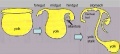
Cloaca

- hindgut region ending at the cloacal membrane
- divided (ventro-dorsally) by the urogenital septum
- ventral - common urogenital sinus
- dorsal - rectum
|
|
Common urogenital sinus
- superior end continuous with allantois
- common urogenital sinus and mesonephric duct fuse (connect)
- differentiates to form the bladder
- inferior end forms urethra
- this will be different in male and female development
Urinary Bladder

- early origins of the bladder at the superior end of the common urogenital sinus
- 8 open inferiorly to the cloaca and superiorly to the allantois
- Septation of the claoca - divides the anterior region to the primordial bladder component from the posterior rectal component.
- associated ureters and urethra
- Ultrasound measurement of the bladder size can be used as a diagnostic tool for developmental abnormalities.
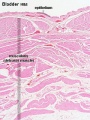
Bladder Structure

Can be described anatomically by its 4 layers from outside inward:
- Serous - the superior or abdominal surfaces and the lateral" surfaces of the bladder are covered by visceral peritoneum, the serous membrane (serosa) of the abdominal cavity, consisting of mesthelium and elastic fibrous connective tissue.
- Muscular - the detrusor muscle is the muscle of the urinary bladder wall.
- Submucosa - connects the muscular layer with the mucous layer.
- Mucosa - (mucus layer) a transitional epithelium layer formed into folds (rugae).
Detrusor Muscle
- The adult detrusor muscle consists of three layers of smooth (involuntary) muscle fibres.
- external layer - fibres arranged longitudinally
- middle layer - fibres arranged circularly
- internal layer - fibres arranged longitudinally
Ureter Development
- Uteric bud origin
- Adult ureter is a thick-walled muscular tube, 25 - 30 cm in length, running from the kidney to the urinary bladder.
- Anatomically two parts the abdominal part (pars abdominalis) and pelvic part (pars pelvina).
- Ureter has three layers: outer fibrous layer (tunica adventitia), muscular layer (tunica muscularis) and mucous layer (tunica mucosa).
- The muscular layer can also be subdivided into 3 fibre layers: an external longitudinal, a middle circular, and an internal longitudinal.
Urethra Development
- Further development of the urinary system varies depending on the sex of the embryo.
- Males - the pelvic urethra forms the membranous urethra, the prostatic urethra and penile urethra. (The sex of the above animation and sections is male)
- Females - the pelvic urethra forms the membranous urethra and the vestibule of the vagina.
Abnormalities

| ICD10 Congenital malformations of the urinary system (Q60-Q64) | ||||
|---|---|---|---|---|
| The International Classification of Diseases (ICD) World Health Organization's classification used worldwide as the standard diagnostic tool for epidemiology, health management and clinical purposes. Includes this section on XVII Congenital Malformations. | ||||
| Q60 Renal agenesis and other reduction defects of kidney
Incl.: atrophy of kidney: congenital infantile congenital absence of kidney
| ||||
| Q61 Cystic kidney disease
Excl.: acquired cyst of kidney (N28.1) Potter's syndrome (60.6)
| ||||
Q62 Congenital obstructive defects of renal pelvis and congenital malformations of ureter
| ||||
| Q63 Other congenital malformations of kidney
Excl.: congenital nephrotic syndrome (N04.-)
| ||||
Q64 Other congenital malformations of urinary system
| ||||
| World Health Organisation. International Statistical Classification of Diseases and Related Health Problems. (1992) 10th Revision (ICD-10). Geneva: WHO ICD-10 - 2016 Online (English) | ||||
| ||||
| ICD10 - Gastrointestinal | Genital | Renal | Integumentary |
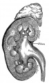
Horseshoe Kidney

|
|
Kidney Vascular
Supernumerary renal arteries


Supernumerary renal vein


Urorectal Septum Malformation
- thought to be a deficiency in caudal mesoderm which in turn leads to the malformation of the urorectal septum and other structures in the pelvic region.
- Recent research has also identified the potential presence of a persistent urachus prior to septation of the cloaca (common urogenital sinus).
Bladder
- absent or small bladder -
associated with renal agenesis.
Bladder Exstrophy

- developmental abnormality associated with bladder development.
- origins appear to occur not just by abnormal bladder development, but by a congenital malformation of the ventral wall of abdomen (between umbilicus and pubic symphysis).
- There may also be other anomolies associated with failure of closure of abdominal wall and bladder (epispadias, pubic bone anomolies).
Ureter and Urethra
- Ureter - Duplex Ureter
- Urethra- Urethral Obstruction and Hypospadias

Hydronephrosis
Polycystic Kidney Disease

- diffuse cystic malformation of both kidneys
- cystic malformations of liver and lung often associated, Often familial disposition
- Two types
- Infantile (inconsistent with prolonged survival)
- Adult (less severe and allows survival)
- Autosomal dominant PKD disease - recently identified at mutations in 2 different human genes encoding membrane proteins (possibly channels)
Wilms' Tumor

- (nephroblastoma) Named after Max Wilms, a German doctor who wrote first medical articles 1899
- most common type of kidney cancer children
- WT1 gene - encodes a zinc finger protein
- Both constitutional and somatic mutations disrupting the DNA-binding domain of WT1 result in a potentially dominant-negative phenotype
- some blastema cells (mass of undifferentiated cells) persist to form a ‘nephrogenic rest’
- Most rests become dormant or regress but others proliferate to form hyperplastic rests
- any type of rest can then undergo a genetic or epigenetic change to become a neoplastic rest
- can proliferate further to produce a benign lesion (adenomatous rest) or a malignant Wilms’ tumour
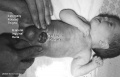
Prune Belly Syndrome
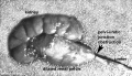
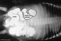
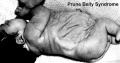
Prune_belly


- lower urinary tract obstruction
- mainly male
- fetal urinary system ruptures leading to collapse and "prune belly" appearance.
Additional Images
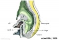
Stage 11 historic Atwell (1930)
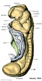
Stage 11 historic Heuser (1930)
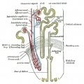
Nephron structure
Nephron physiology
Kidney and adrenal gland (adult)
Endoderm cartoon
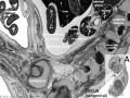
Fetal urogenital region most lateral right
Fetal urogenital region lateral right
Fetal urogenital region medial
Fetal urogenital region midline
Bladder histology

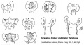
Horseshoe kidney
Hydronephrosis
Renal outflow obstruction
Bladder Exstrophy


References
Textbooks
- The Developing Human: Clinically Oriented Embryology (8th Edition) by Keith L. Moore and T.V.N Persaud - Moore & Persaud Chapter 13 p303-346
- Larsen’s Human Embryology by GC. Schoenwolf, SB. Bleyl, PR. Brauer and PH. Francis-West - Chapter 10 p261-306
- Before We Are Born (5th ed.) Moore and Persaud Chapter14 p289-326
- Essentials of Human Embryology, Larson Chapter 10 p173-205
- Human Embryology, Fitzgerald and Fitzgerald Chapter 21-22 p134-152
Online Textbooks
- Developmental Biology by Gilbert, Scott F. Sunderland (MA): Sinauer Associates, Inc.; c2000 Chapter 14 Intermediate Mesoderm | Figure 14.18. General scheme of development in the vertebrate kidney | Figure 23-23. Mechanism of mesenchymal inductive effect on the ureteric bud | Figure 14.21. Ureteric bud growth is dependent on GDNF and its receptor
- Molecular Cell Biology by Lodish, Harvey; Berk, Arnold; Zipursky, S. Lawrence; Matsudaira, Paul; Baltimore, David; Darnell, James E. New York: W. H. Freeman & Co.; c1999 Reciprocal Epithelial-Mesenchymal Interactions Regulate Kidney Development | Figure 23-21. Embryonic development of the kidney
Reviews
- Quaggin SE, Kreidberg JA. Development of the renal glomerulus: good neighbors and good fences. Development. 2008 Feb;135(4):609-20. PMID: 18184729
- Brenner-Anantharam A, Cebrian C, Guillaume R, Hurtado R, Sun TT, Herzlinger D. Tailbud-derived mesenchyme promotes urinary tract segmentation via BMP4 signaling. Development. 2007 May;134(10):1967-75. PMID: 17442697
- Forefronts Symposium on Nephrogenetics: from development to physiology March 8-11, 2007 Danvers, MA A meeting to synthesize an integrated view of the normal development and function of the kidney from the genetic standpoint.
- Costantini F. Renal branching morphogenesis: concepts, questions, and recent advances. Differentiation. 2006 Sep;74(7):402-21. PMID: 16916378
Search
- Bookshelf intermediate mesoderm | kidney development | renal development | ureteric bud | nephron development | bladder development
- Pubmed intermediate mesoderm | kidney development | renal development | ureteric bud | nephron development | bladder development
Terms
| Renal Terms | ||
|---|---|---|
| ||
|
| ANAT2341 Course Timetable | |||
|---|---|---|---|
| Week (Mon) | Lecture 1 (Mon 1-2pm) | Lecture 2 (Tue 3-4pm) | Practical (Fri 1-3pm) |
| Week 2 (1 Aug) | Introduction | Fertilization | Lab 1 |
| Week 3 (8 Aug) | Week 1 and 2 | Week 3 | Lab 2 |
| Week 4 (15 Aug) | Mesoderm | Ectoderm | Lab 3 |
| Week 5 (22 Aug) | Early Vascular | Placenta | Lab 4 |
| Week 6 (29 Aug) | Gastrointestinal | Respiratory | Lab 5 |
| Week 7 (5 Sep) | Head | Neural Crest | Lab 6 |
| Week 8 (12 Sep) | Musculoskeletal | Limb Development | Lab 7 |
| Week 9 (19 Sep) | Renal | Genital | Lab 8 |
| Mid-semester break | |||
| Week 10 (3 Oct) | Public Holiday | Stem Cells | Lab 9 |
| Week 11 (10 Oct) | Integumentary | Endocrine | Lab 10 |
| Week 12 (17 Oct) | Heart | Sensory | Lab 11 |
| Week 13 (24 Oct) | Fetal | Birth and Revision | Lab 12 |
|
ANAT2341 2016: Moodle page | ECHO360 | Textbooks | Students 2016 | Projects 2016 | |||