AE Practical - Neural Histology: Difference between revisions
| Line 124: | Line 124: | ||
File:Brain_histology_01.jpg|Overview Cortex (mouse) | File:Brain_histology_01.jpg|Overview Cortex (mouse) | ||
File:Brain_histology_02.jpg|Cortex (mouse) | File:Brain_histology_02.jpg|Cortex (mouse) | ||
</gallery> | </gallery> | ||
Revision as of 12:20, 24 September 2012
Introduction

- Draft Page
 This page provides histology support information for central nervous system structure. Disclaimers
|

|
Aims

- Obtain an understanding of the normal histological appearance of selected central and peripheral nervous system tissues namely spinal cord, cerebellum and peripheral nerve.
- To examine unique microscopic characteristics of each of the nervous tissues.
- To introduce the histology and neuropathology associated with cerebral infarction and haemorrhage.
Key concepts

The brain and spinal cord comprise the central nervous system (CNS). The nerves that emerge from the spinal cord and brain to pass to parts of the body are the peripheral nervous tissue (PNS). Nervous tissue, with many interconnections, forms a complex system of neuronal communication within the body and is specialized for detecting stimuli, integrating functions, controlling effectors and higher functions. Nervous tissue consists of cell bodies, cell processes (nerves), and neuroglia (supporting cells).
Neurons
- Structural and Functional units of the nervous system.
These cells (around 12 billion) are responsible for the receptive, integrative, and motor functions of the nervous system. They can generate nerve impulses (irritability), and can transmit these impulses along their processes (conductivity). They range in diameter from 5 to 150 μm and contain 3 parts: a cell body, multiple dendrites and a single axon.
- Cell body (soma, perikaryon) is the region of the neuron containing a large pale-staining spherical, nucleus with a conspicuous nucleolus and perinuclear cytoplasm.
- Dendrites project from the cell body and are specialized for receiving (afferent) stimuli from sensory cells, axons and other neurons which are then transmitted towards the soma.
- Axons arise as a single thin process extending longer distances from the cell body than the dendrite. As with dendrites, the terminals of the axon are branching and terminate in end bulbs (terminal boutons), which come close to another cell and form a synapse.
Peripheral Nerve Fibers
Peripheral nerves are bundles (fascicles) of nerve fibers (axons) surrounded by several CT sheaths. Each bundle contains sensory and motor components.
Myelinated Fibers (1-20μm diameter)
Myelin (rich in lipid) is the membrane of the Schwann cell organized into a spiral sheath that is wrapped several times around the axon. Schwann cells are cells whose cytoplasm contains a flattened nucleus, a small Golgi apparatus, and a few mitochondria. Myelinated fibers are capable of rapid transfer of impulses (touch sensory pathways).
Unmyelinated Fibers (less than 2μm in diameter)
Some axons in the PNS are surrounded by Schwann cells but not wrapped with layers of myelin. They arefound in pain and temperature sensory pathways and motor paths to the viscera.
Practical class activities
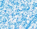
Spinal Cord
Virtual Slide Box (Spinal cord and Spinal cord smear) and Zurich Virtual Slide database (Spinal cord-Thoracic Segment-Luxol Fast Blue, Neutral Red and Spinal cord-Lumbar Segment-Azan and Meninges-Azan).
Identify gray and white matter, central canal (surrounded by ependymal cells), dorsal and ventral horns, meninges (pia, arachnoid and dura mater), subarachnoid space with dorsal and ventral rootlets, blood vessels, a motor neurone with a cell body (soma), nucleus, nucleolus, Nissl granules, an axon with axon hillock area, dendrites, glial cells (oligodendrocytes, astrocytes).
| Spinal cord (Luxol Fast Blue) | |
|---|---|

|

|
- What is the difference between white and gray matter?
| Spinal cord - Grey and white matter | |
|---|---|

|

|
| Spinal cord - Grey matter | |
|---|---|

Grey matter (HE) |

Grey matter (silver) |
- What do Nissl granules represent?
- What is the functional difference between an axon and a dendrite?
- What is the function of ependymal cells?
- What function do the meninges serve and what type of tissue are the meninges made up of?
- What is the function of an oligodendrocyte and of an astrocyte?
Histology Images
Overview
Grey matter
Grey matter
White matter
Ependymal cells


- Spinal Cord: Overview 1 | Overview 2 | Overview animation | Grey matter | Grey matter | Grey matter | White matter | Overview unlabeled | Grey matter unlabeled 1 | Grey matter unlabeled 2 | White matter unlabeled 1 | Ependymal cells unlabeled
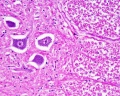
Cerebellum

Virtual Slide Box (Brain/Cerebellum and Cerebellum silver stain) and Zurich Virtual Slide database (Cerebellum silver stain).
- Identify the folia (folds), meninges (pia and arachnoid mater), blood vessels, and white and gray matter. The gray matter is subdivided into 3 distinct layers namely outer molecular, inner granular and middle Purkinje cell layer. Note the processes on the Purkinje cells.
- What does white matter consist of?
- What is the function of the Purkinje fibers?
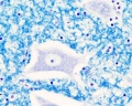
Peripheral Nerve
Virtual Slide Box (Peripheral Nerve) and Zurich Virtual Slide database (Nerve; Goldner and Nerve; Haematoxylin and Eosin)
- Identify fascicles (bundles) of nerves, levels of connective tissue wrappings (epineurium, perineurium, endoneurium), fibroblast nuclei, adipose tissue, blood vessels, myelinated nerve fibers, axons, and Schwann cells.
- What is the function of a Schwann cell and what effect does myelin have on nerve transmission?
- Why do 3 levels of connective tissue wrap nerve fibers?
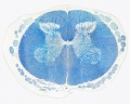
Brain Histology
Overview Cortex (mouse)
Cortex (mouse)


Development
The following information is not part of the current class, but for students interested in issues of normal and abnormal neural development.
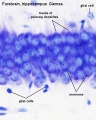
Developing Cortex
|

|
Terms
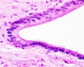
- arachnoid mater - (Greek, arachne = spider + -oeides = form) A meshwork (spider web-like) connective tissue covering of the central nervous system, forms part of the meningial layers. Lies between tough outer dura mater layer and the inner fine pia mater layer. These three connective tissue layers have different embryonic origins: dura is from mesoderm, pia and arachnoid layers are neural crest in origin. The space underlying the arachnoid mater (subarachnoid space) is filled with cerebrospinal fluid.
- artifact - changes and distortions introduced to the normal tissue structure by the histological processing. Common artifacts include: folds (gives the tissue a darker appearance), tears (rips in the tissue can be seen in epithelia), shrinkage when tissues loose mainly liquid through histological processing, and cuts often used in tissue preparation.
- blood-brain barrier - (BBB) formed by endothelial cells of brain capillaries, differing in junctions, transport and glial association from those found in peripheral capillaries. Damage to the BBB can occur following injuries involving ischemia and reperfusion.
- cell body - (soma, perikaryon) is the region of the neuron containing a large pale-staining spherical, nucleus with a conspicuous nucleolus and perinuclear cytoplasm.
- dura mater - (Latin, dura mater = hard mother) The outer tough connective tissue meningial coat of the 3 layers that cover the central nervous system of 3 layers (overlays the arachnoid mater middle layer and pia mater inner layer). These three connective tissue layers have different embryonic origins: dura is from mesoderm, pia mater and arachnoid layers are neural crest in origin.Duramater at the level of the spinal cord is separated from the periosteum of the vertebral canal by an epidural space.
- leptomeninges - (thin meninges) Term referring to just the pia mater and arachnoid mater layers of the meninges.
- meninges - (singular meninx; Greek, meninx = membrane) Anatomical term describing the three connective tissue layers that surround the entire central nervous system (brain and spinal cord). The 3 layers from the central nervous outward are: pia mater, arachnoid mater, and the dura mater. These layers also have differing embryonic origins; dura mater is mesoderm, pia mater and arachnoid are neural crest. The space under the arachnoid layer (subarachnoid space) is filled with cerebrospinal fluid. (More? Image - Meninges cartoon)
- meningococcal disease - (meningitis) Term describing the bacterial infection of cerebrospinal fluid of the spinal cord and brain. Note meningitis can also be caused by a viral or other organism infection. Treatment and outcomes differ for either viral (less severe, resolves without specific treatment) or bacterial (severe, may result in brain damage, hearing loss, or learning disability) infections. (More? Bacterial Infection | Postnatal Development | CDC - meningococcal disease | Medline Plus - Meningitis)
- myelin - (myelin sheath) is the membrane of a glial cell (brain, oligodendrocyte; peripheral nerve, Schwann cell) organized into a spiral sheath that is wrapped many times around the axon.
- neuropil - (neuropile) is the region of nerve fibres (axons and dendrites) with numerous synapses and also glia, with few neural cell bodies.
- pia mater - A fine connective tissue covering of the central nervous system, forms innermost part of the meningial layers. Lies beneath the arachnoid mater and then tough outer dura mater layer. These three connective tissue layers have different embryonic origins: pia mater and arachnoid layers are neural crest in origin, dura is from mesoderm. The space overlying the pia mater (subarachnoid space) is filled with cerebrospinal fluid. The pia mater has close contact with the spinal cord and brain, in the brain it follows down into the sulci and fissures of the cortex. This layer also fuses with the membranous lining of the ventricles (ependyma) forming the choroid plexus.
External Links
External Links Notice - The dynamic nature of the internet may mean that some of these listed links may no longer function. If the link no longer works search the web with the link text or name. Links to any external commercial sites are provided for information purposes only and should never be considered an endorsement. UNSW Embryology is provided as an educational resource with no clinical information or commercial affiliation.
- The Cerebral Circulation. Cipolla MJ. San Rafael (CA): Morgan & Claypool Life Sciences; 2009. Chapter 2 Anatomy and Ultrastructure
- Clinical Methods: The History, Physical, and Laboratory Examinations. 3rd edition. Walker HK, Hall WD, Hurst JW, editors. Boston: Butterworths; 1990. Chapter 55 Cerebrovascular Disease
- Medical Microbiology. 4th edition. Baron S, editor. Galveston (TX): University of Texas Medical Branch at Galveston; 1996. Chapter 96 Microbiology of the Nervous System
- Meningitis CDC - meningococcal disease | Medline Plus - Meningitis
- Neuroscience. 2nd edition. Purves D, Augustine GJ, Fitzpatrick D, et al., editors. Sunderland (MA): Sinauer Associates; 2001. Chapter 1 The Cellular Components of the Nervous System
Glossary Links
- Glossary: A | B | C | D | E | F | G | H | I | J | K | L | M | N | O | P | Q | R | S | T | U | V | W | X | Y | Z | Numbers | Symbols | Term Link
Cite this page: Hill, M.A. (2024, June 21) Embryology AE Practical - Neural Histology. Retrieved from https://embryology.med.unsw.edu.au/embryology/index.php/AE_Practical_-_Neural_Histology
- © Dr Mark Hill 2024, UNSW Embryology ISBN: 978 0 7334 2609 4 - UNSW CRICOS Provider Code No. 00098G