ANAT2241 Endocrine System: Difference between revisions
mNo edit summary |
|||
| (16 intermediate revisions by the same user not shown) | |||
| Line 1: | Line 1: | ||
{{ANAT2241 header}} | |||
==General Objective== | |||
To know the histological and cytological features and hormones of the endocrine glands. | |||
===Specific Objectives=== | |||
# To know the structure of the thyroid and parathyroid glands and the hormones secreted. | |||
# To know the histology of the adrenal cortex and medulla and the hormones secreted by each section. | |||
# To understand the close relationship between the hypothalamus and the pituitary. | |||
# To know the parts of the hypophysis (pituitary). To identify chromophil (acidophil, basophil) and chromophobe cell types of the pars distalis and the hormones associated with each group of cells. To describe the structure of the neurohypophysis and the process of neurosecretion. | |||
# To know the histology of pancreatic islet cells and their hormonal secretions. | |||
==Learning Activities== | |||
Examine the following virtual slides, identify draw and label the following structures. Note the hormone secreted by the various structures and their major function. | |||
Virtual Slides: [https://moodle.telt.unsw.edu.au/mod/book/view.php?id=789982&chapterid=100773 Endocrine System] | |||
== | ==Endocrine Axes== | ||
Many of the endocrine organs act together as part of a signalling axis, beginning with the hypothalamus. There is also a sex-specific axis for female (ovary) and male (testis), both identified as gonad. | |||
<gallery> | |||
File:HPA_axis.jpg|Hypothalamus - Pituitary - Adrenal (HPA) | |||
File:HPT axis.jpg|Hypothalamus - Pituitary - Thyroid (HPT) | |||
File:HPG female axis.jpg|Hypothalamus - Pituitary - Gonad (female) (HPG) | |||
File:HPG male axis.jpg|Hypothalamus - Pituitary - Gonad (male) (HPG) | |||
</gallery> | |||
==Histology== | ==Histology== | ||
Note this support page includes additional endocrine gland histology that may not be covered in the current practical class, or has been covered in previous classes in the course. | |||
===Pineal=== | |||
[[File:Pineal_histology_001.jpg|300px|alt=Pineal_histology]] | |||
Pineal (high power) | |||
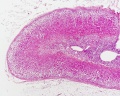
===Thyroid=== | |||
{| | |||
| [[File:Thyroid_histology_001.jpg|300px|alt=Thyroid histology]] | |||
| [[File:Thyroid_histology_002.jpg|300px|alt=Thyroid histology]] | |||
|- | |||
| Thyroid (low power) | |||
| Thyroid (high power) | |||
|} | |||
{{Thyroid histology}} | |||
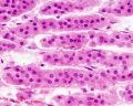
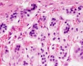
===Parathyroid=== | |||
{| | |||
| [[File:Parathyroid_histology_001.jpg|300px|alt=Parathyroid histology]] | |||
| [[File:Parathyroid_histology_002.jpg|300px|alt=Parathyroid histology]] | |||
|- | |||
| Parathyroid (low power) | |||
| Parathyroid (high power) | |||
|} | |||
===Pancreas=== | |||
The specific cell types within the pancreatic islets cannot be identified by routine histology staining and require specialised immunostaining techniques. | |||
Pancreatic Islet - Endocrine cells from human (fetal and adult) and {{mouse}} (adult) pancreas. | |||
{| | |||
| [[File:Pancreatic islet.png|500px|alt=Pancrea_histology]] | |||
| valign=top| | |||
* <font color=blue>'''Blue'''</font> - nuclear staining (all cell) | |||
* <font color=green>'''Green'''</font> - glucagon (alpha cells) | |||
* <font color=red>'''Red'''</font> - insulin (beta cells) | |||
'''A–C:''' Section of adult mouse pancreas stained for glucagon (green) and insulin (red). | |||
'''D–F:''' Section of an adult human pancreas stained for glucagon (green) and insulin (red). | |||
'''G–I:''' Section of a human fetal pancreas analyzed 4.5 months after transplantation and stained for glucagon (green) and insulin (red). | |||
Nuclear staining (blue) was performed with DAPI. Scale bars: 25 µm. | |||
|} | |||
===Pituitary=== | |||
{| | |||
| [[File:Turkish saddle-17th century.jpg|150px]] | |||
| The shape of the bone base of the {{skull}} surrounding the {{pituitary}} led to the naming '''sella turcica''' (Latin, ''sella'' = saddle; ''turcica'' = Turkish), as when first identified the bony space resembled this saddle shape. | |||
| [[File:Historic-pituitary.jpg|150px]] | |||
|} | |||
{| | |||
! Pituitary - Adenohypophysis | |||
|- | |||
| [[File:Pituitary_histology_001.jpg|300px|alt=Pituitary histology]] | |||
| [[File:Pituitary_histology_002.jpg|300px|alt=Pituitary histology]] | |||
| [[File:Pituitary_histology_003.jpg|300px|alt=Pituitary histology]] | |||
|} | |||
{{Pituitary Histology}} | |||
===Adrenal=== | |||
<gallery> | <gallery> | ||
File:Adrenal histology 001.jpg|Adrenal - Cortex and Medulla | File:Adrenal histology 001.jpg|Adrenal - Cortex and Medulla | ||
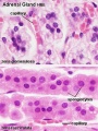
File:Adrenal histology 002.jpg|Adrenal - Cortical Zones | File:Adrenal histology 002.jpg|Adrenal - Cortical Zones | ||
File:Adrenal histology 003.jpg|Adrenal - Zona Reticularis and Medulla | File:Adrenal histology 003.jpg|Adrenal - Zona Reticularis and Medulla | ||
Adrenal histology 004.jpg | |||
Adrenal histology 005.jpg | |||
Adrenal histology 006.jpg | |||
Adrenal histology 007.jpg | |||
Adrenal histology 008.jpg | |||
Adrenal histology 009.jpg | |||
Adrenal histology 010.jpg | |||
Adrenal histology 011.jpg | |||
</gallery> | </gallery> | ||
{{ANAT2241 footer}} | {{ANAT2241 footer}} | ||
Latest revision as of 10:08, 2 August 2019

| ANAT2241 This practical support page content is not part of the virtual science practical class and provides additional information for student self-directed learning purposes. All practical class pages are located on Moodle - ANAT2241 |
General Objective
To know the histological and cytological features and hormones of the endocrine glands.
Specific Objectives
- To know the structure of the thyroid and parathyroid glands and the hormones secreted.
- To know the histology of the adrenal cortex and medulla and the hormones secreted by each section.
- To understand the close relationship between the hypothalamus and the pituitary.
- To know the parts of the hypophysis (pituitary). To identify chromophil (acidophil, basophil) and chromophobe cell types of the pars distalis and the hormones associated with each group of cells. To describe the structure of the neurohypophysis and the process of neurosecretion.
- To know the histology of pancreatic islet cells and their hormonal secretions.
Learning Activities
Examine the following virtual slides, identify draw and label the following structures. Note the hormone secreted by the various structures and their major function.
Virtual Slides: Endocrine System
Endocrine Axes
Many of the endocrine organs act together as part of a signalling axis, beginning with the hypothalamus. There is also a sex-specific axis for female (ovary) and male (testis), both identified as gonad.
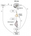
Hypothalamus - Pituitary - Adrenal (HPA)
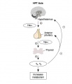
Hypothalamus - Pituitary - Thyroid (HPT)
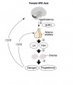
Hypothalamus - Pituitary - Gonad (female) (HPG)
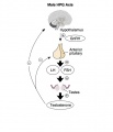
Hypothalamus - Pituitary - Gonad (male) (HPG)


Histology
Note this support page includes additional endocrine gland histology that may not be covered in the current practical class, or has been covered in previous classes in the course.
Pineal

Pineal (high power)
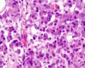
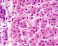
Thyroid

|

|
| Thyroid (low power) | Thyroid (high power) |
- Thyroid Links: low power image | high power image | unlabeled human image | unlabeled sheep image | thyroid
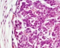
Parathyroid

|

|
| Parathyroid (low power) | Parathyroid (high power) |
Pancreas
The specific cell types within the pancreatic islets cannot be identified by routine histology staining and require specialised immunostaining techniques.
Pancreatic Islet - Endocrine cells from human (fetal and adult) and mouse (adult) pancreas.

|
A–C: Section of adult mouse pancreas stained for glucagon (green) and insulin (red). D–F: Section of an adult human pancreas stained for glucagon (green) and insulin (red). G–I: Section of a human fetal pancreas analyzed 4.5 months after transplantation and stained for glucagon (green) and insulin (red). Nuclear staining (blue) was performed with DAPI. Scale bars: 25 µm. |
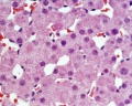
Pituitary

|
The shape of the bone base of the skull surrounding the pituitary led to the naming sella turcica (Latin, sella = saddle; turcica = Turkish), as when first identified the bony space resembled this saddle shape. | 
|
| Pituitary - Adenohypophysis | ||
|---|---|---|

|

|

|
- Pituitary Histology: Pituitary overview | Anterior H&E | Anterior H&E | Anterior labeled | PAS/O Overview | Acidophils | Basophils | Posterior labeled | Posterior unlabeled | Histology Stains | BGD - Endocrine Histology | Pituitary Development
Adrenal
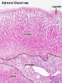
Adrenal - Cortex and Medulla
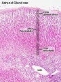
Adrenal - Cortical Zones
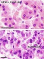
Adrenal - Zona Reticularis and Medulla


Course Links
- Histology Glossary: A | B | C | D | E | F | G | H | I | J | K | L | M | N | O | P | Q | R | S | T | U | V | W | X | Y | Z | ANAT2241 Support | Histology | Histology Stains | Embryology Glossary
| Common Histology Stains | ||||||||||||||||||||||||||||||||||||||||||||||||||||||||||||||||||||||||||||||||||||||||||||||||||||||||||||||||||||||||||||||||||||||||||||||||
|---|---|---|---|---|---|---|---|---|---|---|---|---|---|---|---|---|---|---|---|---|---|---|---|---|---|---|---|---|---|---|---|---|---|---|---|---|---|---|---|---|---|---|---|---|---|---|---|---|---|---|---|---|---|---|---|---|---|---|---|---|---|---|---|---|---|---|---|---|---|---|---|---|---|---|---|---|---|---|---|---|---|---|---|---|---|---|---|---|---|---|---|---|---|---|---|---|---|---|---|---|---|---|---|---|---|---|---|---|---|---|---|---|---|---|---|---|---|---|---|---|---|---|---|---|---|---|---|---|---|---|---|---|---|---|---|---|---|---|---|---|---|---|---|---|
| ||||||||||||||||||||||||||||||||||||||||||||||||||||||||||||||||||||||||||||||||||||||||||||||||||||||||||||||||||||||||||||||||||||||||||||||||
| ||||||||||||||||||||||||||||||||||||||||||||||||||||||||||||||||||||||||||||||||||||||||||||||||||||||||||||||||||||||||||||||||||||||||||||||||
Practical Support
- Pages can be accessed from any internet connected computer.
ANAT2241 Support Links: The Virtual Microscope | Covering and Lining Epithelia | Glandular Epithelia | CT Components | CT Types | Bone, Bone Formation and Joints | Muscle | Nervous | Blood | Eye | Cardiovascular | Respiratory | Integumentary | Gastrointestinal | Gastrointestinal Organs | Lymphatic and Immune | Endocrine | Urinary | Female Reproductive | Male Reproductive | Histology Stains | Histology Drawings | Practicals Health and Safety 2013 | Moodle - 2019
ANAT2241 This practical support page content is not part of the science practical class and provides only background information for student self-directed learning purposes.
Cite this page: Hill, M.A. (2024, June 16) Embryology ANAT2241 Endocrine System. Retrieved from https://embryology.med.unsw.edu.au/embryology/index.php/ANAT2241_Endocrine_System
- © Dr Mark Hill 2024, UNSW Embryology ISBN: 978 0 7334 2609 4 - UNSW CRICOS Provider Code No. 00098G