Endocrine - Pineal Development: Difference between revisions
mNo edit summary |
mNo edit summary |
||
| (38 intermediate revisions by the same user not shown) | |||
| Line 5: | Line 5: | ||
[[File:Pineal gland position.jpg|thumb|Pineal gland position]] | [[File:Pineal gland position.jpg|thumb|Pineal gland position]] | ||
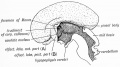
The pineal gland (epiphysis cerebri) has an important role in the sleep/wake daily cycle (circadian), high melatonin plasma levels at nighttime and very low levels at daytime, and reproductive development. The gland is thought to evolutionarily to have been positioned as to be exposed to light, and hence remains a regulator of cyclic rhythms associated with day/night and day length. The pineal hormone (melatonin) has targets both in the nervous system and in many different peripheral tissues. | The pineal gland (epiphysis cerebri) has an important role in the sleep/wake daily cycle (circadian), high melatonin plasma levels at nighttime and very low levels at daytime, and reproductive development. The gland is thought to evolutionarily to have been positioned as to be exposed to light, and hence remains a regulator of cyclic rhythms associated with day/night and day length. The pineal hormone (melatonin) has targets both in the nervous system and in many different peripheral tissues. Melatonin synchronises circadian rhythms acting through the two G-protein-coupled receptors (MT1, MT2) in the suprachiasmatic nucleus of the {{hypothalamus}}. | ||
The embryo and fetus pineal does not produce significant amounts of melatonin, though | The embryo and fetus pineal does not produce significant amounts of melatonin, though has abundant tissue receptors. The maternal pineal gland produces melatonin in the normal circadian fashion and this melatonin can cross both the placenta and blood-brain barrier. In other species, maternal melatonin crosses the placenta into fetal circulation and may provide photoperiodic information during fetal development that influences later postnatal circadian (daily day/night) and seasonal (day length) rhythms. The pineals of non-mammalian vertebrates are photoreceptive, whereas those of mammals do not normally respond to directly light. Pineal blood supply is derived from the posterior cerebral artery choroidal branches and through the pineal recess is bathed in cerebrospinal fluid ({{CSF}}). | ||
The melatonin levels in premature infants is lower and delayed, but not different when calculated from conception date. Other factors such as preeclampsia, growth restriction, and nursery lighting can cause altered rhythm development. The same study has also shown that full-term infants born at home and full-term twins born in the hospital had significantly lower metabolite excretion levels than hospital-born singleton infants at the same ages despite similar body weights.{{#pmid:8636362|PMID8636362}} Allopregnanolone, a pineal neurosteroid, acts on the {{cerebellum}} supporting survival Purkinje cell by suppressing caspase-3 expression.{{#pmid:30339808|PMID30339808}} | |||
Postnatally in humans, the melatonin levels may act on clock gene expression in the pars tuberalis of the {{anterior pituitary}} for photoperiodic switching. Melatonin also has important functions related to the program timing of {{puberty}}. Note that there are many clinical studies investigating the possible role of melatonin in diverse health areas, from oxygen starvation at birth through to neural effects in old age. | |||
Overview | |||
* part of epithalmus - neurons, glia and pinealocytes | * part of epithalmus - neurons, glia and pinealocytes | ||
* pinealocytes secrete melatonin - cyclic nature of activity, melatonin lowest during daylight | * pinealocytes secrete melatonin - cyclic nature of activity, melatonin lowest during daylight | ||
| Line 21: | Line 23: | ||
'''Links:''' {{pineal}} | [[:Category:Pineal|Category:Pineal]] | [[Lecture - Endocrine Development]] | [[Lecture - Head Development]] | |||
{| class="wikitable mw-collapsible mw-collapsed" | |||
! Historic Pineal Papers | |||
{{ | |-bgcolor="F5FFFA" | ||
| [[Paper - On the pineal region in human embryos|1917 Pineal Region]] | [[Paper - The Human Pineal Gland and Pineal Cysts|1932 Pineal Gland and Cysts]] | [[Paper - Development and histogenesis of the human pineal organ (1935)|1935 Pineal]] | [[Paper - Development and histogenesis of the human pineal organ|1937 Human Pineal]] | [[Paper - The nervous and vascular relations of the pineal gland (1940)|1940 Nerve and Vascular Supply]] | |||
|} | |||
<br> | |||
{{Endocrine Links}} | |||
== Some Recent Findings == | == Some Recent Findings == | ||
[[File:Mouse pineal E15 to E21 neuroepithelium 01.jpg|thumb|alt=Mouse pineal E15 to E21 neuroepithelium|Mouse pineal E15 to E21 neuroepithelium{{#pmid:27861587|PMID27861587}}]] | |||
{| | {| | ||
|-bgcolor="F5FAFF" | |-bgcolor="F5FAFF" | ||
| | | | ||
* ''' | * '''Morphology and quantification of {{sheep}} pineal glands at pre-pubertal, pubertal and post-pubertal periods'''{{#pmid:29774950|PMID29774950}} "The pineal gland is a neuroendocrine organ associated with photoperiodic regulation in mammals. The aim of this study was to evaluate the pineal gland at the pre-pubertal, pubertal and post-pubertal periods by means of morphology and stereology. The study examined at total of 24 ovine pineal glands collected from healthy female Akkaraman breed. Thick sections (40 μm) were cut and treated with synaptophysin. ...The melatonin staining density was the highest in the pubertal group. The density of lipofuscin staining was higher in the pubertal and post-pubertal groups." {{Sheep}} | ||
* ''' | |||
* '''Infradian Rhythm of the Content of Secretory Granules in Pinealocyte Cytoplasm in Mice and Rats'''{{#pmid:29931631|PMID29931631}} "The numerical density of secretory granules dense-core vesicles (DCV) in the cytoplasm of pinealocytes of the pineal gland was estimated by transmission electron microscopy in male white mice and Wistar rats. The 3-day biorhythm and lunaphase changes in the DCV content in the perikaryon and the processes of pinealocytes, which are manifested significantly in different seasons of the year, are established. The three-day biorhythm in adult male mice in comparison with younger male rats is not expressed uniformly in different phases of the moon. The in-phase manifestation of infradian biorhythms in different species of animals during the year with an unchanged daily photophase indicates the existence of common external synchronizers for mammals of these biorhythms that are not associated with the light/dark cycle." {{mouse}} | {{rat}} | |||
* '''Cellular Basis of Pineal Gland Development: Emerging Role of Microglia as Phenotype Regulator'''{{#pmid:27861587|PMID27861587}} "The adult pineal gland is composed of pinealocytes, astrocytes, microglia, and other interstitial cells that have been described in detail. However, factors that contribute to pineal development have not been fully elucidated, nor have pineal cell lineages been well characterized. ...The pineal gland begins as an evagination of neuroepithelium in the roof of the {{third ventricle}}. The pineal primordium initially consists of radially aligned Pax6+ precursor cells that express vimentin and divide at the ventricular lumen. After the tubular neuroepithelium fuses, the distribution of {{Pax}}6+ cells transitions to include rosette-like structures and later, dispersed cells. In the developing gland all dividing cells express Pax6, indicating that Pax6+ precursor cells generate pinealocytes and some interstitial cells. The density of Pax6+ cells decreases across pineal development as a result of cellular differentiation and microglial phagocytosis, but Pax6+ cells remain in the adult gland as a distinct population. Microglial colonization begins after pineal recess formation. Microglial phagocytosis of Pax6+ cells is not common at early stages but increases as microglia colonize the gland. In the postnatal gland microglia affiliate with Tuj1+ nerve fibers, IB4+ blood vessels, and Pax6+ cells. We demonstrate that microglia engulf Pax6+ cells, nerve fibers, and blood vessel-related elements, but not pinealocytes. We conclude that microglia play a role in pineal gland formation and homeostasis by regulating the precursor cell population, remodeling blood vessels and pruning sympathetic nerve fibers." | |||
|} | |} | ||
{| class="wikitable mw-collapsible mw-collapsed" | {| class="wikitable mw-collapsible mw-collapsed" | ||
! More recent papers | ! More recent papers | ||
|- | |- | ||
| [[File:Mark_Hill.jpg|90px|left]] {{Most_Recent_Refs}} | | [[File:Mark_Hill.jpg|90px|left]] {{Most_Recent_Refs}} | ||
Search term: [http://www.ncbi.nlm.nih.gov/pubmed/?term=Pineal+Embryology ''Pineal Embryology''] | Search term: [http://www.ncbi.nlm.nih.gov/pubmed/?term=Pineal+Embryology ''Pineal Embryology''] | [http://www.ncbi.nlm.nih.gov/pubmed/?term=Pineal+Development ''Pineal Development''] | [http://www.ncbi.nlm.nih.gov/pubmed/?term=Melatonin+Embryology ''Melatonin Development''] | [http://www.ncbi.nlm.nih.gov/pubmed/?term=Melatonin+Development ''Melatonin Embryology''] | [http://www.ncbi.nlm.nih.gov/pubmed/?term=Melatonin+Receptor ''Melatonin Receptor''] | [http://www.ncbi.nlm.nih.gov/pubmed/?term=Pineal+Hypoplasia ''Pineal Hypoplasia''] | [http://www.ncbi.nlm.nih.gov/pubmed/?term=Pineal+Tumour ''Pineal Tumour''] | ||
|} | |||
{| class="wikitable mw-collapsible mw-collapsed" | |||
! Older papers | |||
|- | |||
| {{Older papers}} | |||
* '''The Lhx9 homeobox gene controls pineal gland development and prevents postnatal {{hydrocephalus}}'''{{#pmid:24647753|PMID24647753}} "Lhx9 is a member of the LIM homeobox gene family. It is expressed during mammalian embryogenesis in the brain including the pineal gland. Deletion of Lhx9 results in sterility due to failure of gonadal development. The current study was initiated to investigate Lhx9 biology in the pineal gland. Lhx9 is highly expressed in the developing pineal gland of the rat with transcript abundance peaking early in development; transcript levels decrease postnatally to nearly undetectable levels in the adult, a temporal pattern that is generally similar to that reported for Lhx9 expression in other brain regions. Studies with C57BL/6J Lhx9 (-/-) mutant mice revealed marked alterations in brain and pineal development. Specifically, the superficial pineal gland is hypoplastic, being reduced to a small cluster of pinealocytes surrounded by meningeal and vascular tissue. The deep pineal gland and the pineal stalk are also reduced in size. Although the brains of neonatal Lhx9 (-/-) mutant mice appear normal, severe hydrocephalus develops in about 70 % of the Lhx9 (-/-) mice at 5-8 weeks of age; these observations are the first to document that deletion of Lhx9 results in hydrocephalus and as such indicate that Lhx9 contributes to the maintenance of normal brain structure. Whereas hydrocephalus is absent in neonatal Lhx9 (-/-)mutant mice, the neonatal pineal gland in these animals is hypo plastic." | |||
* '''Melatonin and stable circadian rhythms optimize maternal, placental and fetal physiology'''{{#pmid:24132226|PMID24132226}} "A number of conclusions naturally evolve from the data summarized in this review: (i) melatonin, of both pineal and placental origin, has essential functions in fetal maturation and placenta/uterine homeostasis; (ii) circadian clock genes, which are components of all cells including those in the peripheral reproductive organs, have important roles in reproductive and organismal (fetal and maternal) physiology; (iii) due to the potent antioxidant actions of melatonin, coupled with its virtual absence of toxicity, this indoleamine may have utility in the treatment of pre-eclampsia, intrauterine growth restriction, placental and fetal ischemia/reperfusion, etc. (iv) the propensity for parturition to occur at night may relate to the synergism between the nocturnal increase in melatonin and oxytocin." | |||
* '''Melatonin as a central molecule connecting neural development and calcium signaling'''{{#pmid:21465271|PMID21465271}} "Melatonin (MEL) is a neuroendocrine hormone secreted by the pineal gland in association with the suprachiasmatic nucleus and peripheral tissues. MEL has been observed to play a critical role in the reproductive process and in the fetomaternal interface. Extrapineal synthesis has been reported in mammalian models during pregnancy, especially by the placenta tissue." | |||
|} | |} | ||
==Development Overview== | ==Development Overview== | ||
| Line 45: | Line 64: | ||
* caudal roof, median diverticulum, epiphysis | * caudal roof, median diverticulum, epiphysis | ||
* Initially a hollow diverticulum, cell proliferation to solid, pinealocytes (neuroglia), cone-shaped gland innervated by epithalamus | * Initially a hollow diverticulum, cell proliferation to solid, pinealocytes (neuroglia), cone-shaped gland innervated by epithalamus | ||
[[Neural - Epithalamus Development|'''Epithalamus''']] consists of the pineal gland and habenular nuclei | |||
{| | |||
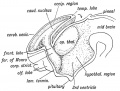
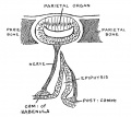
| Fetal Pineal Anatomy{{#pmid:24757681|PMID24757681}} | |||
Superior (dorsal) view of the diencephalic-mesencephalic area of a 3.5-month-old human fetus. | |||
The third ventricle (3 ventr) without pial covering is seen to the right in the micrograph. | |||
The small pineal gland is a small protuberance (arrow) and merging via the broad stalk with the habenula (Ha). Sup col.: superior colliculus. | |||
Bar = 2 mm. | |||
| [[File:Fetal pineal gland 01.jpg|400px]] | |||
|} | |||
==Melatonin== | ==Melatonin== | ||
[[File:Melatonin.jpg|thumb|Melatonin molecular structure]] | [[File:Melatonin.jpg|thumb|Melatonin molecular structure]] | ||
Melatonin synchronises circadian rhythms through two G-protein-coupled receptors (MT1, MT2). Night release from the pineal gland activates melatonin receptors in the suprachiasmatic nucleus of the {{hypothalamus}}, this synchronises light–dark cycle of physiology and behaviour. | |||
* Melatonin is synthesized from the amino acid tryptophan within the pinealocytes. | * Melatonin is synthesized from the amino acid tryptophan within the pinealocytes. | ||
** Serotonin is first acetylated by aryl alkylamine N-acetyltransferase (AA-NAT), then converted to melatonin by acetyl serotonin methyl transferase (ASMT also known as hydroxyindole O-methyltransferase or HIOMT). | ** Serotonin is first acetylated by aryl alkylamine N-acetyltransferase (AA-NAT), then converted to melatonin by acetyl serotonin methyl transferase (ASMT also known as hydroxyindole O-methyltransferase or HIOMT). | ||
* Melatonin release is stimulated by darkness and inhibited by light and is said to have neurological "chronobiotic" properties for resynchronization of sleep and circadian rhythms disturbances. In the periphery, melatonin is also involved in the regulation of several complex cycles: seasonal reproduction, body weight and energy balance. | * Melatonin release is stimulated by darkness and inhibited by light and is said to have neurological "chronobiotic" properties for resynchronization of sleep and circadian rhythms disturbances. In the periphery, melatonin is also involved in the regulation of several complex cycles: seasonal reproduction, body weight and energy balance. | ||
* Melatonin levels can be monitored by urinary excretion of the melatonin metabolite 6-sulfatoxymelatonin (aMT.6S). | * Melatonin levels can be monitored by urinary excretion of the melatonin metabolite 6-sulfatoxymelatonin (aMT.6S). | ||
'''Key Facts''' | |||
* less than 30 min to 60 min - serum half-life of melatonin | |||
* 70% - serum melatonin bound to albumin | |||
* 30% - diffuses in surrounding tissues | |||
* liver - primary metabolism | |||
* kidney - secondary metabolism | |||
===Melatonin Receptors=== | ===Melatonin Receptors=== | ||
The hormone melatonin acts through receptors (high affinity G protein-coupled) embedded in the cell membrane. Three different receptor subtypes have been identified in mammals: MT1 ( | The hormone melatonin acts through receptors (high affinity G protein-coupled) embedded in the cell membrane. Three different receptor subtypes have been identified in mammals: MT1 (MTNR1A) and MT2 (MTNR1B) and a putative binding site called MT3. | ||
* MT1 - expressed in humans in the pars tuberalis of the pituitary gland and the suprachiasmatic nuclei of the hypothalamus. | * [https://www.omim.org/entry/600665 MT1] - ({{Chr4}}q35.2) expressed in humans in the pars tuberalis of the pituitary gland and the suprachiasmatic nuclei of the hypothalamus. | ||
* MT2 - expressed in the retina. | * [https://www.omim.org/entry/600804 MT2] - ({{Chr11}}q14.3) expressed in the retina and brain.{{#pmid:7568007|PMID7568007}} | ||
* MT3 - expressed in many non-mammalian vertebrates in a range of brain areas. | * MT3 - expressed in many non-mammalian vertebrates in a range of brain areas. | ||
:'''Links:''' [http://www.nature.com/nrd/journal/v9/n8/images/nrd3140-f2.jpg Image - melatonergic receptors coupled via Gαi] | :'''Links:''' [http://www.nature.com/nrd/journal/v9/n8/images/nrd3140-f2.jpg Image - melatonergic receptors coupled via Gαi] | ||
==Innervation== | ==Innervation== | ||
The gland is connected to the hypothalamus suprachiasmatic nucleus (SCN) central rhythm generator | The gland is connected to the hypothalamus suprachiasmatic nucleus (SCN) central rhythm generator through a multi-synaptic pathway. | ||
Nerve fibers innervating the mammalian pineal gland originate from perikarya located in the sympathetic superior cervical ganglion, the parasympathetic sphenopalatine and otic ganglia, as well as by nerve fibers originating in the central nervous system. | Nerve fibers innervating the mammalian pineal gland originate from perikarya located in the sympathetic superior cervical ganglion, the parasympathetic sphenopalatine and otic ganglia, as well as by nerve fibers originating in the central nervous system.{{#pmid:12111544|PMID12111544}} | ||
* sympathetic nerves - contain norepinephrine and neuropeptide Y as neurotransmitters | * sympathetic nerves - contain norepinephrine and neuropeptide Y as neurotransmitters | ||
| Line 71: | Line 116: | ||
* trigeminal ganglion - containing substance P, calcitonin gene-related peptide, and pituitary adenylate cyclase-activating peptide | * trigeminal ganglion - containing substance P, calcitonin gene-related peptide, and pituitary adenylate cyclase-activating peptide | ||
== Molecular Development == | |||
{| | |||
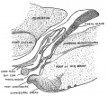
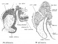
| [[File:Mouse pineal E15 to E21 neuroepithelium 01.jpg|500px]] | |||
Pineal gland {{mouse}} (E15 to E21) | |||
| The pineal gland develops from neuroepithelial cells that express the transcription factor Pax6 and the intermediate filament vimentin. | |||
Panels display confocal microscopy of immunolabeled sagittal sections of rat pineal gland (PG) from embryonic day (E) 15 to E21. | |||
* '''A1-G1''' show expression of Pax6 (green) and vimentin (VIM, red) in pineal precursor cells at low magnification. | |||
* '''A2-G2''' display the same structures at higher magnification. | |||
* '''A3-G3''' and '''A4-G4''' show Pax6 and vimentin expression for each stage of development, respectively. | |||
Pineal organogenesis begins around E15 as an evagination of the neuroepithelium in the dorsal diencephalon that is densely populated by Pax6-expressing cells (green). The developing PG becomes a tubular extension at E16. The orientation of Pax6/VIM+ cells is radial at these stages. At E17 the pineal neuroepithelium begins to fold and fuses at the midline. After fusion of the neuroepithelium, double immunolabeled rosette-like structures are visible in the E18-E21 developing PG. At E21 the PG has developed into a recognizable globular structure. (A1-G1) 20x; scale bar: 75 μm. (A2-G4) 60x; scale bar: 25 μm. PC, posterior commissure. SCO, subcommissural organ. 3v, third ventricle. | |||
|} | |||
* '''Bsx''' - homeobox gene-encoded transcription factor{{#pmid:31808568|PMID31808568}} | |||
* '''Nodal''' - zebrafish required for dorsal convergence of pineal precursors.{{#pmid:17996054|PMID17996054}} | |||
* '''Pax6''' - rat pineal gland from E16, peak expression around E18.{{#pmid:23076630|PMID23076630}} | |||
* '''Fgf8a''' - zebrafish epithalamus acts permissively to promote parapineal fate.{{#pmid:23250206|PMID23250206}} | |||
* '''DARPP-32''' (Dopamine- and cAMP-regulated phosphoprotein of 32 kDa) is involved in the retinal pathway transmitting photic information that resets the circadian clock. | |||
* OTX2, RAX, CRX, PAX4, TBX2B - also identified in development. | |||
'''Links:''' {{molecular}} | |||
==Abnormalities== | ==Abnormalities== | ||
* '''Pineal Hypoplasia''' associated with retinal disease. | * '''Pineal Hypoplasia''' associated with retinal disease. | ||
* '''Pineal Tumours''' in children are associated with abnormal puberty development. | * '''Pineal Tumours''' in children are associated with abnormal puberty development. | ||
==Histology== | ==Histology== | ||
| Line 91: | Line 159: | ||
:'''Links:''' [[:File:Pineal histology 002.jpg|large histology image]] | :'''Links:''' [[:File:Pineal histology 002.jpg|large histology image]] | ||
== References == | == References == | ||
| Line 109: | Line 178: | ||
===Reviews=== | ===Reviews=== | ||
{{#pmid31866317}} | |||
{{#pmid:30502386}} | |||
{{#pmid:30247830}} | |||
{{#pmid:23466884}} | |||
{{#pmid:16021838}} | |||
{{#pmid:15589268}} | |||
{{#pmid:15352385}} | |||
{{#pmid:14561326}} | |||
{{#pmid:9852259}} | |||
===Articles=== | ===Articles=== | ||
{{#pmid:31918071}} | |||
{{#pmid:27861587}} | |||
{{#pmid:25758643}} | |||
{{#pmid:19665543}} | |||
{{#pmid:16988913}} | |||
{{#pmid:16182388}} | |||
{{#pmid:8938291}} | |||
===Search PubMed=== | |||
'''Search Pubmed:''' [http://www.ncbi.nlm.nih.gov/sites/entrez?db=pubmed&cmd=search&term=pineal%20development pineal development] | '''Search Pubmed:''' [http://www.ncbi.nlm.nih.gov/sites/entrez?db=pubmed&cmd=search&term=pineal%20development pineal development] | ||
| Line 139: | Line 222: | ||
==Additional Images== | ==Additional Images== | ||
<gallery> | |||
File:Adult human brain MRI01.jpg | |||
File:Fetal pineal gland 01.jpg | |||
</gallery> | |||
===Historic=== | |||
{{Historic Disclaimer}} | |||
{{Ref-Cooper1932}} | |||
<gallery> | |||
File:Cooper1932-fig01.jpg|Fig. 1. Sagittal midline section through head of 25 mm embryo | |||
File:Cooper1932-fig02.jpg|Fig. 2. Sagittal section through head of 35 mm embryo | |||
File:Cooper1932-fig03.jpg|Fig. 3. Sagittal section through pineal region embryo 3-4 months | |||
File:Cooper1932-fig04.jpg|Fig. 4. Sagittal section through pineal region foetus of 4 months | |||
</gallery> | |||
{{Ref-Keith1902}} | |||
<gallery> | |||
File:Keith1902 fig017.jpg
|Keith1902 Fig 17 | |||
File:Keith1902 fig167.jpg
|Keith1902 Fig 167 | |||
File:Keith1902 fig173.jpg
|Keith1902 Fig 173 | |||
File:Keith1921 fig091.jpg
|Keith1921 Fig 91 | |||
File:Keith1921 fig100.jpg
|Keith1921 Fig 100 | |||
File:Keith1921 fig104.jpg
|Keith1921 Fig 104 | |||
File:Keith1921 fig105.jpg
|Keith1921 Fig 105 | |||
</gallery> | |||
==Terms== | ==Terms== | ||
{{Endocrine terms}} | |||
Latest revision as of 12:56, 5 April 2020
| Embryology - 17 Apr 2026 |
|---|
| Google Translate - select your language from the list shown below (this will open a new external page) |
|
العربية | català | 中文 | 中國傳統的 | français | Deutsche | עִברִית | हिंदी | bahasa Indonesia | italiano | 日本語 | 한국어 | မြန်မာ | Pilipino | Polskie | português | ਪੰਜਾਬੀ ਦੇ | Română | русский | Español | Swahili | Svensk | ไทย | Türkçe | اردو | ייִדיש | Tiếng Việt These external translations are automated and may not be accurate. (More? About Translations) |
Introduction


The pineal gland (epiphysis cerebri) has an important role in the sleep/wake daily cycle (circadian), high melatonin plasma levels at nighttime and very low levels at daytime, and reproductive development. The gland is thought to evolutionarily to have been positioned as to be exposed to light, and hence remains a regulator of cyclic rhythms associated with day/night and day length. The pineal hormone (melatonin) has targets both in the nervous system and in many different peripheral tissues. Melatonin synchronises circadian rhythms acting through the two G-protein-coupled receptors (MT1, MT2) in the suprachiasmatic nucleus of the hypothalamus.
The embryo and fetus pineal does not produce significant amounts of melatonin, though has abundant tissue receptors. The maternal pineal gland produces melatonin in the normal circadian fashion and this melatonin can cross both the placenta and blood-brain barrier. In other species, maternal melatonin crosses the placenta into fetal circulation and may provide photoperiodic information during fetal development that influences later postnatal circadian (daily day/night) and seasonal (day length) rhythms. The pineals of non-mammalian vertebrates are photoreceptive, whereas those of mammals do not normally respond to directly light. Pineal blood supply is derived from the posterior cerebral artery choroidal branches and through the pineal recess is bathed in cerebrospinal fluid (CSF).
The melatonin levels in premature infants is lower and delayed, but not different when calculated from conception date. Other factors such as preeclampsia, growth restriction, and nursery lighting can cause altered rhythm development. The same study has also shown that full-term infants born at home and full-term twins born in the hospital had significantly lower metabolite excretion levels than hospital-born singleton infants at the same ages despite similar body weights.[1] Allopregnanolone, a pineal neurosteroid, acts on the cerebellum supporting survival Purkinje cell by suppressing caspase-3 expression.[2]
Postnatally in humans, the melatonin levels may act on clock gene expression in the pars tuberalis of the anterior pituitary for photoperiodic switching. Melatonin also has important functions related to the program timing of puberty. Note that there are many clinical studies investigating the possible role of melatonin in diverse health areas, from oxygen starvation at birth through to neural effects in old age.
Overview
- part of epithalmus - neurons, glia and pinealocytes
- pinealocytes secrete melatonin - cyclic nature of activity, melatonin lowest during daylight
- inhibit hypothalamic secretion of GnRH until puberty, pineal gland then rapidly regresses.
- other activities - possibly gamete maturation, antioxidant effect, protect neurons?
Links: pineal | Category:Pineal | Lecture - Endocrine Development | Lecture - Head Development
| Historic Pineal Papers |
|---|
| 1917 Pineal Region | 1932 Pineal Gland and Cysts | 1935 Pineal | 1937 Human Pineal | 1940 Nerve and Vascular Supply |
Some Recent Findings

|
| More recent papers |
|---|
 This table allows an automated computer search of the external PubMed database using the listed "Search term" text link.
More? References | Discussion Page | Journal Searches | 2019 References | 2020 References Search term: Pineal Embryology | Pineal Development | Melatonin Development | Melatonin Embryology | Melatonin Receptor | Pineal Hypoplasia | Pineal Tumour |
| Older papers |
|---|
| These papers originally appeared in the Some Recent Findings table, but as that list grew in length have now been shuffled down to this collapsible table.
See also the Discussion Page for other references listed by year and References on this current page.
|
Development Overview
- Neuroectoderm - prosenecephalon then diencephalon
- caudal roof, median diverticulum, epiphysis
- Initially a hollow diverticulum, cell proliferation to solid, pinealocytes (neuroglia), cone-shaped gland innervated by epithalamus
Epithalamus consists of the pineal gland and habenular nuclei
| Fetal Pineal Anatomy[9]
Superior (dorsal) view of the diencephalic-mesencephalic area of a 3.5-month-old human fetus. The third ventricle (3 ventr) without pial covering is seen to the right in the micrograph. The small pineal gland is a small protuberance (arrow) and merging via the broad stalk with the habenula (Ha). Sup col.: superior colliculus. Bar = 2 mm. |

|
Melatonin

Melatonin synchronises circadian rhythms through two G-protein-coupled receptors (MT1, MT2). Night release from the pineal gland activates melatonin receptors in the suprachiasmatic nucleus of the hypothalamus, this synchronises light–dark cycle of physiology and behaviour.
- Melatonin is synthesized from the amino acid tryptophan within the pinealocytes.
- Serotonin is first acetylated by aryl alkylamine N-acetyltransferase (AA-NAT), then converted to melatonin by acetyl serotonin methyl transferase (ASMT also known as hydroxyindole O-methyltransferase or HIOMT).
- Melatonin release is stimulated by darkness and inhibited by light and is said to have neurological "chronobiotic" properties for resynchronization of sleep and circadian rhythms disturbances. In the periphery, melatonin is also involved in the regulation of several complex cycles: seasonal reproduction, body weight and energy balance.
- Melatonin levels can be monitored by urinary excretion of the melatonin metabolite 6-sulfatoxymelatonin (aMT.6S).
Key Facts
- less than 30 min to 60 min - serum half-life of melatonin
- 70% - serum melatonin bound to albumin
- 30% - diffuses in surrounding tissues
- liver - primary metabolism
- kidney - secondary metabolism
Melatonin Receptors
The hormone melatonin acts through receptors (high affinity G protein-coupled) embedded in the cell membrane. Three different receptor subtypes have been identified in mammals: MT1 (MTNR1A) and MT2 (MTNR1B) and a putative binding site called MT3.
- MT1 - (4q35.2) expressed in humans in the pars tuberalis of the pituitary gland and the suprachiasmatic nuclei of the hypothalamus.
- MT2 - (11q14.3) expressed in the retina and brain.[10]
- MT3 - expressed in many non-mammalian vertebrates in a range of brain areas.
Innervation
The gland is connected to the hypothalamus suprachiasmatic nucleus (SCN) central rhythm generator through a multi-synaptic pathway.
Nerve fibers innervating the mammalian pineal gland originate from perikarya located in the sympathetic superior cervical ganglion, the parasympathetic sphenopalatine and otic ganglia, as well as by nerve fibers originating in the central nervous system.[11]
- sympathetic nerves - contain norepinephrine and neuropeptide Y as neurotransmitters
- parasympathetic nerves - contain vasoactive intestinal peptide and peptide histidine isoleucine
- trigeminal ganglion - containing substance P, calcitonin gene-related peptide, and pituitary adenylate cyclase-activating peptide
Molecular Development
|
Pineal gland mouse (E15 to E21) |
The pineal gland develops from neuroepithelial cells that express the transcription factor Pax6 and the intermediate filament vimentin.
Panels display confocal microscopy of immunolabeled sagittal sections of rat pineal gland (PG) from embryonic day (E) 15 to E21.
Pineal organogenesis begins around E15 as an evagination of the neuroepithelium in the dorsal diencephalon that is densely populated by Pax6-expressing cells (green). The developing PG becomes a tubular extension at E16. The orientation of Pax6/VIM+ cells is radial at these stages. At E17 the pineal neuroepithelium begins to fold and fuses at the midline. After fusion of the neuroepithelium, double immunolabeled rosette-like structures are visible in the E18-E21 developing PG. At E21 the PG has developed into a recognizable globular structure. (A1-G1) 20x; scale bar: 75 μm. (A2-G4) 60x; scale bar: 25 μm. PC, posterior commissure. SCO, subcommissural organ. 3v, third ventricle. |
- Bsx - homeobox gene-encoded transcription factor[12]
- Nodal - zebrafish required for dorsal convergence of pineal precursors.[13]
- Pax6 - rat pineal gland from E16, peak expression around E18.[14]
- Fgf8a - zebrafish epithalamus acts permissively to promote parapineal fate.[15]
- DARPP-32 (Dopamine- and cAMP-regulated phosphoprotein of 32 kDa) is involved in the retinal pathway transmitting photic information that resets the circadian clock.
- OTX2, RAX, CRX, PAX4, TBX2B - also identified in development.
Links: molecular
Abnormalities
- Pineal Hypoplasia associated with retinal disease.
- Pineal Tumours in children are associated with abnormal puberty development.
Histology

Adult Histology
- Astrocytes - small dark nuclei
- Pinealocytes - most nuclei present, larger lighter and round nuclei surrounded by a broad rim of light cytoplasm
- Endothelial cells - nuclei in association with the vessels and capillaries traversing the tissue.
- Cytoplasmic processes - "stringy" appearance from both pinealocytes and astrocytes
- Links: large histology image
References
- ↑ Kennaway DJ, Goble FC & Stamp GE. (1996). Factors influencing the development of melatonin rhythmicity in humans. J. Clin. Endocrinol. Metab. , 81, 1525-32. PMID: 8636362 DOI.
- ↑ Tsutsui K. (2019). Kobayashi award: Discovery of cerebellar and pineal neurosteroids and their biological actions on the growth and survival of Purkinje cells during development (review). Gen. Comp. Endocrinol. , 284, 113051. PMID: 30339808 DOI.
- ↑ 3.0 3.1 Ibañez Rodriguez MP, Noctor SC & Muñoz EM. (2016). Cellular Basis of Pineal Gland Development: Emerging Role of Microglia as Phenotype Regulator. PLoS ONE , 11, e0167063. PMID: 27861587 DOI.
- ↑ Bolat D, Kürüm A & Canpolat S. (2018). Morphology and quantification of sheep pineal glands at pre-pubertal, pubertal and post-pubertal periods. Anat Histol Embryol , 47, 338-345. PMID: 29774950 DOI.
- ↑ Gerasimov AV, Kostyuchenko VP, Potapov AV, Varakuta EY, Karpova MR, Sukhanova GA & Logvinov SV. (2018). Infradian Rhythm of the Content of Secretory Granules in Pinealocyte Cytoplasm in Mice and Rats. Bull. Exp. Biol. Med. , 165, 276-279. PMID: 29931631 DOI.
- ↑ Yamazaki F, Møller M, Fu C, Clokie SJ, Zykovich A, Coon SL, Klein DC & Rath MF. (2015). The Lhx9 homeobox gene controls pineal gland development and prevents postnatal hydrocephalus. Brain Struct Funct , 220, 1497-509. PMID: 24647753 DOI.
- ↑ Reiter RJ, Tan DX, Korkmaz A & Rosales-Corral SA. (2014). Melatonin and stable circadian rhythms optimize maternal, placental and fetal physiology. Hum. Reprod. Update , 20, 293-307. PMID: 24132226 DOI.
- ↑ de Faria Poloni J, Feltes BC & Bonatto D. (2011). Melatonin as a central molecule connecting neural development and calcium signaling. Funct. Integr. Genomics , 11, 383-8. PMID: 21465271 DOI.
- ↑ Møller M, Phansuwan-Pujito P & Badiu C. (2014). Neuropeptide Y in the adult and fetal human pineal gland. Biomed Res Int , 2014, 868567. PMID: 24757681 DOI.
- ↑ Reppert SM, Godson C, Mahle CD, Weaver DR, Slaugenhaupt SA & Gusella JF. (1995). Molecular characterization of a second melatonin receptor expressed in human retina and brain: the Mel1b melatonin receptor. Proc. Natl. Acad. Sci. U.S.A. , 92, 8734-8. PMID: 7568007 DOI.
- ↑ Møller M & Baeres FM. (2002). The anatomy and innervation of the mammalian pineal gland. Cell Tissue Res. , 309, 139-50. PMID: 12111544 DOI.
- ↑ Carstensen MB, Hertz H, Bering T, Møller M, Rohde K, Klein DC, Coon SL & Rath MF. (2020). Circadian regulation and molecular role of the Bsx homeobox gene in the adult pineal gland. J. Pineal Res. , 68, e12629. PMID: 31808568 DOI.
- ↑ Aquilina-Beck A, Ilagan K, Liu Q & Liang JO. (2007). Nodal signaling is required for closure of the anterior neural tube in zebrafish. BMC Dev. Biol. , 7, 126. PMID: 17996054 DOI.
- ↑ Rath MF, Rohde K, Klein DC & Møller M. (2013). Homeobox genes in the rodent pineal gland: roles in development and phenotype maintenance. Neurochem. Res. , 38, 1100-12. PMID: 23076630 DOI.
- ↑ Clanton JA, Hope KD & Gamse JT. (2013). Fgf signaling governs cell fate in the zebrafish pineal complex. Development , 140, 323-32. PMID: 23250206 DOI.
Online Textbooks
- Pineal Gland and Cancer-An Epigenetic Approach to the Control of Malignancy: Evaluation of the Role of Melatonin Eurekah Bioscience Collection - Neuropharmacology
- Endocrine changes in puberty Endocrinology -> The gonad
- Second Malignancies Cancer Medicine -> Section 24: The Eye -> 85. Neoplasms of the Eye -> Pediatric Ophthalmic Oncology: Ocular Diseases
- The Action of Melatonin on Experimental in-Vivo Tumors Eurekah Bioscience Collection -> Neuropharmacology -> Pineal Gland and Cancer-An Epigenetic Approach to the Control of Malignancy: Evaluation of the Role of Melatonin -> Effect of Melatonin on Tumor Growth
- Potential Significance of (Patho)Physiological Changes of Melatonin for the Aetiology of Cancer Eurekah Bioscience Collection -> Neuropharmacology -> Pineal Gland and Cancer-An Epigenetic Approach to the Control of Malignancy: Evaluation of the Role of Melatonin
- Effects of Exogenous Melatonin AHRQ Evidence reports and summaries -> AHRQ Evidence Reports, Numbers 61 - 119 -> 108. Mel
Journals
- Journal of Pineal Research Molecular, Biological, Physiological and Clinical Aspects of Melatonin
Reviews
{{#pmid31866317}}
Farias Altamirano LE, Freites CL, Vásquez E & Muñoz EM. (2018). Signaling within the pineal gland: A parallelism with the central nervous system. Semin. Cell Dev. Biol. , , . PMID: 30502386 DOI.
Ilahi S & Ilahi TB. (2018). Physiology, Pineal Gland. , , . PMID: 30247830
Chen YC, Sheen JM, Tiao MM, Tain YL & Huang LT. (2013). Roles of melatonin in fetal programming in compromised pregnancies. Int J Mol Sci , 14, 5380-401. PMID: 23466884 DOI.
Weinert D. (2005). Ontogenetic development of the mammalian circadian system. Chronobiol. Int. , 22, 179-205. PMID: 16021838
Macchi MM & Bruce JN. (2004). Human pineal physiology and functional significance of melatonin. Front Neuroendocrinol , 25, 177-95. PMID: 15589268 DOI.
Barrenetxe J, Delagrange P & Martínez JA. (2004). Physiological and metabolic functions of melatonin. J. Physiol. Biochem. , 60, 61-72. PMID: 15352385
Ekström P & Meissl H. (2003). Evolution of photosensory pineal organs in new light: the fate of neuroendocrine photoreceptors. Philos. Trans. R. Soc. Lond., B, Biol. Sci. , 358, 1679-700. PMID: 14561326 DOI.
Thomas L, Drew JE, Abramovich DR & Williams LM. (1998). The role of melatonin in the human fetus (review). Int. J. Mol. Med. , 1, 539-43. PMID: 9852259
Articles
Barros VRP, Monte APO, Santos JMS, Lins TLBG, Cavalcante AYP, Gouveia BB, Müller MC, Oliveira JL, Donfack NJ, Araújo VR & Matos MHT. (2020). Melatonin improves development, mitochondrial function and promotes the meiotic resumption of sheep oocytes from in vitro grown secondary follicles. Theriogenology , 144, 67-73. PMID: 31918071 DOI.
Ibañez Rodriguez MP, Noctor SC & Muñoz EM. (2016). Cellular Basis of Pineal Gland Development: Emerging Role of Microglia as Phenotype Regulator. PLoS ONE , 11, e0167063. PMID: 27861587 DOI.
Shoja MM, Hoepfner LD, Agutter PS, Singh R & Tubbs RS. (2016). History of the pineal gland. Childs Nerv Syst , 32, 583-6. PMID: 25758643 DOI.
Sun B, Wang D, Tang Y, Fan L, Lin X, Yu T, Qi H, Li Z & Liu S. (2009). The pineal volume: a three-dimensional volumetric study in healthy young adults using 3.0 T MR data. Int. J. Dev. Neurosci. , 27, 655-60. PMID: 19665543 DOI.
Al-Hussain SM. (2006). The pinealocytes of the human pineal gland: A light and electron microscopic study. Folia Morphol. (Warsz) , 65, 181-7. PMID: 16988913
Saito S, Tachibana T, Choi YH, Denbow DM & Furuse M. (2005). ICV melatonin reduces acute stress responses in neonatal chicks. Behav. Brain Res. , 165, 197-203. PMID: 16182388 DOI.
Sumida M, Barkovich AJ & Newton TH. (1996). Development of the pineal gland: measurement with MR. AJNR Am J Neuroradiol , 17, 233-6. PMID: 8938291
Search PubMed
Search Pubmed: pineal development
External Links
External Links Notice - The dynamic nature of the internet may mean that some of these listed links may no longer function. If the link no longer works search the web with the link text or name. Links to any external commercial sites are provided for information purposes only and should never be considered an endorsement. UNSW Embryology is provided as an educational resource with no clinical information or commercial affiliation.
- NIH The Julius Axelrod Papers | The Pineal Gland and the "Melatonin Hypothesis," 1959-1974
- NIH Child Health and Human Development (USA) Pineal Gland and Chronobiology: Regulation of Pineal Function
- University of Cincinnati SURVEY OF ENDOCRINE ORGANS
Additional Images

Historic
| Historic Disclaimer - information about historic embryology pages |
|---|
|
Cooper ERA. The human pineal gland and pineal cysts. (1932)
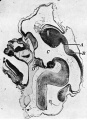
Fig. 1. Sagittal midline section through head of 25 mm embryo
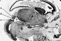
Fig. 2. Sagittal section through head of 35 mm embryo
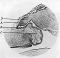
Fig. 3. Sagittal section through pineal region embryo 3-4 months
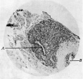
Fig. 4. Sagittal section through pineal region foetus of 4 months


Keith A. Human Embryology and Morphology. (1902) London: Edward Arnold.
Keith1902 Fig 17
Keith1902 Fig 167
Keith1902 Fig 173
Keith1921 Fig 91
Keith1921 Fig 100
Keith1921 Fig 104
Keith1921 Fig 105


Terms
| Endocrine Terms (expand to view) |
|---|
|
| Other Terms Lists |
|---|
| Terms Lists: ART | Birth | Bone | Cardiovascular | Cell Division | Endocrine | Gastrointestinal | Genital | Genetic | Head | Hearing | Heart | Immune | Integumentary | Neonatal | Neural | Oocyte | Palate | Placenta | Radiation | Renal | Respiratory | Spermatozoa | Statistics | Tooth | Ultrasound | Vision | Historic | Drugs | Glossary |
Glossary Links
- Glossary: A | B | C | D | E | F | G | H | I | J | K | L | M | N | O | P | Q | R | S | T | U | V | W | X | Y | Z | Numbers | Symbols | Term Link
Cite this page: Hill, M.A. (2026, April 17) Embryology Endocrine - Pineal Development. Retrieved from https://embryology.med.unsw.edu.au/embryology/index.php/Endocrine_-_Pineal_Development
- © Dr Mark Hill 2026, UNSW Embryology ISBN: 978 0 7334 2609 4 - UNSW CRICOS Provider Code No. 00098G