Respiratory System - Upper Respiratory Tract
| Embryology - 23 Jun 2026 |
|---|
| Google Translate - select your language from the list shown below (this will open a new external page) |
|
العربية | català | 中文 | 中國傳統的 | français | Deutsche | עִברִית | हिंदी | bahasa Indonesia | italiano | 日本語 | 한국어 | မြန်မာ | Pilipino | Polskie | português | ਪੰਜਾਬੀ ਦੇ | Română | русский | Español | Swahili | Svensk | ไทย | Türkçe | اردو | ייִדיש | Tiếng Việt These external translations are automated and may not be accurate. (More? About Translations) |
Introduction

The respiratory system does not carry out its physiological function (of gas exchange) until after birth. The respiratory tract, diaphragm and lungs do form early in embryonic development. In the head/neck region, the pharynx forms a major arched cavity within the phrayngeal arches.
The respiratory tract is divided anatomically into 2 main parts:
- upper respiratory tract - consisting of the nose, nasal cavity and the pharynx.
- lower respiratory tract - consisting of the larynx, trachea, bronchi and the lungs.
Note that some components of the upper respiratory tract development are covered in Smell Development and Head Development.
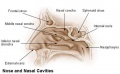
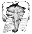
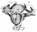
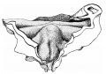
Nasal cavities
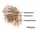
Pharynx
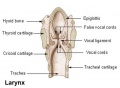
Larynx


Some Recent Findings

|
| More recent papers |
|---|
 This table allows an automated computer search of the external PubMed database using the listed "Search term" text link.
More? References | Discussion Page | Journal Searches | 2019 References | 2020 References Search term: Upper Respiratory Tract Embryology <pubmed limit=5>Upper Respiratory Tract Embryology</pubmed> Search term: Pharynx Development <pubmed limit=5>Pharynx+Development</pubmed> Search term: Larynx Development <pubmed limit=5>Larynx+Development</pubmed> |
Textbooks
- Human Embryology Larson Chapter 9 p229-260
- The Developing Human: Clinically Oriented Embryology (6th ed.) Moore and Persaud Chapter 12 p271-302
- Before We Are Born (5th ed.) Moore and Persaud Chapter 13 p255-287
- Essentials of Human Embryology Larson Chapter 9 p123-146
- Human Embryology Fitzgerald and Fitzgerald Chapter 19,20 p119-123
- Anatomy of the Human Body 1918 Henry Gray 1. The Respiratory Apparatus
Nasal Olfactory and Respiratory Epithelium
Olfactory epithelium

|

|
| Olfactory Epithelium (overview) | Olfactory Epithelium (detail) |
- Olfactory cells
- Sustentacular cells - located mainly in the superficial cell layer of the epithelium (difficult to distinguish from olfactory cells).
- Basal cells - identified by their location in the epithelium.
Epithelium
- Cilia are not visible
- goblet cells are absent from the olfactory epithelium.
Lamina Propria
- olfactory axon bundles (lightly stained, rounded areas) connected to olfactory cells.
- Bowman's glands - (small mucous glands, olfactory glands) function to moisturise the epithelium.
- Nasal Olfactory Histology: overview image | detail image | Smell Development | Histology | Histology Stains
Respiratory Epithelium

|

|
| Respiratory Epithelium (overview) | Respiratory Epithelium (detail) |
- goblet cells
- ciliated cells
- basal cells
Lamina propria
- connective tissue
- cavernous sinusoids - large spaces (empty or filled with red blood cells)
- glandular tissue - mucous glands (green) and muco-serous glands (brownish-green)
Bone
- Lamellae and osteocytes in lacunae.
- Haversian systems are rare or absent.
- Respiratory Histology: Bronchiole | Alveolar Duct | Alveoli | EM Alveoli septum | Alveoli Elastin | Trachea 1 | Trachea 2 | labeled lung | unlabeled lung | Respiratory Bronchiole | Lung Reticular Fibres | Nasal Inferior Concha | Nasal Respiratory Epithelium | Olfactory Region overview | Olfactory Region Epithelium | Histology Stains
Paranasal Sinuses

Paranasal sinuses are thought to develop as "pneumatisation" of bone and out-pocketing of the respiratory nasal epithelium. There are 4 paired sinuses, named by their anatomical location, and lined with respiratory epithelium. The sinuses begins to form at 10 weeks (GA) by primary pneumatisation, later in fetal development secondary pneumatisation occurs enlarging the existing spaces. These sinuses continue to enlarge postnatally.
Note that during development, these are amniotic fluid fluid-filled spaces, therefore pneumatisation (USA, pneumatization) is a misnomer as only postnatally fluid loss forms the air-filled (pneumatic) spaces.
A study of fetal cleft lip and palate[5] showed that sphenoid sinus was still present with variability in morphology compared to normal fetus perhaps due to altered shape and size of the adjacent hypertrophic cartilaginous structures.
Computed tomography measurements from a study of 120 adult (age 18-65 years) maxillary and frontal sinuses.[6]
- mean maxillary sinus volume 15.7±5.3 cm3
- larger in males than in females.
- no correlation between the volume of maxillary sinuses with either age or side.
- mean bone thickness at the canine fossa was 1.1±0.4 mm.
Frontal Sinus
Ethmoid Sinus
A 1997 article[7] based upon study of coronal sections of the heads of 23 human fetuses from 18-mm CR length to 282-mm CR length. The study suggests that the ethmoid sinus forms by: "constriction of the nasal cavity by a pair of turbinal cushions, and evagination from the nasal cavity by proliferation and subsequent disintegration of the nasal epithelium".
Sphenoid Sinus
Data quoted in a 1996 article[8] on the sphenoid sinus.
- 4 month - sphenoid sinuses can be identified.
- at birth - sinus remains small and is little more than an evagination of the sphenoethmoid recess.
- year 3 - invasion of the sphenoid bone is more rapid
- year 7 - sinus has extended posteriorly to the level of the sella turcica.
- year 12 - sphenoid pneumatization reaches its final form and a size equivalent to the adult
- adult - further enlargement into the basisphenoid may occur.
Maxillary Sinus
Developmentally, the maxillary sinus originates in the middle meatus and extends into the ethmoid cartilage.
The data below is from a recent microscopical study of 100 human fetuses from the 9th to the 37th week (GA).[9]
- week 10 - maxillary sinus begins development.
- week 37 - the anterior-posterior diameter has a mean of 4.36 mm; ossification of the medial wall was absent, and the floor was located below the attachment of the inferior turbinate. Septa and recesses were temporarily observed.
- maxillary sinus osmium (opening) was located at the anterior third of the ethmoid infundibulum
- final dimensions were 1.96 mm in length and 0.44 mm in width.
- mean length between the ostium to the lamina papyracea and nasolacrimal duct was 1 mm.
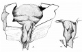
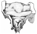
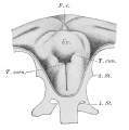
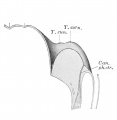
Larynx
The larynx and vocal folds lie at the gastrointestinal tract and respiratory tract separation. Adult functions of this region are associated with breathing, protection and speech. Developmentally, this region goes through the early recanalization process. See the recent review.[2]
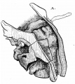
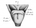
This is a historical pictorial description of larynx embryonic development.[10]
- 1910 - Larynx Development
embryo 5 mm
embryo 6 mm
embryo 6.6 mm
embryo 7.5 mm
embryo 8.5 mm
embryo 12 mm
embryo 22 mm


The following gallery shows the development of the human larynx from the embryonic to fetal period.[11]
- 1912 - Larynx Development
8mm embryo
8-9 mm embryo 28 to 29 days larynx entrance
8-9 mm embryo 28 to 29 days section
15-16 mm embryo 40-42 days larynx entrance
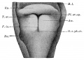
30 mm embryo larynx entrance
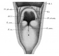
16/23 cm male fetus larynx entrance
29/43 cm male fetus larynx entrance


Movies
The animations below allow a comparison of early and late embryonic lung development. Compare the size and relative position of the respiratory structures and their anatomical relationship to the developing gastrointestinal tract.
| Early embryo (stage 13)
3 dimensional reconstruction based upon a serial reconstruction from individual Carnegie stage 13 embryo slice images. | |
| Late embryo (stage 22)
3 dimensional reconstruction based upon a serial reconstruction from individual embryo slice images Carnegie stage 22, 27 mm Human embryo, approximate day 56. |
- Links: Movies
References
- ↑ <pubmed>26119728</pubmed>| Cell Rep.
- ↑ 2.0 2.1 Lungova V & Thibeault SL. (2020). Mechanisms of larynx and vocal fold development and pathogenesis. Cell. Mol. Life Sci. , , . PMID: 32253462 DOI.
- ↑ Mori M, Mahoney JE, Stupnikov MR, Paez-Cortez JR, Szymaniak AD, Varelas X, Herrick DB, Schwob J, Zhang H & Cardoso WV. (2015). Notch3-Jagged signaling controls the pool of undifferentiated airway progenitors. Development , 142, 258-67. PMID: 25564622 DOI.
- ↑ Hines EA, Jones MK, Verheyden JM, Harvey JF & Sun X. (2013). Establishment of smooth muscle and cartilage juxtaposition in the developing mouse upper airways. Proc. Natl. Acad. Sci. U.S.A. , 110, 19444-9. PMID: 24218621 DOI.
- ↑ Smith TD, Siegel MI, Mooney MP, Burrows AM & Todhunter JS. (1997). Formation and enlargement of the paranasal sinuses in normal and cleft lip and palate human fetuses. Cleft Palate Craniofac. J. , 34, 483-9. PMID: 9431465 <0483:FAEOTP>2.3.CO;2 DOI.
- ↑ Sahlstrand-Johnson P, Jannert M, Strömbeck A & Abul-Kasim K. (2011). Computed tomography measurements of different dimensions of maxillary and frontal sinuses. BMC Med Imaging , 11, 8. PMID: 21466703 DOI.
- ↑ Monteiro VJ & Dias MP. (1997). Morphogenic mechanisms in the development of ethmoidal sinuses. Anat. Rec. , 249, 96-102. PMID: 9294654
- ↑ Antoniades K, Vahtsevanos K, Psimopoulou M & Karakasis D. (1996). Agenesis of sphenoid sinus. Case report. ORL J. Otorhinolaryngol. Relat. Spec. , 58, 347-9. PMID: 8958546 DOI.
- ↑ Nuñez-Castruita A, López-Serna N & Guzmán-López S. (2012). Prenatal development of the maxillary sinus: a perspective for paranasal sinus surgery. Otolaryngol Head Neck Surg , 146, 997-1003. PMID: 22267494 DOI.
- ↑ Frazer JE. Development of the larynx. (1910) J Anat. 44: 156-191. PMID 17232839
- ↑ Template:Ref-GrosserLewisMcmurrich1912
Reviews
Mirilas P. (2011). Lateral congenital anomalies of the pharyngeal apparatus: part III. cadaveric representation of the course of second and third cleft and pouch fistulas. Am Surg , 77, 1257-63. PMID: 21944636
Daniel SJ. (2006). The upper airway: congenital malformations. Paediatr Respir Rev , 7 Suppl 1, S260-3. PMID: 16798587 DOI.
Pohunek P. (2004). Development, structure and function of the upper airways. Paediatr Respir Rev , 5, 2-8. PMID: 15222948 DOI.
Articles
Teul I, Slawinski G, Lewandowski J, Dzieciolowska-Baran E, Gawlikowska-Sroka A & Czerwinski F. (2010). Nasal septum morphology in human fetuses in computed tomography images. Eur. J. Med. Res. , 15 Suppl 2, 202-5. PMID: 21147652
Martinez-Ten P, Adiego B, Perez-Pedregosa J, Illescas T, Wong AE & Sepulveda W. (2010). First-trimester assessment of the nasal bones using the retronasal triangle view: a 3-dimensional sonographic study. J Ultrasound Med , 29, 1555-61. PMID: 20966466
Asaumi R, Sato I, Miwa Y, Imura K, Sunohara M, Kawai T & Yosue T. (2010). Understanding the formation of maxillary sinus in Japanese human foetuses using cone beam CT. Surg Radiol Anat , 32, 745-51. PMID: 20490493 DOI.
Search PubMed
Search Pubmed: Upper Respiratory Tract Development | Upper Respiratory Tract Embryology
Terms
- nasal concha - (turbinate) Within the nasal cavity the narrow, long and curled bone shelf increasing the nasal surface area and partially separating into smaller cavities. (concha = resemble a shell)
Additional Images

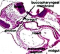
stage 11 foregut

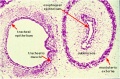
week 4 early respiratory endodermal bud

week 4 later respiratory endodermal bud
Stage 22 trachea
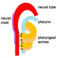
Head arches cartoon
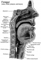
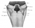
Pharynx
Nasal cavities
Pharynx
Larynx


- part of foregut development
- anatomically the nose, nasal cavity and the pharynx
- the pharynx forms a major arched cavity within the pharyngeal arches
- Larynx Image Links: All cartilages of the larynx | Epiglottis cartilage | Thyroid cartilage | Cricoid cartilage | Arytenoid cartilage | Larynx ligaments anterior | Larynx ligaments posterior | Larynx sagittal section | Larynx and upper trachea | Larynx entrance | Larynx interior | Larynx muscular attachments | Larynx muscles 1 | Larynx muscles 2 | Larynx muscles 3 | Cartilage Development | Respiratory System Development
External Links
External Links Notice - The dynamic nature of the internet may mean that some of these listed links may no longer function. If the link no longer works search the web with the link text or name. Links to any external commercial sites are provided for information purposes only and should never be considered an endorsement. UNSW Embryology is provided as an educational resource with no clinical information or commercial affiliation.
Glossary Links
- Glossary: A | B | C | D | E | F | G | H | I | J | K | L | M | N | O | P | Q | R | S | T | U | V | W | X | Y | Z | Numbers | Symbols | Term Link
Cite this page: Hill, M.A. (2026, Haziran 23) Embryology Respiratory System - Upper Respiratory Tract. Retrieved from https://embryology.med.unsw.edu.au/embryology/index.php/Respiratory_System_-_Upper_Respiratory_Tract
- © Dr Mark Hill 2026, UNSW Embryology ISBN: 978 0 7334 2609 4 - UNSW CRICOS Provider Code No. 00098G