Paper - Retrogressive Changes in the Fetal Vessels and the Suspensory Ligament of the Liver
| Embryology - 27 Apr 2024 |
|---|
| Google Translate - select your language from the list shown below (this will open a new external page) |
|
العربية | català | 中文 | 中國傳統的 | français | Deutsche | עִברִית | हिंदी | bahasa Indonesia | italiano | 日本語 | 한국어 | မြန်မာ | Pilipino | Polskie | português | ਪੰਜਾਬੀ ਦੇ | Română | русский | Español | Swahili | Svensk | ไทย | Türkçe | اردو | ייִדיש | Tiếng Việt These external translations are automated and may not be accurate. (More? About Translations) |
Meyer AW. Retrogressive changes in the fetal vessels and the suspensory ligament of the liver. (1914) Amer. J Anat. 477-521.
| Historic Disclaimer - information about historic embryology pages |
|---|
| Pages where the terms "Historic" (textbooks, papers, people, recommendations) appear on this site, and sections within pages where this disclaimer appears, indicate that the content and scientific understanding are specific to the time of publication. This means that while some scientific descriptions are still accurate, the terminology and interpretation of the developmental mechanisms reflect the understanding at the time of original publication and those of the preceding periods, these terms, interpretations and recommendations may not reflect our current scientific understanding. (More? Embryology History | Historic Embryology Papers) |
Retrogressive Changes in the Fetal Vessels and the Suspensory Ligament of the Liver
From the Division of Anatomy of the Medical School of Stanford University
Twenty-Six figures (1921)
Introduction
The formation of the ligamentum teres and the persistence of the suspensory ligament of the liver in some mammals and not in others are closely related to some of the questions discussed elsewhere (Meyer ’14). In text books, manuals and some monographs on the anatomy of the domestic and other animals it is usually stated that the thickened caudal border of the suspensory ligament of the liver represents the obliterated umbilical vein or round hepatic ligament. Krause (’84) writing on the anatomy of the rabbit adds, that it may occasionally remain patent. Reighard and Jennings (’O1) state that in the cat “The ligamentum teres is the thickened free caudal border of the suspensory ligament. It is the remains of the fetal umbilical vein.” Ellenberger and Baum (’91) writing on the dog, state however, that the suspensory ligament merely contains remnants of the umbilical vein (Ligamentum teres) and Martin (’02—’04) in his Lehrbuch der Anatomic der Haustiere says “In dem rechten Einschnitte (of the liver) ist die Gallenblase mit ihrem Gange eingebettet, in dem linkem Zieht sich. das Ligamentum teres hinein bis zur Nabelvenengrube.” Chaveau (’90) also states that “At its (the suspensory ligament’s) free border is a fibrous cord (the round ligament) formed by the obliteration of the fetal umbilical vein,” and Milne-Edwards (’60) in the “Lecons sur la physiologic et l’anatomie comparee” declared that “On designe sous le nom de ligament rand le cordon fibreux qui resulte de l’atrophie de la veine ombilicale et qui loge dans l’epaisseur de oe repli pres de son bord libre.”
These quotations it seems also represent the consensus of present opinion in this matter and yet anyone who has carefully observed the anatomy of the round and suspensory ligaments of the liver in the domestic animals must have been impressed with the fact that both are often entirely absent in comparatively young animals and at least partially even if not entirely so, in most very old animals. Moreover, it is more than probable that the statements quoted are applicable only to the young of some of the domestic animals. In several dogs three to twelve months old only a vestige of the round ligament could be found in the youngest animals and the suspensory ligament was already reduced to a very small triangular fold which represented approximately but 2 per cent of its original extent. With some slight qualifications this statement also applies to sheep, and to a somewhat less extent also to cats, guinea pigs and rabbits. In most of these animals the suspensory ligament at the time of birth usually extends, of course, along the ventral abdominal wall as far as the umbilicus and contains the umbilical vein in its free margin.
As has been stated elsewhere by the writer, the intra—abdominal portion of the umbilical vein in the sheep cannot and does not retract at time of birth, but remains filled with unclotted blood save in its contracted portion in the immediate vicinity of the umbilicus where it contains a small clot. But soon after birth the umbilical vein is freed from its attachment to the abdominal wall as a result of degenerative changes and then undoubtedly undergoes a delayed though a more rapid retraction and regression after the manner described by Robin (’60), Haberda C96) and Baumgarten (’77) in the case of the vein, and as is true especially in case of the arteries in man. That this retraction and regression takes place comparatively rapidly is well shown by the fact that it had resulted in almost complete disappearance of the umbilical vein or so-called round ligament, in lambs 5 to 7 weeks old. Since degeneration of the round and suspensory ligaments are closely associated the condition of both these structures as found in this lamb and in dogs will be described with some detail.
Instead of a continuous suspensory ligament extending from the umbilicus to the region of the coronary ligament only a fine thread-like strand of peritoneum about 1 mm. wide extended from about the midpoint of the ventral surface of the liver to the abdominal wall about 4 cm. cranial to the umbilicus. Between the latter point and the umbilicus there was a small fold of peritoneum. The peritoneum in this region covered a small quantity of extra-peritoneal fat and was somewhat wrinkled but entirely smooth within a distance of 2 cm. of the umbilicus. Along the abdominal -wall cranial to this strand and on the diaphragm, a fine, free fringe of peritoneum which become gradually wider could be seen very plainly and finally ran into a small triangular suspensory ligament from the dorsal extremity of the caudal margin of which another fold of peritoneum about half a centimeter wide extended to the central end of the above mentioned fine strand. Between the latter and the suspensory ligament there was a large oval opening bounded by the ventral abdominal wall, the diaphragm, suspensory ligament, the liver and the above—mentioned narrow strand. From the dorsal or hepatic attachment of the latter a narrow fringe of peritoneum also extended caudally along the ventral surface of the liver to a small funnel-shaped fossa, or pit in the substance of the liver from the bottom of which projected a small, pointed, conical teat about one centimeter long and three millimeters wide at the base. Because of its appearance and location, this at once suggested the remnant of the umbilical vein. That it was actually such was confirmed later by gross and microscopic examination and it was interesting that this remnant still had a small conical lumen which was in communication with the left portal vein.
The suspensory ligament measured only 2.5 cm. along the hepatic surface and along the diaphragm and had a free cresentic border about 3 cm. long. It is evident, of course, that it and the fine strand as well as the narrow fringes represented only small remnants of the original extensive structure which had undergone almost complete degeneration so early in the life of the animal. Hence it is clear that we have here a very interesting and instructive stage in the degeneration and obliteration of both the umbilical vein and the suspensory ligament which would undoubtedly have been complete a few weeks later. Consequently then, since the only remnant of the suspensory ligament in young sheep is a very small triangular fold and since even this small remnant disappears practically entirely in older animals it is evident that neither it nor the round ligament can be said to exist in adult sheep for both disappear completely during the first months of life. Evidently then the umbilical vein never becomes ligamentous although fibrous transformation may begin, but retracts and degenerates completely and the suspensory ligament becomes fenestrated and undergoes practically total destruction. It is apparent and interesting, however, that by far the greater portion of the suspensory ligament disappears very rapidly and that a very small triangular cranial portion may persist much longer or even permanently.
What is true of the sheep, also holds for the dog for in no case could a trace of either of these ligaments be found in mature or old animals. Moreover, in a dog approximately a year old there was not even an indication of the above-described teat-like remnant of the umbilical vein attached to the left portal as found in the sheep, and the suspensory ligament had already been reduced to a very small triangular. structure only about 1 cm. broad which again represented only the most craniodorsal portion of the fetal structure.
Likewise in dogs approximately two months old there was only a vestige of the umbilical vein in the form of a short tag and only a small triangular remnant of the suspensory ligament. The latter measured 1.3 cm. along the diaphragmatic surface, 1.1 cm. on the hepatic and 9 cm. along its free caudal border. Hence the original ligament can be said to have degenerated almost completely. There was no round ligament whatever and the umbilical vein had retracted and degenerated so completely that nothing remained of it save a small teat or process 3.5 mm. long and 1.5 mm. thick which projected from the wall of the eft portal vein exactly in the position in which the similar remnant had been found in the lamb. Hence it is evident that the processes of obliteration of the round and suspensory ligaments in the dog are wholly comparable to those in the sheep and that they occur with approximately equal rapidity.
A similar condition was also found in a dog about four months old. In this animal the one remnant of the umbilical vein was a fine strand about 5 mm. long and 1 to 1.5 mm. thick Which was attached to and lay on the ventral wall of the portal vein at its bifurcation. The suspensory ligament too was somewhat smaller than in the above animal. Moreover, on one of several dogs only seven weeks old both the suspensory ligament and the umbilical vein had completely disappeared save for a very fine filament of peritoneum which extended from a point opposite the tip of the ensiform cartilage to the left portal vein. At the proximal or hepatic, end of this filament a small, short remnant of the umbilical vein was still present. In the other dog only a small filament 2.5 cm. long which was partly fibrous, was still attached to the left portal, and the suspensory ligament had completely disappeared save for a fine strand of peritoneum which was suspended from the ventral abdominal wall near the tip of the xiphoid process. However, that considerable variation exists in the time of the disappearance of the suspensory ligament, at least, is shown by the fact that in two dogs about one and one-half years old the suspensory ligament which was still present, began at a point opposite the middle of the xiphoid process. Yet the umbilical vein had wholly disappeared in both these animals. The findings in two pups three weeks old and especially in two new-born dogs are in marked contrast to those in the last two animals. In these four pups the suspensory ligaments had completely disappeared except for a very small falciform portion directly ventral to the vena cava. In both the new-born animals the umbilical veins ran directly from the left portal to the umbilicus and was completely isolated. Hence it is evident that whenever this condition exists the formation of a round ligament in the caudal border of the suspensory ligament had never included the vein or had already degenerated before birth, the latter was isolated and hence could no more form a ligament after becoming detached from the umbilicus than the omphalomesenteric or hypogastric vessels can do so after these have become detached, retracted and degenerated, unless a permanent instead of a temporary attachment were subsequently acquired. Moreover, from the findings in these two new-born dogs it seems not at all unlikely that the two pups of the seventh week in whom no remnant of the suspensory ligament could be found were also instances in which degeneration and regression of the suspensory ligament had already begun at the time of birth.
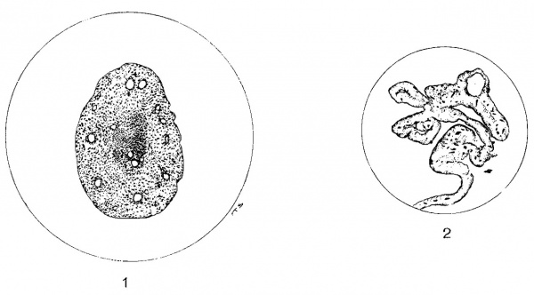
Even in a pup 91 hours old marked changes were plainly evident in the distal portion of the umbilical vein as shown in figure 1. In this case portions of the lumen of the vessel are filled with connective tissue containing some blood vessels. The degenerating musculature which has undergone embryonic regression has lost its characteristic arrangement and staining qualities and also contains numerous blood vessels but can still be recognized as such. This vessel and these changes will be discussed fully below.
Fig. 1 Degenerating umbilical vein of a lamb 91 hours old. X142.
Fig. 2 Plicated caudal border of the suspensory ligament of a rabbit. In some portions these folds are fused. X275.
The degeneration and disappearance of the suspensory ligament and the umbilical vein in the cat occur much slower than in the dog and sheep. In the oldest cats examined no remnant of either structure could be found, however, and in cats one—half to a year old, and occasionally in young kittens, it was not uncommon to find the suspensory ligament more or less fenestrated, a fact which can undoubtedly be correllated with its degeneration or disappearance. Not uncommonly the free caudal border of the suspensory ligament was rolled up as a scroll suggesting the presence of an unusually large round ligament but upon closer microscopic and macroscopic examination not a remnant of the vein nor a fibrous substitute for it could be found and the thickened border of the suspensory ligament was composed of folds of peritoneum or of loose connective tissue. This rolled up or pleated condition of the free caudal border occasionally seen was especially illustrated in the caseof some rabbits and guinea-pigs as shown in figure 2.
In several rabbits over a year old from a third to a half of the suspensory ligament was still preserved although no remnant of the round ligament could be found, but in a guinea-pig of the same age the suspensory ligament was completely preserved from a point opposite the xiphoid process, although the round ligament could not be detected beyond the ventral surface of the liver.
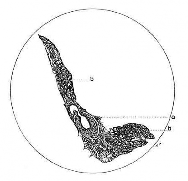
That the rolled up or plicated border of the suspensory ligament can easily simulate the round ligament is excellently illustrated by the behavior of a special fold of the suspensory ligament which is occasionally reflected towards the gall bladder, to the right of the main ligament and which not infrequently contains a vein (fig. 3). When such is the case this reflection has a free border exactly similar to that of the main ligament and if one were to judge from gross appearance one would be compelled to conclude that there are two instead of but one round ligament in these cases for this vein is sometimes very thick-walled as in this instance. In rabbits the condition of both the round and suspensory ligaments is very similar. In guinea pigs they persist relatively longer.
Fig. 3 Drawing of the unrolled border of an accessory free fold from the suspensory ligament to the gall bladder of the dog. The large thick-walled vein is shown at a point where one of its branches joins. a, vein; 1), fat. X42.
Because of the great difficulty in obtaining cats, rabbits and guinea-pigs of definitely known ages the observations on them have been much fewer and hence any conclusions drawn from them might require modification as a result of a more comprehensive series of observations. There is no question, however, that in all these animals the umbilical vein ultimately disappears first near the umbilicus, i.e., centripetally and not centrifugally as Robin and Herzog stated it does in man. It is also clear that the reason that the round ligament in most of the domestic animals usually seems to begin at the tip of the Xiphoid is that the vein has retracted and degenerated and is forced against the Ventral abdominal wall between the umbilicus and xiphoid by the narrow regressing suspensory ligament in whose crescentic border it comes to lie, although in doing so it manifestly must take a more roundabout course than before. This is, no doubt, due to the fact that after its initial retraction the vein which is enclosed in the caudal border of the suspensory ligament is drawn cranially as a result of pre- or post-natal changes. Moreover, it is not at all unlikely that as a result of these factors the free caudal border of the ligament also becomes markedly concave instead of remaining straight or approximately so. The entire absence of a fibrous substitute for the umbilical vein in the sheep, dog, etc., between the umbilicus and the xiphoid process must, it seems to me, be due to the retraction of the vein and would seem to suggest very strongly that Haberda is mistaken when he says that the amount of retraction determines the length of the fibrous filament. Moreover, it is not at all improbable that the tension exerted by the retracting and degenerating suspensory ligament is one of the factors responsible for the late retraction of the umbilical vein for it is scarcely conceivable that the contractile power of the degenerating musculature of the vessel would itself be sufficient to bring about this late retraction. Indeed, the absence of the round ligament or umbilical vein in the region between the umbilicus and the tip of the xiphoid process must be due to a comparatively rapid retraction and degeneration of the distal portion of the vein and it is especially significant that no fibrous remnant can as a rule be found between the umbilicus and the caudal extremity of the umbilical vein of animals, which is at all comparable to the fibrous remnants found between the umbilicus and the distal extremities of the hypogastric arteries in man. According to Robin, the adventitia of the arteries remains behind during the process of the delayed retraction in man and if it were also left behind in the later delayed retraction in non-ruminants one could suppose that the fine, fibrous, filaments between the hypogastric arteries and the umbilicus, in man, might have such an origin, but as will be seen later, such an assumption is unnecessary.
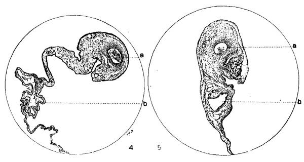
The rate of degeneration of the suspensory ligament and especially of the umbilical vein, in cats seems to be determined very largely by the previous existence or by the genesis of venous radicles which pour their blood into its caudal extremity. Such a condition was noticed very commonly in cats and guinea—pigs but never in dogs and sheep, although the degenerating umbilical vein may rarely acquire a secondary attachment in these animals also. It is a rather remarkable thing that after becoming detached at the umbilicus, the retracted umbilical vein not infrequently receives several exceedingly minute venous radicles which lie immediately extra-peritoneally at the ventral surface of the thick fold of fat constantly present in cats between the umbilicus and a point opposite the base of the xiphoid process. These venous radicles become gradually larger proximally and can sometimes be seen to unite and to join the tapering extremity of the unpaired retracted umbilical vein at a point opposite the middle of the Xiphoid process. This peculiar relationship was occasionally very evident because all the vessels beginning with the finest veins even, were very full of blood and the umbilical vein could be seen Without the least difficulty to empty into the left portal. It might at first thought seem probable that this main vein is a para—umbi1ical or Burow’s vein instead of the true umbilical vein, but microscopic as well as gross examination shows this not to be the case. Moreover, it would be remarkable that all trace of the umbilical vein should have disappeared in a kitten but ten Weeks old, for example. This transformation of the major portions of the umbilical veins into an integral, even if but a temporary, part of the peripheral Venous system is particularly significant in its bearing upon the degeneration and disappearance of the vein and the subsequent formation of a round ligament. Occasionally also a large lymphatic trunk (figs. 4 and 5) several millimeters in caliber runs parallel and adjacent to the vein in the caudal border of the suspensory ligament directly cranial to the Vein. The unusual size, distension and beauty of this trunk which joins the larger lymphatic vessels at the hilus of the liver makes it very conspicuous and it is evident that its presence and that of a converted and actively functioning umbilical vein must have a very important bearing on the time of disappearance of the vein and of the suspensory ligament. That the lumen of the umbilical vein may be preserved longer where branches are located was well established by Baumgarten and by Kirchbach (’99) for man.
In case of the kitten two weeks old the umbilical vein had completely lost its characteristic walls although the lumen was still about .75 mm. in size and full of blood. The Walls were composed only of an endothelial lining bounded by connective tissue and there was an entire absence of non-striated muscle even Within a half centimeter of the left portal. Nevertheless, that this vein was the original umbilical vein is shown by the entire absence of any remnant or vein in the caudal border of the suspensory ligament. Moreover, the total disappearance of the umbilical vein at so early a day in cats is, of course, very unlikely.
Apparently then, in these animals the retraction and degeneration of the suspensory ligament takes place pari passu with that of the umbilical vein and is dependent upon and determined by the retrogressive changes in the vein to a certain extent at least. That these processes are more or less independent of each other, however, is well illustrated by two dogs over a year old in which the suspensory ligaments were well preserved While the umbilical vein had disappeared completely. A similar condition is also not uncommonly found in old rabbits, cats and guinea-pigs. On the other hand, in the dog and sheep in which the umbilical vein becomes totally isolated and never has any permanent connections with the peripheral veins it degenerates very rapidly. But in the cat, guinea—pig, rat and rabbit in which the vein is not isolated and in which the suspensory ligament degenerates much slower, it occupies a relation similar to that of the hypogastric arteries in the same animals. In these animals it is also exceedingly common to find one or more small veins running roughly parallel to and in the immediate vicinity of the degenerating umbilical vein. The largest of these is usually plainly visible to the naked eye but the rest are generally of microscopic size only. Although injections of these vessels were not made yet from observation with the unaided eye and from a study of sections of the ligaments they apparently correspond to the para—umbilical veins of Sappey rather than to the vein of Burow. The largest of these veins which does not join the degenerating umbilical vein is generally plainly visible because it is filled with blood. In the dog there frequently are also a large number of microscopic veins in the adventitia of the degenerating umbilical vein which join the latter. These seem to be most numerous at the distal extremity of the Vessel where the lumen is frequently multiple, and arise from the vasa vasorum.
Fig. 4. Round and suspensory ligaments of the cat. a, remnant of the umbilical vein; 1), a large lymphatic. X97.
Fig. 5. A section of the suspensory and round ligaments of the cat. a, remnant of the umbilical vein; b, lymphatic. X79.
The behavior of the hypogastric arteries in these animals was quite different from that of the vein for they retract quite early and usually become attached to the apex of the bladder. They had already retracted in young rats whose eyes had not yet opened and could be traced peripherally only as far as the apex of the bladder. In a rat approximately three months old no trace of them could be found microscopically in this region. In cats eleven to twelve days old they have usually begun to retract although the urachus is often and the umbilical vein is always, still attached to the umbilicus at this time. It is also true, no doubt, that the time of separation of the arteries is dependent upon the time of sloughing of the cord, which undoubtedly varies as much in animals as Weckerling’s (’08) extensive analysis showed it does in the human infant. In a dog one week old the vessels were still firmly attached and there was no indication of a beginning retraction. These observations confirm those of Robin although I cannot corroborate his observation that the retraction of the arteries is always simu1taneous. This is perhaps never the case if one of them becomes infected, for the infected Vessel remains attached longer. But even excluding infections it is not at all likely that the two arteries always rupture simultaneously.
In a few instances in cats it was also noticed that the retracting arteries and urachus drew the vein caudally across the internal surface of the umbilicus. Such occurrences were well described and illustrated in Robin’s excellent investigation.
Robin stated that the obliterated hypogastric arteries usually do not remain attached to the summit of the bladder in ruminants and carnivora. As far as sheep are concerned, Robin’s statement is confirmed, but in dogs and cats they are almost always attached to the urachus or to a cicatricial formation at the very apex of the bladder. The same statement holds for young rats, guinea—pigs and rabbits although the minute size of the vessels in these animals makes it much more difficult to observe and trace them with the unaided eye. Besides, it is not rare to find the distal ends of the vessels more or less coiled or tortuous shortly after they become detached from the umbilicus or even much later in the sheep. Hence it seems strange that a secondary attachment is acquired to the apex of the bladder. This attachment which is a fibrous one is no doubt due to the fact that before the vessels become detached, the bladder and urachus occupy a position between the converging hypogastric arteries. Hence as the bladder becomes distended they are forced firmly against its sides and may even infold them. After becoming detached the atrophy or retraction of the lateral folds of the peritoneum in which the arteries lie draws them or at least holds them, in intimate contact with the lateral walls and the conical apex of the bladder and the degenerating urachus. Were it not for this fact it would seem to follow that the detached free ends of the hypogastric arteries would be retracted passively more and more with each successive dilatation of the bladder until they reached a point far from the apex. After they become attached to the latter further retraction is, of course, impossible and the further obliteration or disappearance of these vessels must hence be due to atrophy, degeneration and to a fibrous transformation in loco. Since, in contrast with the vein the atrophic changes in the arteries take’ place very gradually it is apparent that the condition in which the Vessels are found depends much upon the age of the animal. In old dogs, for example, the degenerated and transformed arteries usually cannot be seen near the apex of the bladder unless the latter is distended and then only as very fine, fibrous, often more or less discontinuous cords which gradually become somewhat thicker as the base of the bladder is approached. As already stated, instead of being attached to the apex of the bladder the free ends of the retracted hypogastric arteries in the lamb usually lie more or less curled up in a wide and loose fold of peritoneum at the sides of the bladder just lateral to its apex. This position is probably very largely due to the fact that they retract actively immediately after birth and not simultaneously with the urachus. Moreover, they lie in broad, loose peritoneal folds instead of being intimately associated with the bladder and urachus and with each other for ten or twelve days or more, before retraction can occur and this later retraction is a very gradual and not a sudden and extensive one.
The fact that the arteries which retract earlier degenerate much later than the vein, especially in the dog and sheep, has already been mentioned and some of the factors involved have been suggested. A further factor it seems to me lies in the fact that after becoming attached to the urachus and the apex of the bladder the arteries are alternately stretched and relaxed with each successive distension and evacuation of the bladder. While this stretching is a purely passive process the relaxation may nevertheless be accompanied by a certain amount of active contraction of the vascular musculature the effect of which may be retarded atrophy and degeneration.
In rabbits twelve days old the hypogastric arteries and urachus had already retracted completely and their free ends met at the apex of the bladder while in a rabbit one year old they could not be detected except in the region at the base of the bladder because they had atrophied so completely.
In several cats one year old they were still well—preserved and firmly adherent to the apex of the bladder but in two animals only three months old they could, on the contrary, scarcely be detected and could be traced only a little beyond the base of the bladder. Often, however, when the atropic fibrous substitute is invisible in the contracted state of the bladder it can easily be detected upon distension and it is evident that their presence or absence in the lateral surfaces and the apex of the bladder is probably largely if not wholly dependent upon the fact as to whether or not they gain a secondary attachment to the degenerating urachus.
Since, the sudden marked retraction which occurs in the hypogastric arteries of the lamb can not occur at all in the dog, cat, rabbit and guinea pig in which only a gradual retraction takes place some days or weeks after birth, it would be possible to construct a series beginning with man, in whom retraction is evidently slight, inconstant and always delayed and ending with the sheep and other ruminants in which it is immediate and practically complete a few hours after birth. It has seemed to me that as far as man is concerned the slight amount of the late retraction and the slowness of it, may be due in part at least to the somewhat different relations of the arteries and the bladder to the abdominal wall and peritoneum. In the domestic animals the urinary bladder is practically an intra-peritoneal organ while in man it is extra—peritoneal. Hence in man the hypogastric arteries lie between the peritoneum and the transversalis fascia for a comparatively long distance. Moreover, because of the different position which it occupies in man, evacuation of the distended bladder cannot assist much in the retractions of the vessels in the infant. Moreover, the descent of the bladder from the region of the umbilicus is much more gradual even if finally more pronounced when the adult condition is reached. It is of doubtful value and validity, to be sure, to compare postnatal degenerative processes in animals born in such widely varying states of maturity, which have such varying life cycles and which grow at still more widely varying rates, nevertheless the fact remains that in spite of these differences very similar processes occur at somewhat different times in all animals. The differences that exist are of degree rather than of kind.
It is not at all uncommon to find a small amount of blood near the free ends of the torn hypogastric arteries in the lamb but such was, of course, never the case in animals in which the vessels are ruptured extra—abdominally and in which only a delayed retraction occurs. The presence of extensive clots Within the vessels was rare in the lamb but common in the other animals for in them an effective obliteration of the lumina Was probably made difficult by the fact that the vessels remained attached to the umbilicus thus making contraction of the intraabdominal portions more difficult. Since the presence of clotted or unclotted blood must of necessity, prevent rapid obliteration of the lumen of a vessel and delay retrogressive changes in it it does not seem probable that the presence of clots could aid much in preventing hemorrhage as Gmelin thought.
Since the ends of the retracted hypogastric arteries in lambs, often projected freely into the peritoneal cavity rupture of the peritoneum must, of course, have taken place. Since the evacuation of the bladder takes place at intervals before these vessels become detached Without producing this lesion and since the end of only one of the retracted vessels may project intra—peritoneally in the newborn lamb, it does not seem likely that the combined force resulting from the retraction of the detached arteries and the contracting bladder is sufficient to produce a rupture of the overlying peritoneum. It is true that in the lamb the immediate, sudden elastic recoil of the arteries may be associated With or even stimulate, the evacuation of the bladder which usually occurs soon after or even during birth, but these combined forces could scarcely rupture the peritoneum. Hence it seems quite obvious that the latter is torn as a. result of the outward traction produced at the time of rupture of the cord. This conception is also in accord with the statement of Henneberg that the arteries tear intra-abdominally although as previously stated I am inclined to believe that rupture in ruminants occurs extra-abdominally but in portions of the vessels which previous to traction an-d rupture were intra—abdominal.
Since the observations here reported were observed largely incidentally no comprehensive detailed microscopic study of the minute changes which accompany the processes of retraction, atrophy and degeneration of the umbilical vein and hypogastric arteries were made. However, quite a number of specimens from several species were examined and from these it is evident that the microscopical picture of the so-called obliteration of the umbilical vessels in the domestic animals, save rarely, is not practically concluded a few weeks after birth as Kirchbach asserts is true of the ductus venosus and vena umbilicalis in man. Kirchbach’s statement in contradicted also by Haberda. Attention has already been called to the fact that, as a rule, the umbilical vein in both the dog and sheep disappears totally in the course of a few months. From examination of the remnants of the veins found before this time and from other facts stated above it is obvious that in the animals examined, this obliteration, atrophy, degeneration and absorption is invariably centripetal and not contrifugal as Robin says is the case in man. The free distal extremities of the filaments representing the remnants of the umbilical vein which were still attached to the left portal, were composed of vascular ill-preserved, fibrous connective tissue enclosed in peritoneum except in case of one lamb in which bundles of muscle fibers were preserved even in the degenerating tip. Farther proximally, i.,e. nearer the left portal, a small remnant of the umbilical Vein remained, but still farther centrally a remnant of the lumen was also present, and finally a portion of the original vessel with well-preserved walls somewhat reduced in size could be recognized. In case of the lamb about 5 to 7 weeks old the small degenerating remnant of the vein which was approximately 2 cm. long contained a macroscopic lumen for about half its extent but the intima was poorly preserved and absent in places. There was no proliferation of the endothelium and an elastica interna was not noticed in Van Gieson stains. The media which had undergone a. hyaline degeneration, was represented only by a granular detritus in some places and small degenerating nuclei were accumulated in it near the lumen. There was no distinct adventitia and the fibrous connective tissue which surrounded and invaded the irregular degenerating mass also seemed to be in process of degeneration.
The distal ends of the veins in dogs and cats were practically in the same condition although in some cases the original lumen had become multiple by being encroached upon by the folded degenerating media. It not infrequently contained some erythrocytes in a fair state of preservation and occasionally the adventitia was very vascular where it was well—preserved. Numerous small Vessels were also found in the degenerating media and not infrequently these communicated with the original lumen to which they ran more or less parallel. No cellular infiltration such as described by Haberda for man, Was seen and the Whole picture was that of a degeneration rather than that of an obliterative endarteritis. In fact, aside from the presence of the small blood Vessels in the connective tissue the impression is usually that of a passive rather than of an active process and it is difficult to see Why a purely temporary complete substitution of fibrous connective tissue should occur in the Veins only to be absorbed as soon as formed. Such a complete substitution of fibrous tissue for the vein may occur, to be sure, in animals in Which a true ligamentum teres hepatis is formed, but not, of course, in those in Which it never comes into existence because of early resorption of the vein. Nevertheless, that a partial transformation may take place even in these animals has already been stated regarding the umbilical Vein from a dog 91 hours old a portion of which is represented under higher magnification (figs. 6 and 7). Although this animal was less than four days old the lumen o:f the peripheral portion of the vein which was still attached to the umbilicus, is completely obliterated by a slightly vascular connective tissue and the degenerating media has become Vascularized. The muscle fibers have lost their outlines and specific staining reactions and are represented by a degenerating mass containng irregular nuclei. Somewhat farther centrally, i.e., nearer the liver, this vascularization is much more evident as shown in figure 7 and the fibrous connective tissue filling the lumen has more the appearance of newly formed tissue. Still farther centrally the regular, reduced filumen which contains blood is surrounded by a Well-preserved intima and a better preserved though degenerating musculature (fig. 8). In the case of this dog then the peripheral portion of the lumen of the vein became completely obliterated through connective tissue formation while its musculature was degenerating and being penetrated by numerous Vessels arising from the vasa vasorum, in spite of the fact that the Vessel was to undergo rapid degeneration and absorption.
Fig. 6 Central portions of umbilical vein of a dog 91 hours old, showing the lumen obliterated by vascular connective tissue surrounded by the degenerating media. X275.
The presence of such profound changes so soon postnatum confirms the observation that embryonic regression of the umbilical Vein sometimes begins before birth. In several instances it was noticed, for example, that the cross—section of the media of the umbilical vein lacked all the characteristics of plain muscle and looked not unlike a syncytium. Such appearance could, of course, be explained only by prenatal degenerative changes or by the supposition that the musculature of the wall of the vein sometimes fails to reach maturity during fetal life — a supposition which would, however, fail to explain the early fibrous transformation above mentioned. Moreover, the surprising fluctuations in the duration of pregnancy which have been observed would seem to make the occurrence of pre-natal degenerative changes not impossible.
Plate 1
Explanation of Figures
Fig. 7. A different portion of the obliterated vein shown in figure 6. X630.
Fig. 8. Umbilical vein of the dog; same as figures 6 and 7 but somewhat farther centrally. X450.
Fig. 9. Portion of wall of umbilical Vein of dog 45 hours old, showing process of degenerating media extending into the lumen. X750.
Fig. 10. From the same vessel as figure 9, showing process formation, vacuolation beneath the endothelium and vascularization of the process. X750.
Fig. 11. Omphalomesenteric vessel from a dog 44 hours old, showing degenerating endothelium and media and some ill-preserved blood in the lumen. X750.
In another dog but 45 hours old the conditions were quite different for the lumen of the vein which was patent contained uncoagulated blood except in the immediate neighborhood of the umbilicus. Nevertheless, degenerative changes had already appeared and were particularly evident in the musculature especially immediately beneath, i.e., external to the endothelium. The cell outlines were lost and the media was represented by a uniformly ill—preserved syncytial mass which easily took an acidophile stain. The nuclei were very large, vesicular, and irregular in outline and arrangement. The intima was quite well preserved in most places but in others the cells which were irregular in form had become elongated, the nuclei were large and swollen and large Vascuoles were found between and especially below the cells of the intima. The most striking thing, however, is the occurrence of projections of club-shaped processes into the lumen as represented in figures 9 and 10. In the case of figure 9 this projection was still capped by the degenerating endothelial cells on which erythrocytes lay. The same is true of the similar though larger and longer projection represented in figure 10 which is taken from the same vessel somewhat farther peripherally. In this case the elongation and vacuolation of the endothelial cells some of which have been cast off, are well seen but the honey-combed process is still capped by some of them.
Since no series of vessels taken from pups only several hours apart in age were examined it is impossible to say whether this process formation is a constant phenomenon in the degenerating vein in the dog. It was not observed in the sheep and before discussing the significance of these facts I desire to call attention to changes observed in the hypogastric arteries and the omphalomesenteric veins. In case of two of the latter which were still attached to the abdominal wall in a dog 91 hours old similar degeneration of the endothelium and the musculature was present but the formation of processes was not observed. As shown in figure 11 the intima which is absent in places, has undergone degeneration although the lumen still contains blood. figure 12 shows a section of the accompanying omphalomesenteric vessel apparently in a more advanced stage of degeneration, for here the whole lumen is obliterated. by the large cast-off endothelial cells and fibrin which form a network completely filling it and simulating endothelial proliferation and migration. In the case of both these vessels the musculature has undergone considerable degeneration, and readily takes an acidophile or special connective stain, making it difficult to distinguish the connective tissue which is, of course, also a purely temporary constituent.
Fig. 12. Transverse section from an omphalomesenteric vessel of a dog 44 hours old. Lumen filled with desquamatcd endothelium and a fibrin network. X750.
In case of the hypogastric artery of the same dog no process formation is present and the intima has completely disappeared over a considerable extent of the distal portion. The lumen which is not lined by endothelium although containing blood is surrounded and encroached upon by a wide band of fibrous tissue in some of the large meshes of which isolated irregular degenerating nuclei lie. However, as shown in figure 13 there are no signs of endothelial proliferation or of infiltration and the outer layers of the media are quite well—preserved. At the extreme distal portion of the vessel all of the musculature is intact and the lumen has its characteristic shape. Slightly farther proximally it contains some blood, the intima is absent and Where the blood lies in contact with the wall the meshwork of connective tissue extends out into it and contains newly formed vessels (fig. 14). The rest of the lumen is bounded by the circularis of the media for no elastica interna is present in this portion. Where the blood which is not formed into a thrombus, fills the whole lumen this proliferation of connective tissue is evident over the entire circumference and in some places the lumen is entirely pervaded by it. The relations of this connective tissue to that in the media are so clear that one cannot doubt that it is a direct continuation and remnant of that which was contained between the muscle bundles of the media and that some of the fenestra in the network previously contained muscle-fiber bundles which have degenerated and have been absorbed.
That these degenerative and obliterative changes are not invariable or constant in occurrence, however, is shown by a specimen of a hypogastric artery taken from a dog about a year and a half old as shown in figure 15. In this case all the constituents of the wall of the vessel including the endothelium and the elastica interna are still present and well-preserved although the connective tissue is greatly increased in amount especially at the periphery. The vessel is gradually becoming transformed by the proliferation of the adventitial and inter—fascicular connective tissue which is always present in comparatively large quantities in vessels from mature fetuses. Farther centrally (fig. 16) in the Vessel——i.e., nearer its origin—there is but little connective tissue near the lumen which is surrounded by poorly preserved endothelium, a partial elastica interna and a very Well-preserved media which on one side is encroached upon markedly and replaced by fibrous tissue.
The conditions as found in this dog are quite different from those found in the hypogastric arteries of a lamb three and one half weeks old a section of which is represented in figure 17. In this case there are portions of the vessel in which the lumen is completely surrounded by a thin layer of fibrous tissue (a) which varies considerable in thickness but which lies internal to the elastica interna (b). Although the endothelium is absent in places the musculature is well—preserved where it is not invaded by the connective tissue, and contains many elastic fibers (c). In the more distal portion the vascular fibrous connective tissue plugs the whole lumen of the vessel and serial sections toward the tip show that it did not enter from without through the free extremity. The intima is preserved in some places and the connective tissue then lies between it and the elastica interna. Where the intima is absent it lies in long strands in the lumen. The connective tissue internal to the elastica :'s directly continuous in some places with that external to it which lies between the muscle bundles of the media. Near the extremity of the vessel the inner layers of the media are encroached upon and entirely replaced by the vascular connective tissue which plugs the entire lumen. Hence, I am inclined to believe that the process of obliteration in this lamb is entirely comparable to and represents a later stage than those found in the hypogastric arteries of the dog as represented in figures 13 and 14.
From these things it is evident that obliteration of the hypogastric arteries in the dog and sheep may take place in at least two ways. In one case the degenerating musculature is replaced by fibrous tissue which encroaches upon it and upon the lumen from the periphery toward the center and in the other the lumen is encroached upon and the surrounding musculature displaced by connective tissue arising from within after degeneration and desquamation of the intima and proliferation of vessels from the vasa vasorum. It is evident of course that whenever this transformation takes place from without, i.e., from the periphery, the lumen and its bounding intima as well as the inner layer of "the media, may be preserved very much longer than in the other case and it is also evident that both processes of obliteration may be present at the same time.
Plate 2
Explanation of Figures
Fig. 13. Inner layers of Hypogastric artery from :L dog; 91 hours old. X410. Van Uieson stain. Connective tissue, black; muscle and blood, yellow.
Fig. 14. From a hypogastric artery from :1 dog; 91 hours old, showing; the obliteration of the lumen through vascular connective tissue. X630.
Fig. 15. Portion of the wall of at hypogastric artery of :1 dog 1:} years old. X62. Conneetive tissue, black; muscle, brown.
Fig. 16. Portion of the Vessel shown in figure 15 but somewhat more centrally. X630. Van Gieson stain. Connective tissue, black; muscle, yellow.
Fig. 17. A section of the inner two-fifths of the wall of it hypogastric artery of :1 lamb 5% weeks old. Note the extremely folded elasticu interna, the absence of an intima, and the of :1 layer of fibrous tissue between the latter and 1h(- elnstioa as well as numerous (‘l.‘lSTi(' fibers in the media. X620.
From the facts observed especially in the umbilical veins of the dog and sheep, it is apparent that similar processes are initiated in them but that they cannot extend beyond the initial stage because of the rapid degeneration and absorption of these vessels. In the cat, rabbit and guinea-pig, on the other hand, in which the disappearance of the umbilcal veins is a much slower process a corresponding and complete fibrous transformation may take place.
So far as the umbilical vein and hypogastric arteries of these animals are concerned no evidence Whatever for the origin of connective tissue from endothelium has been obtained. Merkel also (’03) insists that the endothelial cells do not take part in the organization of a thrombus, and asserts that Baumgarten was mistaken when he concluded that an actual proliferation of the endothelial cells takes place. Nevertheless, in the absence of corroborative facts it is not denied that Haberda was mistaken when he declared that in man “Die our Obliteration der Nabelgefasse fiihrenden Veranderungen bestehen in einer zu Bindegewebe sich metamorphosierenden Wucherung des Gefassendothels der sich an den Nabelenden der Geféisse eine von der Nabelwunde ausgehende entziindliche Zelleninfiltration hinzugesellt.” This proliferation which is said to take its origin from the endothelium of the obliterating vessels was described by Thoma, confirmed by Baumgarten, emphasized by Hauptmann and in a measure corroborates the observations of Mall (’02 and ’12) and of Kling (’02) to the effect that reticulums may arise from endothelium.
A very interesting phenomenon was noticed in the extra-abdominal portion of an umbilical vein of a lamb which died during labor. In the case of this lamb a portion of the vein contained blood which Was in process of coagulation. Upon microscopical examination the lumen which was fairly regular save for small ridges was seen to be dilated and filled with blood containing strands of fibrin and a fibrin network in some places. The whole vessel was well-preserved, an elastica interna was present, and the endothelium was intact save in some places where the cells had become detached or where fibrin strands were in contact with the wall. In these places the endothelial cells had wandered far out into the lumen along the strands of fibrin. In some places there were small accumulations of endothelial cells and not infrequently the presence of protoplasmic processes made them quite irregular in form. In places where a number of these cells were found in a fibrin network the appearance roughly simulated that of an embryonic connective tissue. These endothelial cells were all well-preserved and the only cells in process of degeneration were some of the blood cells contained in the clot. The musculature also was well-preserved except in two areas on approximately opposite sides of the lumen where the outlines of the inner fibers were obliterated and their place taken by irregular lumps of hyaline material which stained very intensely with hematoxlyn and suggested degenerative changes. No evidences of mitoses were present in the endothelium and there was no cellular infiltration.
From What I have seen I am inclined to believe that the presence or absence of blood in the lumen of a vessel is of considerable importance, for in not a single instance were early obliterative processes observed in the empty contracted and. retracted distal extremities of the hypogastric arteries. It is: worth recalling in this connection that Haberda thought that the more rapid obliteration of the arteries is due to the fact that the thrombus in them is less extensive and that the walls of the vessels are in contact. I There is considerable variation, however, in the rate of the complete contraction of the vessels for in a dog which died of inanition 44 hours after birth, the vein was practically empty and quite Well contracted throughout its extent while the arteries were markedly distended with blood. But in another——a still-bom pup from the same litter—~the conditions were exactly the contrary. In the lambs examined the hypogastric arteries werealways empty save for. small clots or unclotted blood here and there. The vein, on the contrary, was always filled with uncoagulated blood except for the presence of a small clot in the beginning of its unpaired portion and my findings in this regard were entirely comparable to those of Pollot (’09) in the ductus arteriosus in which the retrogressive changes were usually not accompanied by thromoses.
Since the retrogressive changes in the umbilical vein of the cat, rat, rabbit and guinea—pig are so much slower and take place in direct relation to and not largely or wholly independent of those in the suspensory ligament as is the case in the dog, a somewhat different condition might be expected to exist. This is the case for in these animals the vein persists much longer throughout a portion of its extent, and not infrequently a fibrous structure which may somewhat rightly be termed a ligament is substituted for it (figs. 18, 19 and 20). However, as already stated, so far as the vein retracts or degenerates completely it is not replaced by a fibrous cord and hence the process of degeneration in this, the distal portions, is from all appearances identical, as a rule, with that in the dog and the sheep. In the more proximal portion of the umbilical vein a roughly cylindrical cord composed of loose vascular fibrous connective tissue of hyaline appearance which is sometimes markedly canalized is often found in cats, rabbits and guinea—pigs. In cats from eleven days to one year old this thickening in the caudal border of the suspensory ligament is usually due to the presence of a band of well preserved fibrous connective tissue in whose proximal or intrahepatic portion a vessel which can be recognized as remnant of the umbilical vein is usually present (figs. 4, 5, 18, 21). But in its distal or hepaticfiportion near the xiphoid process, no such remnant can be detected as a rule in cats only six months old and occasionally no _trace whatever of the umbilical Vein was found in any portions of the caudal border of the suspensory ligament examined in cats one year old (figs. 19, 20, 21, 22, 23). Indeed, in one instance such was the case in a eat only six weeks old and it is worthy of special notice in this connection that in the specimens from these and still younger cats, eleven days old, the picture was always that of a pure degeneration without the least indication of an endothelial proliferation and that in those instances where remnants of the lumen were found the endothelium was one-layered and well-preserved in places.
Fig. 18. Proximal portion of the degenerating umbilical vein from a. kitten fourteen days old. With higher magnification the original lumen is clearly recognizable. a, remnant of original vein; b, connective tissue wall. X142.
Fig. 19. Transverse section of the free caudal border of the suspensory ligament of a out about six weeks old. It contains no recognizable remnant of the umbilical vein, is quite vascular and composed of very loose fibrous connective tissue bounded by peritoneum. X97.
Fig. 20. Transverse section of the peripheral portion of what seemed to be a round ligament in a kitten seven days old. There is no trace of the umbilical vein in the very fibrous connective tissue. X142.
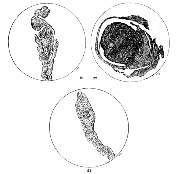
Fig. 21. Distal extremity of round ligament from a kitten 24 days old. No remnant of the umbilical vein is present in the vascular connective tissue. X142.
Fig. 22. Round ligament of a kitten 14 to 18 days old. No recognizable 1'eminant of the umbilical Vein is present. The ligament is composed of a very loose connective tissue not unlike embryonic connective tissue which looks gelatinous. X54.
Fig. 23 Transverse section from the free caudal border. of suspensory ligament of the cat, containing a. large vein with nothing but an endothelial wall bounded by connective tissue. This vein bears no resemblance to the remnant of the umbilical vein. X142.
The fibrous connective tissue which had replaced or encroached upon the vein varied from a poorly preserved dense fibrous connective tissue to a very loose embryonic tissue which because of its gelatinous appearance suggested in the gross even, the umbilical cord to a laboratory assistant (figs. 19, 20, 22). It seldom contained any recognizable remnant of the lumen of the original vein as stated by Kirchbach for the round ligament of man. When the original lumen was preserved it had always become much reduced and often multiple, as a result of folding, as described by Baumgarten in man. In some cases the tissue surrounding the remnant of the vessel was very loose and vascular and the irregular outlines of the periphery of the wall of the degenerating veins generally suggested that it was enclosing the vein rather than that newly-formed elements of intimal origin were actively transforming the vessel from within. Moreover, the inner portion of the media was often better preserved and the lumen of the vessel often contained some erythrocytes while the outer portions of the media, on the contrary, were degenerated. However had this process of degeneration been observed at closer intervals in stages of a few days, e.g., it is not at all unlikely that a certain amount of cellular infiltration, as described by Baumgarten and Haberda in the neighborhood of the umbilicus in man, might have been noticed in cats as well.
The most striking thing about the microscopical structure of the umbilical vein in the new born lamb is the variability in the amount and distribution of the longitudinal muscle fibers of the media. In its extra-abdominal or cordal portion the structure of the vein was often found practically identical with that of the distal extremities of the retracted arteries. The lumen of the contracted vessel may be more or less stellate, crescentic, or a mere slit not infrequently shaped like the printed capital I. There is a single-layered endothelium. An elastica interna was observed several times in sections stained with Van Gieson and with orcein and fuchsin. The media contains longitudinally disposed muscle fibers which are somewhat irregularly distributed, and between which elastic tissue is found. However, none of the longitudinal fibers are found grouped in bundles, among the fibers of the much thicker circularis as is the case in the arteries as a rule. The fibers of the circularis which are arranged rather loosely especially at the periphery, lie in concentric layers, those in the peripheral layers being more definite. No true adventitia can be said to exist although a number of small blood vessels are occasionally found in the small amount of cordal tissue that still surrounds the extraabdominal portion of the torn veins.
The specimens of the intra-abdominal contracted portions of the paired veins which were examined have a very similar structure except that almost no circularis was present and that the fibers are interlacing. But the unpaired portion contrasts strongly with the former and the extra-abdominal portion. The unpaired portion is usually only partly collapsed and not contracted. Its walls are folded slightly and differ from the more distal portions in the entire absence of the longitudinal musculature, and in the occurrence of great variations in the thickness of the circularis. Although composed of much more closely apposed fibers the uncontracted circularis seems unusually thin for the size of the vessel and is surrounded by a fairly definite and vascular adventitia. In describing the umbilical vein in the human fetus Herzog stated that the wall of the vein is formed only by a well-developed media composed of fibrous connective tissue in which non-striated muscle is distributed very irregularly. Such an extremely irregular arrangement of the muscle fibers was seen in the umbilical vein of a dog but two days old and also in the hypogastric arteries of a newborn dog. In the first specimen the muscle was so loosely and irregularly disposed that it looked not unlike loose fibrous connective tissue and the adventitia which contained a number of small paraumbilical veins was thin and absent altogether in some places. Interlacing muscular fibers were also found in both the paired and unpaired portions of the vein of one lamb, however, and this interlacing is not infrequently so extensive that no regularity in the arrangement of the fibers of the media into circularis and longitudinalis can be distinguished. The non-striated muscle fibers spoken of by Herzog are said to be disposed longitudinally or transversely, those extending in both directions lying adjacent and interlacing with each other. Herzog found no elastica interna and no elastic tissue present in man and Hauptmann reported similar results for the vein in the cord of the colt. The last author also stated that a strongly developed adventitia Was present in the developing artery but at no stage in the development of the vein which was said to be in loosest connection with the surrounding tissues.
It is interesting that Haberda stated that all authorities are agreed that elastic tissue is sparse in the extra—abdominal portion of the hypogastric arteries and that even in their intra-abdominal portions less is found than in other arteries. Lochmann, on the contrary, who also worked with human material found an elastica interna present in all vessels and reported the formation of a new media in one case. However, Henneberg insists that Lochman’s methods were not wholly reliable.
Although no special methods for demonstrating the presence of elastic tissue were used in every instance the presence of numerous elastic fibers in the distal extremities of several of the retracted hypogastric arteries of the sheep was unquestioned in specimens stained with Van Gieson even. They were not noticed regularly in sections of vessels from other animals stained in this way, however,- and nothing that suggested the presence of a complete elastica interna was noticed save in some sections. Although occasionally very evident, it was seldom co—extensive with the perimeter of the vessel but was noticeable on'y in small areas. Moreover, since the elastica interna was usually perfectly evident in sections stained with Van Gieson, in very much smaller arteries that lay in the nighborhood, it is to be doubted whether it is invariably present in the peripheral portions of the hypogastric arteries even. Although allowance must be made for the effect of degenerative changes this supposition is further confirmed by the use of special stains such as Unna’s orcein and fuchsin and Weigert’s elastic tissue stain. In a specimen from a lamb 5% weeks old so stained a suprisingly welldeveloped and extremely folded elastica interna which was double in some places lay in the media quite close to the lumen of the hypogastric artery but separated from the latter by a distinct although thin layer of fibrous connective tissue. As shown in figure 17 (b) this elastic membrane was folded so strongly in some places as to form a large accumulation of loops or coils somewhat similar to, though more pronounced than, those pictured by Henneberg in the extra—umbilical portion of the vein of a human fetus. In addition to this very evident elastic membrane numerous very long, tortuous and thick elastic fibers (0) were also distributed throughout the media in far larger numbers than found by Henneberg in the human hypogastric vessels.
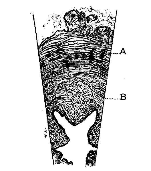
Aside from possible specific differences the more pronounced folding of the elastica interna and the greater tortuousness and thickness of the elastic elements in these specimens as compared with those found by Henneberg in man may, of course, be due to the fact that the latter used specimens from comparatively younger individuals. The extremely folded condition of the elastica in these atrophic vessels and in contracted vessels as well, seems to suggest that the range of contractility and of elasticity of the intima is a rather limited one. Hence when this limit is exceeded by the contracting and atrophying musculature of the media it is folded and re-folded passively. Since the elastica is apparently well-preserved after the musculature and intima have already begun to degenerate it can usually be stained without difficulty by special methods although the failure to detect it in all routine Van Gieson stains might seem to indicate that it disintegrates earlier than the media. It is particularly evident in sections stained with an aqueous solution of fuchsin and mounted in glycerine or by the orcein-fuchsin method of Unna, both of which methods also reveal numerous tortuous elastic tissue fibers scattered throughout the media. Most of the hypogastric arteries examined had a structure in marked contrast to that of the unpaired portions of the umbilical vein. However, their structure is identical with that of the unpaired vein in so far as no elastica interna was usually noticed, in the presence of a single-layered endothelium and a similar adventitia. But no part of the hypogastric arteries except the distal portions were found wholly without longitudinal muscle fibers. However, the longitudinal layer varied greatly in thickness and distribution. This variation in thickness Lochman ascribed to the differences in contraction of the vessel in man. Most of the longitudinal fibers lay internally near the endothelium but various—sized bundles were found scattered about irregularly in the innermost portions of the circularis and not infrequently good-sized bundles were also found in the outer layers. In addition to this peculiarity in distribution of the longitudinal fibers, a peculiar, coarse radial striation or better lamination as shown in figure 24 was occasionally seen in the circularis of the arteries but never in that of the vein. This striation which was not present in all portions of the cross—sections was due to local accumulations of loosely-arranged circularly-disposed bundles of muscle fibers. Since these local accumulations lay approximately opposite each other they gave rise to the above mentioned coarse radial striations. Moreover, the fibers of the circularis were usually rather loosely disposed and the concentric strands separated from each other by a fine collaginous fibrous tissue.
Fig. 24. Transverse section of the hypogastric artery of a new born lamb, showing the radial striation of the circularis. A, circularis, B, longitudinalis. X97.
The lumina of the arteries as in case of the paired portions of the umbilical veins, were roughly stellate. This characteristic of the lumen was produced by a projection into it of thick Welts or ridges or discontinuous longitudinal folds which however did not concern only the longitudinal fibers of the media although the contraction of these fibers, Would, of course, result in an increase in their caliber in a direction at right angles to the lumen and hence would form folds which would have to encroach upon it or produce a dilatation of the vessel. The latter was, however, prevented by the active contraction of the circularis which tended to reduce the size of the lumen or even to obliterate it entirely and hence compress the longitudinal fibers. Since the latter need more room folding of the wall would hence seem to be the natural and inevitable result. It is interesting that Strawinski, Von Hoffman and Herzog have described similar ridges in the unretracted hypogastric arteries in man. Hauptmann also found similar ridges in the noncontracted artery of the horse and states that they “may be as prominent as those described by Strawinski in. man.” Attention must be called to the fact, however, that there are apparently two kinds of ridges for Hauptmann attributed those found in the horse to large irregularly arranged, branched intimal cells 200 to 300;» in size, which are surrounded by dense fibrous tissue and lie directly under the endothelium. Bucura, on the contrary, concluded that the longitudinal ridges are due to contraction of the longitudinal muscle fibers, while Pollot asserted that the ridges and folds which project into the lumen of the ductus arteriosis are undoubtedly not produced by a contraction but are an expression of the unequal development of the constituents of the walls of the vessels. Moreover, Pollot supports this opinion by results obtained from experiments on the aorta of guinea pigs. These consisted of the treatment of excised portions of the aorta with adrenalin, a procedure which failed to produce the ridges. But since the retracted and contracted hypogastric arteries and the umbilical veins in whose walls few longitudinal fibers were present also contain Welts or ridges, and moreover, since they are absent in the non-contracted vessels or portions containing a large clot it is evident that they can be due to a contraction even if not necessarily to a contraction of the longitudinal musculature alone as Henneberg concluded. Since the walls of contracted vessels as ordinarily seen in histological preparations are sinuous or folded more or less I cannot believe that what was seen in the retracted and contracted umbilical vessels is anything but an exaggeration of the same phenomenon and that both kinds of fibers when present are factors in their production for the circular fibers are very evidently even if not primarily, concerned. Moreover these ridges can be seen in degenerating umbilical vessels which are entirely free in their distal portions and the Varying shape and size of these welts makes it impossible to attribute them solely to a varying development of the longitudinalis. Ridges or Wiilste due to proliferation of the intima were never observed although their occurrence in the ductus arteriosis is not therefore denied.
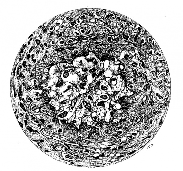
A peculiarity often seen in cats two to four months old or older, and more rarely in young dogs, is the presence of one or more extremely fine long peritoneal bands about half a millimeter thick, extending from one portion of the mesentery to another and surrounding a number of coils of the small intestine. Taking their caliber into consideration these bands are unusually strong although fastened only at their ends and are apparently always bloodless. Since it is only occasionally that one end of these filaments is fastened to the peritoneum near the umbilicus it did not seem very probable at first that they were remnants of the omphalomesenteric vessels but since such delicate filaments were never observed in very young or old cats such an origin seemed not at all an improbable one. Hence two such strands from a cat ten weeks old, were examined microscopically but neither filament contained anything which could be recognized as a remnant of the omphalomesenteric vessels. Both were composed of fibrous connective tissue (fig. 25) containing one or two veins and several smaller arteries. By comparison it will be noticed that the structure of these cords is very similar to that of the temporary round ligament of some cats, rabbits and guinea-pigs. However, since these fine filaments are occasionally eight to fifteen centimeters long in adult animals, such an origin would seem possible only on the supposition that considerable growth or elongation takes place in the vessels or filaments. Nevertheless, since the latter were observed in various stages of transformation such an origin becomes a highly probable one. This supposition was fully confirmed by a case in which one of the omphalomesenteric vessels was found attached to the apex of the bladder between the extremities of the hypogastric arteries in a cat about one year old. The end attached to the bladder had undergone fibrous transformation but the proximal portion still contained a remnant of the original lumen and
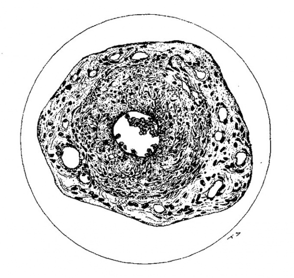
Fig. 25. Obliterated omphalomesenteric vessel from a kitten 18 to 26 days old. No remnant of the original lumen is visible and the whole structure is composed entirely of a vascular connective tissue. X275.
communicated with the superior mesenteric vein near the central end of the latter. In a second case, that of a eat one and a half to two years old a long, fine vessel containing blood throughout its entire extent was found with similar attachments. This vessel which ran among the coils of intestine likewise joined the superior mesenteric vein near its central end. It was quite uniform in diameter, about three—fourths of a millimeter thick, and took its origin in three fine veins on the sides and ventral surface of the apex of the bladder. Just cranial to the latter this surviving omphalomesenteric vein which had secured a secondary attachment was attached to the abdominal wall by an isolated fold of peritoneum 2.5 cm. wide and 2 cm. long although it was wholly free throughout the rest of its course. After the death of the animal as the vessel cooled, the blood was slowly forced out into the superior mesenteric vein and the vessel then appeared only as a fine whitish cord. From these findings and from the similar behavior of the degenerating umbilical Vein in dogs and sheep in which it not infrequently obtains a secondary attachment to the prominent fold of fat between the xiphoid and umbilicus, it is not only evident that the fine fibrous filaments frequently found in cats have such an origin. Moreover, it is evident that their persistence after birth is largely determined by the fact and more especially by the fact that they not infrequently come into relation with the systemic veins. It is also possible that the constant traction exerted upon them as a result of peristalsis in the contained coils of intestines is of some significance in this connection. Whenever they obtain connection with the peripheral veins the original lumen is preserved for a considerably longer time and may be wholly intact even after the musculature has become clearly degenerated. It is also evident that the arteries must undoubtedly degenerate first or earlier at least, for it is inconceivable that they could form part of the systemic circulation even if they obtained a peripheral attachment. That this is true is shown abundantly by the fact that in every case the persisting vessels containing blood were veins and not arteries. Moreover, no one has described a similar relationship of the hypogastric arteries which because of their large size and much later disappearance might be assumed to establish such a relationship far easier than the much smaller and far more functionally transient omphalomesenteric arteries.
Strangely enough such fibrous strands were never noticed in rabbits and no remnant of the omphalomesenteric vessels could be found in young rabbits more than 12 days old, while in guinea pigs they are usually present at the age of three months or more. In the few newborn dogs examined not more than two strands of omphalomesenteric vessels were found and these were often twisted about each other so as to form a single strand which could be separated, however, as far as the lymph nodes at the root of the mesentery. If the two strands were distinct one of them always ran farther caudally and it was this one in which blood could usually be seen. The other strand was shorter, firmer, whiter and looked more like an artery. Upon microscopical examination it was found that the single strand usually contained two vessels one of which might have the characteristics of a vein and the other that of an artery or they might be indistinguishable. In case of the second isolated strand which extends farther caudally the only vessel contained in it looked like a vein. Injection of these from the superior mesenteric vein, in cats one week old was easily accomplished.
Robin called attention to the presence of blood in the omphalomesenteric vessels in cats forty—eight hours after birth, described the course of the vessels and added that in some cats there might be three instead of only two vascular strands each containing two veins and two arteries. According to Robin the omphalomesenteric vessels of the cat detach themselves at the end of one month but as stated above, it is beyond question that not infrequently these degenerating vessels can be found attached to the umbilicus as late as six months and one year or more after birth.
Upon microscopical examination these undetached vessels were usually found to have fairly well-preserved walls with a distinct lumen. No elastica interna was observed and the musculature which had undergone regression was composed of indistinct circularly disposed fibers only. A definite adventitia was not present for it merged into the surrounding thick layer of fibrous connective tissue which was enclosed by peritoneum (fig. 26). The lumen was large, circular or elongated inycross section" and contained some well-preserved erythrocytes as a rule. Each strand contained but a single large vessel and two vessels in each strand as reported by Robin were never seen except in regions where the two separate strands had joined. In case of the presence of three strands three vessels would, of course, be present here.
The apparent failure of the omphalomesenteric Vessels to be completely transformed into fibrous cords before their complete disappearance after becoming detached is probably due to the presence of degeneration and absorption similar to that which occursin the umbilical vein in the dog and sheep. It is also interesting that no proliferation of the endothelium was noticed and that the musculature was comparatively well—preserVed so long after the vessels were of any functional value to the organism. In some cases (fig. 12) the endothelium had been cast off, however, and plugged the small lumen. The cells which had undergone degenerative changes were not infrequently quite clearly outlined, somewhat stellate and formed a meshwork by the union of their processes. In other cases the lumen which contained well-preserved erythrocytes, and the bounding endothelium was well-preserved but the musculature had become so degenerated that it was difficult to recognize it as such. It had become syncytial and had lost its distinctive staining qualities besides being markedly vascularized at the periphery where it was partly or wholly replaced by connective tissue. Similar degenerative changes were also observed in the intima.
Fig. 26. Omphalomesenteric vessel from a pup 91 hours old. The arrangement of the inner portion of the wall suggests that of the original vessel. The lumen contains well-preserved erythrocytes. The outer lighter portion is composed of very loose fibrous connective tissue. The endothelium is intact in this portion. X275.
Summary and Conclusions
- The umbilical arteries of ruminants very likely rupture extra-abdominally but at points which were intra-abdominal previous to traction.
- Complete and immediate contraction of the arteries and of the extr-abdominal portions of the veins is made possible by the semi-fluid consistency of Wharton’s jelly in these animals.
- The suspensory and round ligaments of the liver are purely fetal structures in both the dog and the sheep. They degenerate very early in life and a true round ligament never exists in them.
- Portions of both of these structures persist somewhat longer in cats rabbits, and especially in guinea-pigs and rats in which a more or less permanent round ligament is formed but neither ligament was ever seen in old cats and rabbits.
- Degeneration and disappearance of the umbilical vein proceed centripetally and remnants of the original lumen are not necessarily preserved in the more or less temporary round ligament which may be formed in some of these animals.
- The omphalomesenteric veins persist for an unusually long time after birth, especially in cats and they and the degenerating umbilical vein - except in the dog and sheep - may come into relation to the peripheral venous system. In case of the omphalomesenteric veins the establishment of this relationship is always preceded by detachment at the umbilicus and secondary attachment elsewhere. The detached degenerating umbilical vein of the dog and sheep may also obtain such an attachment but it never comes into similar relationship with the peripheral venous system.
- No thickening or proliferation of the endothelium of the intima was observed in the umbilical and omphalomesenteric veins or in the hypogastric arteries of any of these animals and thrombosis was not a factor in the process of obliteration.
- Fibrous transformation of the hypogastric arteries is due to degeneration of the media accompanied or followed by an ingrowth of connective tissue which is directly continuous with the pre-existing, sub-intimal, intra-medial or adventitial connective tissue.
- The presence of an elastica interna is subject to some variation but it is usually easily demonstrable even in the extraabdominal portions of the arteries and veins of the sheep in which the media contains many elastic fibers.
- Embryonic regression of the media of the umbilical vein occasionally begins before birth and a budding or streaming of the syncytial-like media into the lumen was also observed.
Literature Cited
Since this article was completed by September 26, 1913, the last year's literature is not referred to.
1 BAUMGARTEN, P. 1877 Uber das offenbleiben fotaler Gefasse. Centralbl f. d. med. Wissensch.
2 CHAVEAU, A. 1890 The comparative anatomy of the domesticated animals. New York.
3 ELLENBERGER, W ., u. BAUM, H. 1891 Anatomie des Hundes. Berlin.
4 HABERDA, A. 1896 Die fijtalen Kreislaufwege des Neugeborenen. Wien.
5 KIRCHBACH, O. 1899 Das Verhalten der Vena umbilicalis und des Ductus venosus Arantii nach der Geburt. Inaug. Dissert. Konigsberg.
6 KLING, C. A. 1902 Studien fiber die Entwickelung der Lymphdriisen beim .Menschen. Arch f. mikr. Anat., Bd. 63.
7 KRAUSE, W. 1884 Anatomie des Kaninchens. Leipzig.
8 Mall FP. Development of the connective tissues from the connective syncytium. (1902) Amer. J Anat. 1(3): 329-366
9 Mall FP. On the development of the human heart. (1912) Amer. J Anat. 13: 249-298.
10 MARTIN, P. 1902 Lehrbuch der Anatornie der Haustiere, Stuttgart.
11 MERKEL, H. 1903 Die Betheiligung der Gefasswand an der Organization des Thrombus mit besonderer Beriicksichtigung des Endothels. Venia docendi. Erlangen.
12 MEYER, A. W. 1914 Some observations and considerations on the umbilical structures of the new born. Am. Jour. Obstetrics, vol. 15.
13 MILNE-EDWARDS, H. 1860 Lecons sur la physiologie et l’anatomie compareé de l’homme et des animaux. Paris.
14 P6LLoT, W. 1909 Histologischer Bau und Riickbildung des Ductus arteriosus Botalli. Inaug. Dissert. Heidelberg.
15 REIGHARD, J ., and JENNINGS, H. S. 1901 Anatomy of the cat. New York.
16 ROBXN, CH. 1860 Memoire sur la retraction, la. cicatrication et l’inflammation des vaisseux ombilicaux et sur le systeme ligamenteux quileur succede. Memoires de l'acad. Roy. de Med., T. 24.
17 WECKERLING, G. 1908 Uber die Abhangigkeit der Zeit des Abfalls des Nabelschurrestes von der Art der Abnabelung, der Behandlung der Nabelwunde und einigen anderenMo menten. Dissert. Heidelberg}
| Historic Disclaimer - information about historic embryology pages |
|---|
| Pages where the terms "Historic" (textbooks, papers, people, recommendations) appear on this site, and sections within pages where this disclaimer appears, indicate that the content and scientific understanding are specific to the time of publication. This means that while some scientific descriptions are still accurate, the terminology and interpretation of the developmental mechanisms reflect the understanding at the time of original publication and those of the preceding periods, these terms, interpretations and recommendations may not reflect our current scientific understanding. (More? Embryology History | Historic Embryology Papers) |
Cite this page: Hill, M.A. (2024, April 27) Embryology Paper - Retrogressive Changes in the Fetal Vessels and the Suspensory Ligament of the Liver. Retrieved from https://embryology.med.unsw.edu.au/embryology/index.php/Paper_-_Retrogressive_Changes_in_the_Fetal_Vessels_and_the_Suspensory_Ligament_of_the_Liver
- © Dr Mark Hill 2024, UNSW Embryology ISBN: 978 0 7334 2609 4 - UNSW CRICOS Provider Code No. 00098G


