Paper - Report on an Anencephalic Embryo
| Embryology - 28 Apr 2024 |
|---|
| Google Translate - select your language from the list shown below (this will open a new external page) |
|
العربية | català | 中文 | 中國傳統的 | français | Deutsche | עִברִית | हिंदी | bahasa Indonesia | italiano | 日本語 | 한국어 | မြန်မာ | Pilipino | Polskie | português | ਪੰਜਾਬੀ ਦੇ | Română | русский | Español | Swahili | Svensk | ไทย | Türkçe | اردو | ייִדיש | Tiếng Việt These external translations are automated and may not be accurate. (More? About Translations) |
Frazer JE. Report on an anencephalic embryo. (1921) J Anat. 56(1): 12-9. PMID 17103933
| Historic Disclaimer - information about historic embryology pages |
|---|
| Pages where the terms "Historic" (textbooks, papers, people, recommendations) appear on this site, and sections within pages where this disclaimer appears, indicate that the content and scientific understanding are specific to the time of publication. This means that while some scientific descriptions are still accurate, the terminology and interpretation of the developmental mechanisms reflect the understanding at the time of original publication and those of the preceding periods, these terms, interpretations and recommendations may not reflect our current scientific understanding. (More? Embryology History | Historic Embryology Papers) |
Report on an Anencephalic Embryo
BY J. Ernest Frazer, F.R.C.S.,
St Mary’s Hospital
The specimen which forms the subject of this report is a human embryo measuring some 17 mm., and presenting the condition of anencephaly. The stage of development, however, places the embryo among those of about 27 or 28 mm., and the details of its structure, apart from the abnormal regions, are quite comparable with those in a normal 28 mm., which I have consequently adopted as the standard by which the specimen is judged. I need not go into these particulars, and it will be enough, I think, to say that the specimen is one of anencephaly in a human embryo near the end of the second month; so far as I am aware, no record exists of a similar specimen of this age.
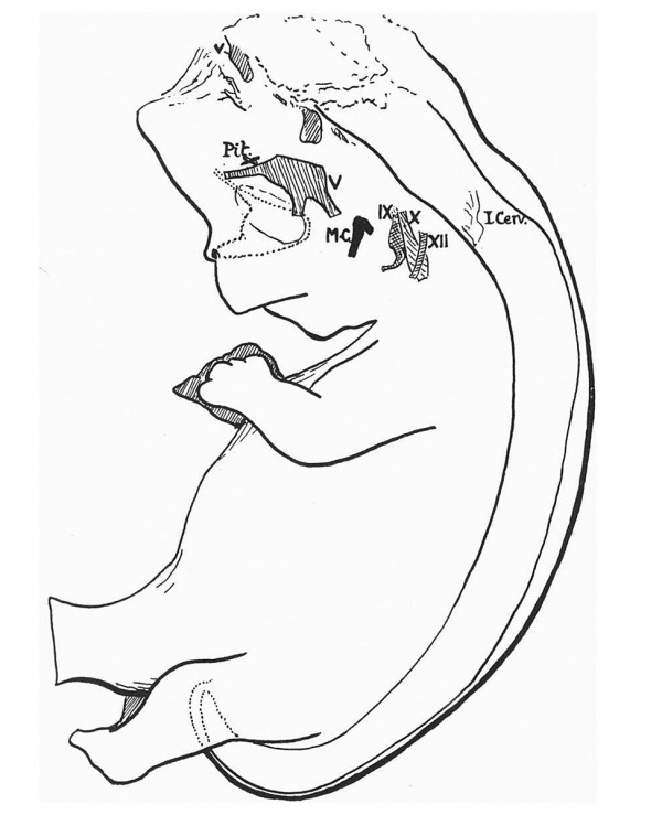
An outline reconstruction of the embryo is shown in fig. 1. It can be seen that, with the exception of the head, the general appearance of the embryo exhibits nothing unusual. It was divided into 10 μ serial sections, and these have been examined with a View to the discovery of any concomitant abnormalities.
(a) Urinogenital System. The anal end of the gut reaches the surface between the hinder extremities of the inner genital folds. The Mullerian ducts run down normally as far as the place where they cross over the Wolffian ducts, i.e. about the pelvic brim. Here, on the left side, the former duct apparently runs into the latter, while the right Mullerian duct disappears, though it cannot be definitely traced into the Wolflian duct. It may be remarked that examination of the genital glands suggests that the embryo is a male, whereas the 28 mm. “control” is female.
(b) Alimentary and Respiratory Systems. With the exception of the minor displacement of the terminal situation already noticed, nothing abnormal was found. The proximal limb of the U-loop was partly in the abdomen, and displaced above the distal limb and mesentery, but this was, I think, evidently accidental and referable to the preparation of the specimen: otherwise, the loop was normally in the umbilical sac.
(c) Circulatory System. Nothing abnormal found except in the head.
(d) Skeletal System. The cartilaginous skeleton appears normal, though there is a suggestion that the neural arches in the lumbar region, and perhaps elsewhere, are not quite as “thick” as in the 28 mm. sections. The impression of being thinner applies to all the tissues lying dorsal to the spinal cord, but there is absolutely no indication of any giving way or stretching to be observed.
(e) Special senses. The inner ear has reached a stage of development similar to that in the normal 28 mm. specimen, but the whole organ is rather smaller: the cells are probably smaller also, and, in the region of the proximal part of the cochlear duct, they are destroyed or absent on the left side, possibly injured during sectioning. The tubo-tympanic region is normal. The eyes are present, and present no evident abnormalities in the sections,but models show that the line of the choroidal fissure can be still appreciated, although the fissure itself is of course closed. It also seems likely that the formation of nerve-fibres is largely inhibited in these eyes, but no methods of special staining were used.
Fig. 1. Linear reconstruction of anencephalic embryo. The shaded areas in the head indicate certain nerve positions. V, the fifth nerve and its attached root; IX, X, XII are corresponding nerves. Some nerve remnants lie along the ventral surface of the elongated hind-brain stem. Pit,., marks the situation of the pituitary rudiment. M.C. is the upper end of Meckel’s cartilage.
The nasal cavities are only apparently abnormal in their shape: this is projected on the drawing in fig. 1 by a dotted line, giving a somewhat triangular outline. The upper angle is prolonged into a small tube of epithelium, drawn out to reach the meningeal level of the skull-base. The anterior nares lead into the cavities through elongated canals. The palate folds lie above the tongue, not in contact with each other: it is probable, I think, that this position had been acquired after death.
Spinal Cord
This appears to be quite normal, similar in size and structure to that in the 28 mm. embryo, and extends to the tail region, as shown in the figure. The spinal nerves also call for no comment; the position of the first cervical nerve is indicated in the figure, and it may be said that in this case there was a tendency to separation of the nerve filaments from the cord.
Head and Brain
The brain is represented (see figure 1) by an elongated portion of the hind-brain, ending fairly abruptly above, and by a pituitary body (X in the figure) and optic stalks and eyes. The pituitary body is smaller than in the 28 mm. specimen, but is almost as well developed, presenting a posterior lobe with the antero-lateral cornua growing up on each side of its stalk. There are cellular outgrowths from the lower part of the glandular portion, and the infundibulum is broken and ragged. The two pituitary organs are shown in fig. 2. The optic stalks begin in front of, and lateral to, the pituitary, from a small chaotic mass of quoridam brain-tissue, and run outwards and downwards to the eyes: they are hollow for some distance.
All the cranial nerves are present in position below the base of the skull. Some of them are indicated in fig.‘ 1, where the shaded projections give the positions of the trigeminal ganglion (V), ninth, tenth, and eleventh nerves. Traced proximally, all the nerves reach the meninges, where they come to an end, evidently torn: it may be pointed. out here that the optic nerve does the same, and the olfactory fibres, quite apparent in their lower parts, are lost in the meningeal level with the upward tube-like prolongations of the nasal cavities. On comparison of the nerves with each other, there is found a marked difference between the third nerve and other nerves in the same sections of the orbit, the former nerve being ‘little more than a vacuolated framework: this, for want of a better term, might conveniently be called degeneration. Muscles supplied by the nerve did not appear to be changed. No “degeneration” was found in the fourth nerve, and no change was apparent in the facial after giving off the chorda tympani—in fact no change could be certainly made out in any cranial nerve other than the third.
A space exists above and behind these broken ends of the cranial nerves on the meningeal aspect of the skull-base, and separates them from the remnant of the hind-brain. This space has evidently resulted from the straightening out of the hind-brain, which should, of course, occupy this region of the cranial cavity by its pontine flexure. The cavity presents occasionally in the sections a certain amount of débris, which in some cases is evidently nervous, and in others no doubt is meningeal.
The only part of the brain which remains~with the exception of the infundibular remnant and the basal débris—is the lower portion of the hind- brain. Reference to fig. 1 will show that the spinal cord is carried up to the head and becomes continuous there with an elongated column of nervous tissue which is directed upwards and forwards, ends somewhat abruptly above, and is surrounded dorso-laterally in its upper part by a broken mass of folded layers which are evidently remnants of the roof-plate of the rhombencephalon: a well-formed choroid plexus[1] is recognisable as being made by the dorsal and upper part of these layers. The neural cavity begins to show a dorsal broadening shortly after passing into the skull, and, some little distance further up, opens out rapidly, so that this part of the brain-stem is widely open dorsally when it has extended up half the distance shown in the figure. The open “ventricle” is, of course, roofed by the layers already referred to, but the attachment of these to the stem is only occasionally visible, possibly owing to changes in this upper end during preparation.
The whole structure is related, on its ventral aspect at any rate, to membranes in which are vessels and remnants of nerves; These nerves cannot be traced into the stem, consist of groups of fibres and ganglionic masses, and may run through several slides. No indication is found, however, to point to the different nerves represented, and it may be said at once that none of the lower cranial nerves can be found attached to the stem. These remains of nerves are represented in the figure by a few shaded areas along the ventral side of the main stem, and among them is a large ganglionic mass lying above the trigeminal ganglion: it does not seem, however, to be a portion of that ganglion, and its real value, like that of the other remains, is doubtful.
When we come to the upper end of the main stem, however, there is, on each side, a large nervous attachment ventro-laterally, and this can be recognised (V in fig. 1) with certainty as the superficial origin of the fifth nerve. The recognition was made first from its size and general relations to the other parts, and was clinched by examination of its deep strands and relations within the stem.
It is only a short stump on each side, and has apparently been carried away from its ganglion by extension of the main stem. The stem comes to an end - a. little distance above the situation of this nerve stump.
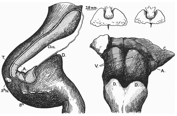
Miss I. C. Mann made a reconstruction model for me of this piece of the brain-stem, which confirms the short account just given. The dorsal view of the model, however, is of sufficient interest, I think, to merit further investigation. It is shown, after removal of the folds of the roof plate, in fig. 2, with a drawing of the hind-brain of the 28 mm. embryo, seen from the left. Dealing first with the normal specimen, the dorsal portions of the spinal cord are seen to end in the dorsal tubercles D (paired), and in front of this the widely open rhombencephalic cavity is covered by the broad roof-plate, of which the attached edge only is shown.‘ Within this cavity the dorsal lamina of the front limb of the pontine flexure (metencephalon) is thickening as a cerebellar plate (Cbm).
Caudal to this, just behind the angle of the flexure, is a small but well defined area, A, in evident connection with the auditory nerve, while the paired prominences of the myelencephalic floor of the cavity lie caudal to these auditory areas. The positions of origin of the fifth, eighth, ninth and tenth nerves are also shown. Turning now to the abnormal brain, the dorsal tubercles are recognised at once at D, and the open region above these is the rhombencephalic cavity, the floor of which is exposed as a result of removal of the roof- plate. But the rounded and prominent masses seen in this floor, V, are not metencephalic, such as would be their value if seen from a dorsal view of such a brain as that of the 28 mm. specimen: they are the floor masses of the myelencephalon, brought up above the level of the dorsal tubercles as a result of the straightening out of the flexure. Thus, cranial and lateral to them, we recognise the auditory area A, from which a groove extends upwards and inwards and probably marks the line of the flexure.
Fig. 2. On the left is a side View of the hind-brain of the 28 mm. embryo. On the right a dorsal View of the brain stump in the anencephalic specimen. The small figures above this are from reconstructions of the pituitary bodies in these two embryos. A, the auditory area; 0', the everted cerebellar plate; Gbm, the same plate in normal position; D, the dorsal tubercles in both brains; V, the ventral laminae of the myelencephalon; and T, the approximate level of destruction.
Further up and further out, the dorsal lamina or cerebellar ridge (C) is present in part on one side, while it is folded in on the other side. This reading of the condition present is in accord with the previous description of the origin of the fifth nerve, which is now seen to bear exactly similar relations to the various parts of the hind-brain in the two specimens. We thus reach the conclusion that the brain-stem of the anencephalic embryo comes to an end at a level which might be represented on the normal 28 mm. reconstruction by the line T, drawn through the lower part of the metencephalon.
Inferences to be drawn from the conditions in this embryo which have been described are that there has been a secondary separation of the cranial nerves from their attachments to the brain, and that the remnant of the brain, free from controlling forces holding it down in position, is no longer bent but has straightened itself and grown up in a direct line with the cord. I do not think there can be reasonable doubt that a brain existed: the presence of eyes, of a neural pituitary outgrowth, of third and fourth nerves, and incidentally of the nervous débris found in the cranium, indicate this very clearly. If, then, we start with the conception of a brain being present, it follows that this must have undergone destruction at a later period, and I think that this catastrophe might reasonably be referred to primary and secondary causes. We must, I think, assume the previous presence of the brain, but we cannot and ought not assume that this brain was normal. The present specimen gives no hint about the nature of that fundamental abnormality, but no doubt the tendency would be to place it in the dorsal laminae or roof-plate, or, taking what is only an inter-current effect, to assume the existence of an embryonic hydrocephalic condition. As just said, the specimen does not appear to give any clue to this, and it remains open to anyone to maintain his own views as to this primary fault. The ventral laminae were evidently able to form third and fourth nerves: further than that it does not seem possible to go in drawing inferences as to their condition, except that they appear perfectly good, so far as they exist, in the stump of the hind-brain.
But whatever the nature of the primary fault, it is clear that it must result in destruction of the continuity of the brain-stem at some point or points. In the present specimen I have the impression—to which I will refer again—that this destruction of the continuity of the tube occurred in the fore-brain, about the region of the rudiments of the corpora mammillaria: it is easy to formulate theories connecting this situation with deficiencies in dorsal and cerebral development. With this necessary solution of the tube, it seems to me that the action of the primary cause may be considered to have reached its acme and now, by virtue of this primary result, the secondary causes come into play and complete the destruction of that part of the brain affected by them.
Some years ago (Lancet, 1916) I described the pituitary region of the brain as the most “fixed” place within the skull, and referred the formation of the mid-brain flexure to the forward growth of the hind-brain acting against this fixed point. I have no wish to put forward any far-reaching morphological argument on this point, which I simply advance for what has always seemed to me to be its mechanical value: the definite adhesion of stomodaeal and neural ectoderm at this place, and their association with the upper end of the buccopharyngeal membrane, Seessels’ pocket (when present), and the adherent notochord, seem to me to give a guarantee of fixation, supported by every sagittal section, which must influence the shape of a brain growing up against its fixation here. The brain, growing more rapidly than the skull-base, piles itself up into curves, so to speak, and these then owe their existence, mechanically, and at least in part, to this fixation of the fore-brain. So, if the continuity of the brain is destroyed behind this fixed region, there is not the mechanical fixation or point of resistance against which the growing brain can produce or maintain curves, and the result is that there is a tendency for the stem to become straight behind the site of the destruction. This is seen in the straight stump left in this specimen, and would reasonably be certainly expected to occur in the mid-brain curve, if the destruction takes place anterior to this. But the straightening out of these curves involves rupture of all structures which pass between them and fixed points in their neighbourhood, and thus we come to secondary rupture of nerves and vessels.
So far as concerns the nerves, there are at least two possible explanations of the nerves being found broken off short at the meningeal level—they may have been ruptured by the straightening out of the curves, or they have been left free as a result of destruction of their sites of origin, without being previously separated from the stem. In this connection it is interesting to consider the hind-brain. Here we find the great fifth nerve evidently torn and yet with its torn root still in position in the brain, and the lower nerves, also torn, have certainly suffered this fate before the destruction of the brain-stem. Hence we can conclude that the rupture of nerves as a result of straightening of the brain can certainly take place, and is probably easier to accomplish than we perhaps imagine, influenced no doubt largely by our experience of adult nerves. This is suggestive, but of course not conclusive, for the third and fourth nerves: we can leave the matter there for the moment, and turn now to the vessels.
The internal carotids are large vessels, of normal appearance and running a normal course up to the origin of the ophthalmic arteries. These branches are of normal size, and the carotids go on after their origin for a little distance, and then abruptly come to an end, with open months. It is evident that they have been torn across, and it would seem that this is a result of the same processes which have tom the nerves. The vertebral arteries, lying in the meningeal tissues on the ventral side of the hinder stump of the brain, join here to form a basilar artery, and this is drawn out on the ventral surface of the stump, practically to its upper end: probably this is the end of the artery, but its division could not be absolutely demonstrated, as the membranes get very ragged at the upper end. This system is composed of vessels of good size, giving off good-sized branches. If we can judge from the remains of the carotid and vertebral systems, there was nothing wrong with these to account for the primary failure in development, but they have been secondarily affected as a result of the primary failure.
The position of the rupture of the carotids seems to me to suggest that the solution of continuity of the brain-stem took place not very far away—in fact, it was an important factor in forming my opinion that this destructive lesion occurred just above the infundibular outgrowth, the other considerations having mainly to do with the structure of this part and its connections with cerebral and other dorsal derivatives. A rupture in this place fits in well with the conditions that were found, Want of fixation of the brain-tube behind the situation leading to rupture of nerves, and failure of blood supply leading to disappearance of tissue. In accordance with this is the fact that the pituitary and eyes, whose vascular supplies remain, are still present, while the cerebral system supplied by the carotids above this level has utterly disappeared: also the hind—brain remains as far as the vascular supply is present and then ends abruptly, although there is nothing in the structure, examined microscopically, to distinguish this from any other hind—brain of the same stage and at the same level.
A reconstruction of the skull-base showed that it was comparable in development with the 28 mm. skull in its post-pituitary portion, except that the otic capsules showed a tendency, perhaps, to stand up more from the level of the floor, suggesting that there had not been any hind-brain structures lying on this floor for some little time: but, in front of the infundibular region, the base was narrowed and shortened, giving the impression of belonging to an earlier stage, although its cartilaginous and other details were in keeping with those of the “control.” The eyes projected out for a considerable distance beyond the side levels of the front part of the skull. The frontal wall of the cranium was folded back sharply over the front of the base, and turned down here, ending in a thinned edge; there were no indications of the occipital sinking so noticeable in older anencephalics.
To sum up, this specimen appears to me to suggest that, although the original and primary cause of anencephaly may be some local defect, limited perhaps to the fore—brain region, or may be of different nature in different cases, secondary causes of the condition ultimately found may also occur: that these secondary conditions result from rupture of the brain-stem and consequent loss of fixation of the front end of this growing stem: that as a result of this the curves tend to straighten out: that consequently the nerves and vessels connected with it give way: and that the brain tissue, as a result of the vascular starvation, breaks up and disappears up to the limits of the remaining blood supply. The specimen does not appear to offer any explanation of the nature of the primary fault.
Notes
- ↑ It is commonly stated that the choroid plexuses are formed in the fourth month. This is not correct: they begin to appear at or just after the middle of the second month, and grow steadily after this. When I speak of the plexus as “well-formed” in this specimen, I am of course referring to its proper stage of development.
Frazer JE. Report on an anencephalic embryo. (1921) J Anat. 56(1): 12-9. PMID 17103933
Cite this page: Hill, M.A. (2024, April 28) Embryology Paper - Report on an Anencephalic Embryo. Retrieved from https://embryology.med.unsw.edu.au/embryology/index.php/Paper_-_Report_on_an_Anencephalic_Embryo
- © Dr Mark Hill 2024, UNSW Embryology ISBN: 978 0 7334 2609 4 - UNSW CRICOS Provider Code No. 00098G


