Paper - Development and variation of the nerves and the musculature of the inferior extremity and of the neighboring regions of the trunk in man
| Embryology - 28 Apr 2024 |
|---|
| Google Translate - select your language from the list shown below (this will open a new external page) |
|
العربية | català | 中文 | 中國傳統的 | français | Deutsche | עִברִית | हिंदी | bahasa Indonesia | italiano | 日本語 | 한국어 | မြန်မာ | Pilipino | Polskie | português | ਪੰਜਾਬੀ ਦੇ | Română | русский | Español | Swahili | Svensk | ไทย | Türkçe | اردو | ייִדיש | Tiếng Việt These external translations are automated and may not be accurate. (More? About Translations) |
Bardeen CR. Development and variation of the nerves and the musculature of the inferior extremity and of the neighboring regions of the trunk in man. (1906) Amer. J Anat. 6:259–390.
| Historic Disclaimer - information about historic embryology pages |
|---|
 |
Development and Variation of the Nerves and the Musculature of the Inferior Extremity and of the Neighboring Regions of the Trunk in Man

By
Professor of Anatomy, University of Wisconsin, Madison.
With 10 Plates And 7 Text Figures. (1906)
Introduction
In a previous article in this journal (Bardeen and Lewis, 01), an outline was given of the early development of the limbs, body-wall and back in the human embryo. Lewis subsequently, 01, gave a more detailed account of the development of the arm, and I have recently, 05, described at some length the development of the spine and of the skeleton of the leg. The purpose of the following paper is a more detailed account of the development of the nerves and musculature of the leg and of the neighboring regions of the trunk and a consideration of the relation of developmental conditions to variations found in the adult. The embryological studies have been based chiefly on embryos belonging to the collection of Professor Mall of the Johns Hopkins University, who kindly placed them at my disposal. The statistical studies of nerve variation are based upon charts drawn from specimens in the dissecting rooms of the Johns Hopkins University and at the University of Wisconsin.
A. Outline of the Development of the Muscles and Nerves of the Inferior Extremity
I. General Features
For a description of the development of the external form of the limbs and of the chief features which characterize the earlier stages in the internal difierentiation, reference may be made to the three papers mentioned above. The posterior limb-bud is first seen as a massing of the mesenchyme at the posterior extremity of the Wolffian ridge, usually opposite the 21st to the 26th spinal segments. This mesenchyme arises in part from the axial mesenchyme, in part possibly from the somatopleure. There is no good evidence that in the mammals the myotomes contribute directly to it. On the contrary the myotomes are sharply marked off by a limiting membrane from the mesenchyme of the limb-bud until this has become extensively developed. Afterwards this limiting membrane disappears, but there is little likelihood that cells derived from the myotomes then wander any considerable distance into the limb-bud. See Bardeen, 00. A capillary network connected with the umbilical artery and the cardinal vein is formed in the limb-bud at an early period. Somewhat later nerves extend into the limb. At the same time the mesenchyme begins to be differentiated into skeletal, muscular and dermal regions. During the development of the limb it shifts distally so that the distal margin of the limb-bud is brought opposite the 27th and 28th, and sometimes also the 29th, spinal segments. As this occurs, bundles of nerve fibres from these more distal spinal segments extend into the limb-bud to contribute to the posterior nerves of the limb. In the adult the most distal nerve to contribute to the nerves of the limb varies from the 26th to the 29th, but is most commonly the 28th. (Bardeen and Elting, or). The number of spinal nerves contributing to the chief nerves of the limb varies from six to nine, but is usually seven or eight (Op. cit.). These variations are in all probability associated with variation in position of the limb-bud to the spinal axis during embryonic development.
The development of the main nerve trunks of the limb may be called the primary stage of nerve development and the associated variation in origin of the nerves, primary variation. As opposed to this primary development and primary variation We may call the growth which distributes the nerves within the limb the secondary stage of development and the variation there found secondary variation. During the primary period the spinal nerves send fibre bundles by direct paths to certain cutaneous areas and muscular anlages. During the secondary period the cutaneous nerves extend over the surface of the limb from the areas to which they are first distributed and the muscle anlages become differentiated into specific muscles to each of which nerve branches are given.
II. Primary Period of Nerve Development
The general structural relations at the period when the nerves begin to extend into the limb-bud are shown in Plate I, Figs. 1 and 2. In Fig. 1 are shown the right limb and the distal half of the trunk from the 17th (9th thoracic) to the 29th (4th sacral) spinal segments in Embryo 2 (length 7 mm., age 26 days). The limb-bud lies opposite the 21st to the 26th spinal segments. The coelom extends to a point opposite the 26th segment, but in the region of the limb it does not extend so far dorsally as in the thoracic region. In the figure several of the myotomes of the left side, the axial mesenchyme, the aorta, the left cardinal vein, the intestines and the uro-genital organs are not shown. A portion of the right cardinal vein and a portion of the right umbilical artery are represented, reduced in size for the sake of clearness. The umbilical artery curves about the distal extremity of the coelom. From the umbilical artery a branch passes into the limb-bud. Veins pass from the limb-bud into the cardinal vein. The blood-vessels of the limb exist at this time in the form of an irregular plexus.
The second, third and fourth lumbar nerves may be seen sending spreading bundles of nerve fibres into the dense tissue of the limb, dorsal to the cardinal vein. They extend, however, for no considerable distance into the limb-bud. The myotomes end abruptly near the base of the limb-bud.
Plate II, Fig. 1, represents the tissue differentiation in a section through the posterior limb-buds of Embryo 2. At the left the bud is shown cut through an area near the distal extremity of the coelom. At the right the cut is more dorsal and extends through the tips of the lumbar spinal nerves.
In Plate I, Fig. 2, are shown the right limb and the posterior half of the trunk from the 26th (8th thoracic) to the 30th (5th sacral) spinal segments in a slightly older embryo (163, length 9 mm.). Bundles of nerve fibres from the five lumbar and first two sacral nerves have become anastomosed into a plexus from which in turn four nerves have sprung. These represent the femoral, obturator, tibial and peroneal nerves. Within the limb the central mesenchyme, near the axis of the embryo, has become condensed. This condensed mesenchyme represents the femur and hip bone of the adult limb. In the drawing the outline of this sclerogenous tissue is made diagrammatically sharp. The femoral portion of the skeletal mass fades gradually into the undifferentiated mesenchyme of the distal portion of the limb. It is this skeletal mass which seems to divide the bundles of nerve fibres into the four main divisions which constitute the origin of the four chief nerves of the limb. The main artery and vein of the limb are represented at a reduced scale. The border vein at this period is well developed (see also Fig. 0, Plate III of the article by Bardeen and Lewis, 01).
The differentiation of the tissue of the limb-bud, first noticed in a condensation of tissue in the region corresponding to where the femur projects against the hip girdle, is quickly followed by further changes. Externally there becomes visible a differentiation of the limb into footplate, crus and thigh, while within the limb-bud the further development of the skeleton is marked by condensation of tissue, scleroblastema, to form the anlage of the skeleton of the foot, leg, thigh and hip girdle. About the scleroblastema is a myogenous zone, the mg/oblastema, composed of a slightly less dense tissue. In Embryo CIX, length 11 mm., this zone is best marked in the region of the hip (Plate II, Fig. It is not clearly defined in the foot region. Between the myoblastema and the ectoderm lies a zone of less condensed tissue, the dermoblastema.
The chief nerves of the limb extend into the myoblastema. This is not a homogeneous layer. On the contrary from the time of its formation regions which represent the anlages of muscles or groups of muscles may be more or less clearly distinguished from regions which represent intermuscular spaces. In Plate III, Figs. 1 and 2, an attempt has been made to outline the muscle masses which represent the anlages of future muscle groups in Embryo 109, length 11 mm. It is impossible to do this with exactness because the various regions are indefinitely bounded.
In this embryo the pelvic portion of the skeleton consists of a central region continuous with the head of the femur. From this central acetabular portion spring iliac, ischial and pubic processes. The femur is short and thick. The tibia and fibula are fairly definitely outlined, the foot-plate less definitely so.
The main nerve trunks have grown for a considerable distance into the limb. From them several of the chief muscular and cutaneous branches have sprung. The figures show these branches fairly well. In addition to the intrinsic nerves of the limb the anterior and posterior border nerves are also represented.
In Fig. 1 it may be seen that the myotomes in the region of the body wall have fused to form the anlage of the abdominal musculature. The lower margin of this extends distally about to the 21st spinal (1st lumbar) nerve. In Fig. E, Plate V of the article by Bardeen and Lewis, O1, it is represented slightly too short. From the ventro—posterior extremity of the abdominal musculature a somewhat indefinitely differentiated band of tissue may be followed to the pubic process of the pelvic girdle.
A slight communicating branch connects the twelfth thoracic with
the first lumbar nerve. The main portion of this latter nerve extends
forward on the internal surface of the distal margin of the anlage of
the abdominal musculature and gives off a lateral, “iliae,” branch.
Ventrally the nerve divides into branches which represent the hypogastric
and inguinal nerves. The 1st lumbar nerve also gives off a branch which
passes to the lumbar plexus.
The obturator nerve arises from the first four lumbar nerves, passes
through the obturator notch of the hip girdle and divides into two main
divisions. Each of these terminates in a differentiated mass of tissue,
the more anterior of which represents the adductor longus and brevis
and the gracilis muscles, the more posterior, the obturator portion of the
adductor magnus and possibly also the obturator externus muscle.
The tibial nerve arises from the fourth and fifth lumbar and first three sacral nerves. From it branches pass to muscle masses representing the obturator internus, quadratus femoris, hamstring, and the superficial and the deep posterior crural musculature. Distal to the tibial nerve the posterior cutaneous nerve of the thigh and the pudendal and caudal nerves may be seen.
In Fig. E, Plate V of the article by Bardeen and Lewis, or, the urachus was represented much foreshortened in order to reveal the muscle masses of the leg. In. Fig. 1, Plate III, the urachus is outlined in its true position as seen directly from the side.
In Plate III, Fig. 2, the genital an.d lumbo-inguinal nerves are seen passing ventro-laterally from the junction of the 1st and 2d lumbar nerves. The femoral nerve is seen passing outwards over the region of
the acetabulum. It is surrounded laterally by the iliopsoas muscle mass and terminates in the quadriceps femoris muscle mass. From it arise lateral and anterior cutaneous branches, a branch which passes to the sartorius muscle mass, and the saphenous nerve.
The peroneal nerve arises from the 4th and 5th lumbar and first two sacral nerves, gives off branches for the anlages of the superior gluteal, inferior gluteal, short head of the biceps and peroneal muscle masses and terminates in the anterior crural muscle mass.
An idea of the relations of the main nerves as they enter the limb in Embryo CIX may likewise be gained from Plate III, Fig. 3. The pelvis, the abdominal and dorsal musculature, the lining of the body cavity, the border nerves and the main nerve trunks of the limb are here represented as viewed from in front. The femur and the main nerve trunks are shown cut in a plane somewhat distal to the head of the femur. The division of the main nerve trunks into separate branches for individual muscles is schematic.
III. Muscle Differentiation
At the period under consideration several possibilities of muscle differentiation must be considered.
1st. — The tissue which represents the muscle masses just mentioned may extend into the limb-bud with the nerves and become differentiated as the muscle branches are given off. The fact that Harrison, 04, has shown that in the tadpole muscle differentiation may take place when no nerves are developed makes this possibility highly improbable.
2d. — The ingrowth of the nerves and the development of muscle branches may cause a “precipitation” of pre-muscle tissue about these branches. This likewise is rendered improbable by Harrison’s experiments.
3d. — Muscle differentiation begins in specific regions. Under normal conditions this differentiation begins simultaneously with the ingrowth of the nerves into the limb. Muscle branches extend into the differentiating musculature, owing perhaps to some specific attraction exerted upon the growing nerves. This seems on the whole to be the most probable course of development. The considerable variation shown in the origin and distribution of the nerves to the muscles renders it not improbable that their ingrowth is due in part to some special attraction exerted by the developing musculature upon the growing nerves, and variously responded to by the latter.
The paths opened up for the growth of the nerves to the muscles are, however, at first not as a rule in regions in which muscle tissue is to be differentiated, but in intermuscular areas. Thus the chief nerve trunks usually grow along paths which lie between main muscle groups. As the muscles of these various groups become differentiated the main nerve trunks of each muscle group are distributed in the septa which separate the individual muscles and finally after a nerve has entered the muscle for which it is destined it is usually distributed at first in the coarser intramuscular septa. During the early stages of development, however, the true muscle tissue cannot be sharply distinguished from the tissue which is to make up the skeletal framework of the muscle. For this reason it often appears as though the nerve to a muscle plunged at once into the midst of muscular tissue.
At a slightly later stage of development than that of Embryo CIX the differentiation of muscular tissue from the skeletal framework of the musculature is much better marked than in that embryo. Thus in Embryo 144, length 14; mm., the individual muscles of the thigh may many of them be clearly distinguished (Plate II, Fig. 3). It may be seen in this embryo that although muscle differentiation in a given muscle is most clearly marked in the region where the respective nerve has come in contact with or has entered the muscle, the differentiation is not limited to this area but extends for a considerable distance toward the skeletal areas to which the muscle is to be attached. It is probable, however, that the differentiation of a given muscle begins as a rule in a region which corresponds with the site of entrance of the chief nerve of that muscle. In Plate II, Fig. 3, several nerves and muscles are shown. The nerve to the gracilis muscle shows especiallylclearly. From this region the gracilis muscle may be traced in successive sections toward the pelvis and toward the tibia. The entrance of the inferior gluteal nerve into the gluteus maximus muscle also shows well in the figure. The two parts of the adductor magnus muscle, the obturator and sciatic portions, are shown near the site of entrance of the respective nerves. The semi-tendinosus muscle and the two heads of the biceps are shown cut at some distance from the site of entrance of nerves. About the two divisions of the sciatic nerve there is some dense tissue which probably does not, however, represent muscle tissue.
It is to be noted that during these earlier stages of muscle differentiation the muscle anlages are often connected at one extremity, less frequently at both extremities, with the skeletal anlages to which the muscle is subsequently attached. The tendons of the muscles are developed in continuity with the anlages of the muscles. As a rule the differentiation of the longer tendons begins in the vicinity of the muscle bellies and gradually extends toward the skeletal attachments.
In a considerably older embryo, 145 CXLV, length 33 mm. (Plate II, Fig. 4), differentiation of the muscles is much further advanced. Not only the muscles but also the fasciculi are separated by a large amount of connective tissue. This shows especially well in the gluteus maximus muscle. The main branches of the nerves of the muscle may be followed in the larger intramuscular septa, the smaller branches in the smaller intramuscular septa. I have elsewhere described the intramuscular growth of nerves in the mammals (Bardeen, oo and 03). It is of interest to note that after muscle differentiation is well under way there is relatively a much greater amount of connective tissue in the musculature of the embryo than in that of the adult.
After the stage of development exhibited by Embryo CIX the conditions within the limb become so complex that they can be better followed by tracing through the development of specific groups of nerves and muscles than by attempting to picture all the details of each successive stage of differentiation of the whole limb. In order, however, that the relations of specific groups of nerves and muscles to the general structural condition of the limb may be followed we shall first briefly describe the relations of the peripheral nervous system to the skeleton at two important stages of development.
IV. Outgrowth of the Nerves
In Embryo 144 (length 14 mm.) the main nerve trunks are well developed as far as the foot. The relations of the nerves to the spinal column, abdominal musculature, skeleton of the limb and the surface of the limb are represented in Plate IV, Figs. 1 and 2. The 12th thoracic nerve sends a communicating branch to the first lumbar and from this latter arise the hypogastric and inguinal branches.
| Plate 4 | |
|---|---|

|

|
| Plate 4-1 | Plate 4-2 |
From the first lumbar nerve a branch is also given off to the lumbar plexus. From the 1st and 2d lumbar nerves arise genital and lumbe-inguinal branches. The femoral and obturator nerves arise from the 1st, 2d, 3d, and 4th lumbar nerves and give off the branches shown in the figures. The sciatic nerve, which arises from the 4th and 5th lumbar and first three sacral nerves, is composed for the greater part of its course of separate peroneal and tibial nerves. The various muscular and cutaneous branches are labeled in the drawing.
In Embryo XXII, length 20 mm. (Plate V, Figs. 1 and 2) , the various nerves mentioned are much more highly developed than in Embryo 144. This difference of development is especially to be noticed in the feet. The figures indicate sufficiently well the relations of the nerves to the skeletal apparatus, the skin and the abdominal musculature.
| Plate 5 | |
|---|---|

|

|
| Plate 5-1 | Plate 5-2 |
A noteworthy fact brought out by these figures is that the cutaneous nerves are distributed at first to the anterior, distal and posterior margins of the embryonic limb, While the dorsal and ventral regions of the limb are given up to the differentiation of musculature.
Having thus considered in brief outline the more general features in the development of the muscles and nerves of the posterior limb we shall take up in turn a more specific study, first, of the development of the cutaneous nerves and then of that of the muscles.
B. Development and Variation of the Cutaneous Nerves
Grosser and Frohlich, 02, have given a good account of the development of the cutaneous nerves of the trunk. I have been unable to find any specific account of the embryonic development of the cutaneous nerves of the limbs, although the work of Sherrington, Head, and others on the segmental distribution of these nerves makes it of interest to inquire whether or not embryonic conditions can help to explain the phenomena these authors have described. In the following section the embryonic development and the variations in distribution of specific groups of nerves are first described and then the more general facts disclosed by this study are briefly reviewed.
I. Anterior Border Nerves
a. Development
When the nerves begin to enter the limb-bud this lies, as pointed out above, usually opposite the five lumbar and first sacral nerves (Plate I, Fig. 1). The posterior margin of the developing body-wall and the anterior margin of the limb-bud usually overlap opposite the 21st segment. The nerves arising from the 21st spinal (list lumbar) nerve are therefore true border nerves, being in part distributed to the abdominal wall and in part ‘to the limb. The 20th and 22d spinal nerves (12th thoracic and 2d lumbar) also usually contribute to a greater or less extent to both regions, the 20th contributing to the cutaneous supply of the leg, the 22d slightly to the extreme margin of the abdominal musculature.
In Embryo 109, length 11 mm. (Plate III, Figs. 1, 2 and 3), the border nerves are beginning to extend toward the skin. At this stage the oblique and the rectus muscles of the abdomen are beginning to be differentiated. The transversus muscle has not yet appeared. Between the ventro-anterior margin of the pubis and the ventro-caudal angle of the difierentiating abdominal musculature a slight thickening of the mesenchyme represents the beginning of the tendon of the rectus and of the inguinal ligament. A considerable interval exists between the distal margin of the abdominal musculature and the anlage of the iliac crest. The musculature lies near the peritoneal cavity, while the crest is in the mesenchyme lateral to this cavity. Between body cavity and crest lies the femoral nerve with its branches (Fig. 3). From the first and second lumbar nerves the iliohypogastric and inguinal and the genital branch of the genito-femoral extend ventrally between the coelomic wall and the distal margin of the developing abdominal musculature. From the common trunk of the iliohypogastric and inguinal nerves a lateral branch, the “ iliac,” extends toward the skin in an area considerably anterior to the ilium. The lumbo-inguinal nerve and the lateral and anterior cutaneous branches of the femoral extend toward the anterior margin of the limb-bud.
- The term “genital” nerve is here used in preference to “spermaticus externus.”
In a slightly older embryo, 144, length 14 mm. (Plate VI, Fig. 1) differentiation of the abdominal musculature has proceeded much further. The external oblique muscle is a thin sheet, somewhat wrinkled in the specimen. In the figure merely its origin from the lower ribs is shown. It extends distally into a sheet of mesenchyme which is thickened at its distal border into an embryonic inguinal ligament (lig. ing.). This latter extends from an anterior mesenchymatous process of the ilium toward the pubis. Ventrally it becomes continuous with the blastema of the pubic crest. Beneath the external oblique lies the internal oblique muscle. Distally this is connected by a mesenchymatous membrane with the inguinal ligament. In the figure merely the costal and inguinal portions of the muscle are shown.
The transversus muscle is differentiated immediately beneath the peritoneal membrane. It is not clear whether the material of the transversus musculature is derived from the coelomic lining or from the myotomes. If from the latter the tissue wanders along the peritoneum from the region of the ribs.
At this early stage the anlage of the processus vaginalis may be seen in the form of a thickened mass of tissue which is continued from the plica gubernatrix through the internal oblique muscle and the aponeurosis of the external oblique above the inguinal ligament to the junction of the thigh with the trunk. Here it spreads out into processes which extend on the one side toward the mid line of the body, on the other toward the femur.
Between the transversus musculature and the internal oblique run the main trunks of the thoracico—abdominal nerves. The ilio—hypogastric and inguinal nerves pierce the internal oblique muscle and the aponeurosis of the external oblique much as in the adult. The iliac branch of the ilio-hypogastric, however, pierces the oblique muscles in a region anterior to its relative adult position. This is also the case in Embryo 22, length 20 mm., Plate V, Fig. 1. Beyond the region of the inguinal nerve the coelomic wall, backed by a thickened membrane representing the transversalis fascia, curves medially while the oblique musculature takes a somewhat lateral direction toward the inguinal ligament. Between the two is a space in which lie the femoral nerve, its proximal branches and the anlage of the ilio-psoas muscle. The genital branch of the genito-femoral nerve follows along the coelomic wall almost parallel with the hypogastric and inguinal nerves but converging toward the latter. The point “X” in the figure represents a region where later the peritoneal wall will be pushed laterally over -the ilio-psoas muscle so as to cover this and be brought in contact with the iliac crest. The lumbo-inguinal nerve passes out beneath the inguinal ligament in the vicinity of the femoral artery. It probably represents a lateral branch of the genito-femoral considered as the ventral division of a typical spinal nerve.
Ventrally the genital nerve, usually after anastomosing with the inguinal, passes along the vaginal process through the aponeurosis of the external oblique and over the inguinal ligament to the thigh. It is interesting to note that this development considerably precedes the descent of the testicle.
In Plate VI, Fig. 2, the border nerves of Embryo 22, length 20 mm., are pictured. It is somewhat diflicult to trace with certainty the border nerves in this embryo, but the figure is believed to illustrate approximately the actual relations. While in Embryo CXLIV a consider- able interval separates the anlage of the iliac crest from the distal margin of the abdominal musculature, in Embryo XXII the crest is much further developed and at the same time has been rotated toward the dorsal portion of the distal margin of the oblique abdominal musculature. This at the sa.me time has extended distally and become attached to the iliac crest. Meanwhile the peritoneal wall has bulged laterally so that the fascial extension of the transversus muscle covers the ilio-psoas muscle in the region of the pelvis and the transversus muscle has formed its pelvic attachments. The main trunks of the border nerves have been brought by these changes into relations which closely resemble those characteristics of the adult. Adult conditions are reached by some further relative shifting of parts and by the growth of the nerves within the areas for which they are destined.
The segmental relations of the border nerves may be best understood by comparing the position of the pelvic girdle when the nerves first ex- tend toward the skin with the condition brought about by the shifting of the girdle. See Plates III, IV, V and VI. In Embryo CIX, the stage in which the nerves first extend toward the skin, the border nerves arise from the spinal nerves in the following order: iliohypogastric, inguinal, genital and lumbo-inguinal. As these nerves grow forward there takes place a rotation of the base of the limb medially, ventrally and posteriorly. At the same time the spinal column becomes straightened and the limb- bud as a whole descends posteriorly. The pubis is carried from a point opposite the 21st '(12th thoracic) segment to a point opposite the 26th, and at the same time the posterior margin of the ilium is usually brought to lie opposite the 26th and 27th vertebra to which it becomes attached. The two pubes are carried forward ventrally until they are united by the symphysis pubis.
As the pubis rotates ventrally and posteriorly the inferior portion of the abdominal wall is extended in a corresponding direction. The ven-
(text to be added)
Figures and Plates
Fig. 2
Fig. 3
Plate 1
Plate 2
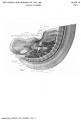
Plate 3-1
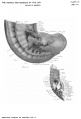
Plate 3-2
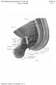
Plate 4-1
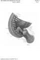
Plate 4-2
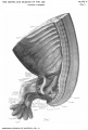
Plate 5-1
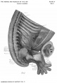
Plate 5-2
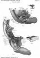
Plate 6


Cite this page: Hill, M.A. (2024, April 28) Embryology Paper - Development and variation of the nerves and the musculature of the inferior extremity and of the neighboring regions of the trunk in man. Retrieved from https://embryology.med.unsw.edu.au/embryology/index.php/Paper_-_Development_and_variation_of_the_nerves_and_the_musculature_of_the_inferior_extremity_and_of_the_neighboring_regions_of_the_trunk_in_man
- © Dr Mark Hill 2024, UNSW Embryology ISBN: 978 0 7334 2609 4 - UNSW CRICOS Provider Code No. 00098G