Paper - A contribution to the morphology and development of the mammalian liver
| Embryology - 26 Apr 2024 |
|---|
| Google Translate - select your language from the list shown below (this will open a new external page) |
|
العربية | català | 中文 | 中國傳統的 | français | Deutsche | עִברִית | हिंदी | bahasa Indonesia | italiano | 日本語 | 한국어 | မြန်မာ | Pilipino | Polskie | português | ਪੰਜਾਬੀ ਦੇ | Română | русский | Español | Swahili | Svensk | ไทย | Türkçe | اردو | ייִדיש | Tiếng Việt These external translations are automated and may not be accurate. (More? About Translations) |
Bradley OC. A contribution to the morphology and development of the mammalian liver. (1908) J Anat. 43: 1-42. PMID 17232788
| Historic Disclaimer - information about historic embryology pages |
|---|
| Pages where the terms "Historic" (textbooks, papers, people, recommendations) appear on this site, and sections within pages where this disclaimer appears, indicate that the content and scientific understanding are specific to the time of publication. This means that while some scientific descriptions are still accurate, the terminology and interpretation of the developmental mechanisms reflect the understanding at the time of original publication and those of the preceding periods, these terms, interpretations and recommendations may not reflect our current scientific understanding. (More? Embryology History | Historic Embryology Papers) |
A Contribution to the Morphology and Development of the Mammalian Liver
By O. Charnock Bradley, M.D., D.Sc.
Royal Veterinary College, Edinburgh.
As is the case with all the organs of the body, the liver of man received much earlier and more detailed attention at the hands of the morphologist than did the corresponding organ of mammals in general. Thus the earliest accounts of the comparative anatomy of the liver were based upon its form and characters in the human subject. But it has come to be recognised that the human liver, in common with the majority of human organs, has undergone a high degree of specialisation during the process of evolution, with the result that it cannot be regarded as a type suitable for descriptive purposes, but must, rather, be itself compared with more primitive and generalised forms.
It is now generally agreed that the so-called lobes of the human liver are arbitrary divisions of the organ made for convenience of description, and are not, correctly speaking, of comparative morphological value. In human anatomy a striking anisuse of the term “lobe” occurs in. the expression “quadrate lobe.” The “quadrate lobe” is merely a quadrilateral area on the visceral surface of the liver, bounded on the left by the longitudinal fissure and on the right by the depression for the gall-bladder. It is not detined by true fissures; for the so-called longitudinal or umbilical fissure, as will be shown later, is not the same in character, or in origin, as the real fissures of the liver of mammals in general. Moreover, though too much importance must not be attached to the relative size of any given area, it may, nevertheless, be pointed out that the “quadrate lobe” is not of constant dimensions even in the human subject; and its homologue in the Mammalia as a whole is of remarkable variability. Rolleston (1) has shown that, in man, it may be absolutely large, and as much as twice the size of the remainder of the under surface of the liver; whereas, on the other hand, it may be quite rudimentary even where no pathological lesion exists.
The right and left lobes of the human liver, as described, correspond to four lobes of the comparative anatomist. It has been shown beyond question that the differentiating fissures have disappeared, a series of livers from animals standing close to man in the zoological scale demonstrating the process of disappearance. Occasionally man reverts to a more primitive condition, and some or all the fissures reappear. Among examples of such reversion is one recorded by Parsons (2), where a human liver had two lobes to the right and two to the left.
The only parts of the liver of man which can be said to have retained their independence in any degree are the Spigelian and caudate lobes. Even these are no longer decided, though occasionally they may be well marked and distinct, as has been shown by Thomson (3). As bearing on phylogeny, as well as on ontogeny, it is of interest to note that they are of proportionally greater size in the foetus than in the adult (cf. Thomson (4) ).
In spite of several hypotheses by which it has been sought to explain the absence of lobes in man and other mammals, the question of the true factor or factors still affords ground for discussion. Duvernoy (5) was of opinion that the character of the food, and the correlated conformation of the stomach, should be regarded as having a profound influence on the formation of fissures and lobes. Others have held a like opinion. As an illustration of the effect of a large and complicated stomach in the direction of reduction of hepatic fissures, the ruminants have been cited. Beyond question the ordinary ruminants have a compact liver; but, even in closely allied species with stomachs of relatively equal size, there is a certain measure of difference in the presence or absence and in the relative depth of fissures and in the size of lobes. The domestic ruminants, to seek no further, while possessing the same gastric peculiarities, have livers readily distinguishable. The aberrant ruminants, the camels, have a stomach no less complex than that of the more ordinary members of the same order, and yet there are two very well-developed fissures dividing the _ liver into three lobes (cf. Lesbre (6) ).
In the horse the stomach is small, but its place is taken physiologically by the enormous large intestine. The hollow abdominal organs of the horse are hardly less ponderous than those of ruminants; still the liver has not become compacted, but has all the fissures associated with the typical mammalian organ.
Whatever influence the character of the alimentary canal may have on the liver of mammals in general, man and the anthropoid apes form an obstacle in the way of anyone wishful to enunciate a universal law. From man down through the anthropoids to the lower monkeys all grades of lobation may be demonstrated. It is obvious, then, that some factor other than the conformation and disposition of the stomach and intestines must play a part in the obliteration of fissures. Keith (7) has considered the position and mode of fixation of the liver as factors in the lobulation in Primates. He is of opinion that the erect posture has caused the disappearance of fissures in man and the anthropoid apes. As the result of the assumption of an upright position, the dorsal mesentery has acquired a more complicated mode of attachment consequent upon the removal of the support formerly afforded to the liver by the ventral wall of the abdomen. Correlated with the more extensive fixation of the liver to the dorsal abdominal wall is the disappearance of fissures. This hypothesis, unfortunately, does not take cognisance of the fact that a practically undivided liver is met with outside the Primates in mammals in which the posture is prone and where the dorsal mesentery has undergone no coniplicated modification.
Keith also observes that, in the anthropoid apes, the abdomen as well as the thorax has become widened at the expense of the dorso-ventral diameter, and that this has had its effect upon the form of the liver. The difference in the shape of the thorax and abdomen of the anthropoids and the lower monkeys as a whole is undoubted ; but the present writer (8) has pointed out that the predominance of the lateral over the dorso-ventral diameter is not confined to the anthropoid apes. It is also demonstrable in some, at least, of the Old World monkeys; and in-these there has been practically no alteration of the liver from the more generalised mammalian type. But even more important in this connection is the fact that in some New World monkeys (eg. Lagothrix humboldtt) the dorso-ventral flattening of the trunk is decidedly pronounced without there having been any effect whatever upon the hepatic fissures. In Humboldt’s woolly monkey the lobes are as distinct as, or possibly more distinct than, in Cercocebus fuliginosus, an Old World monkey with a trunk of somewhat the same relative diameters as in the average mammal.
Ruge (9) also has endeavoured to explain the differences in liver conformation as being due to modifications in the form of the trunk and to a flattening of the dome of the diaphragm. The same objections to this hypothesis might be urged. In the Lagothrix the diaphragm was found to be much flatter than in the sooty mangabey.
Rex (10), whose communication will be discussed in greater detail later, considers that the arrangement of the blood-vessels within the liver is to be regarded in association with the form of the organ as a whole; but he scarcely seeks to explain the absence or presence of fissures as being dependent upon vessels. Homologous regions of the liver in different animals may have homologous vessels whether fissures are present or not.
It is safest to conclude that no single hypothesis, as yet propounded, is in harmony with all the known facts relative to the shape of the mammalian liver and the presence and depth of its fissures. No doubt the form and volume of the abdominal viscera, the habitual attitude of the animal, the relative dimensions and degree of mobility of the trunk, all play a part in producing a correlated moulding of the liver; but, so far as our present knowledge goes, it may be reasonably assumed that no single factor is sufficient to account for all the varied modifications met with in the examination of a series of organs derived from different mammalian orders.
Consequent upon the lack of a recognised type with which the liver of different mammals might be compared, great confusion prevailed in the writings of descriptive anatomists at the beginning of last century. It is © no easy task to make the description of one writer fit in with the account of a contemporaneous, or nearly contemporaneous, author. The works of Cuvier and Meckel may be cited as examples of classics illustrating the great want of uniformity in the treatment of the same subject.
In 1835 Duvernoy (5) expressed himself as dissatisfied with the method, or rather lack of method, followed by comparative anatomists of that day. The discord which prevailed in the number of lobes assigned to the liver in general works and in monographs he describes as remarkable. In order to produce descriptive harmony, he suggested a uniform method. In all animals, he said, there is a principal lobe, to which may be added a right and a left lobe, placed either to the sides of the principal lobe or behind it. The principal lobe itself may be divided into two or three portions. As a further complication, the liver may possess a right and a left lobule attached to the base of the corresponding lobes or to the principal lobe.
The human liver, according to Duvernoy, consists of a principal lobe only, with a prominence equivalent to the right lobule. It therefore lacks a right lobe, a left lobe, and a left lobule. The so-called right and left lobes, admitted by the greater part of writers on human anatomy, but rejected by some, are no more than two portions of the principal lobe, separated, but only on the convex surface of the liver, by the falciform ligament.
In conclusion, Duvernoy discusses the reasons for differences in the form of the liver in different classes of animals. He inclines to the opinion that the character of the food and the associated conformation of the stomach have an influence in determining the external features of the liver. In man, where the rule would not apply, he seeks refuge in the supposition that it may be the erect attitude which is responsible for the peculiar form of the organ.
The question of the number, nomenclature, and homology of the lobes was reconsidered in 1861 by Rolleston (11). He was probably the first to make use of the falciform ligament as an important landmark in defining territories in animals other than man. The suspensory ligament is attached to the suspensory lobe, which, he says, is very frequently tritid, the ligament having one lobule to its left, subequal to a second lobule to its right —the central suspensory. The fossa for the gall-bladder forms the right boundary of the central suspensory lobule, thus separating it from the right lobule of the suspensory lobe which extends from the cystic indentation to the free right edge of the entire lobe. To the left of the suspensory lobe is another, rarely, if at all, deeply incised or indented—the left lobe, whereas a right lobe is divided into three secondary lobules—the right kidney lobule, the swperior right lobule, and the lobulus Spigelit.
Rolleston’s suspensory lobe is clearly equivalent to Duvernoy’s lobe principal, and his left suspensory lobule to the lobe principal gwuche ; but Rolleston strikes a blow at the pre-eminent importance attached by Duvernoy to the lobe principal when he confesses himself inclined to consider that the left suspensory: lobule (lobe principal gauche of Duvernoy) is wholly lost in such livers as those of man and the ruminants.
To Rolleston was ascribed by Flower the credit of having been the first to recognise the importance of the caudate lobe (right kidney lobule), which had previously been confounded with the Spigelian lobe of man by many anatomists. It is questionable if such credit is rightly bestowed; for we have seen that Duvernoy recognised and figured a distinct right lobule attached to the right lobe.
Owen (12), in 1868, criticised Duvernoy’s view that the human liver is to be regarded as homologous with the principal lobe of quadrupeds; or, conversely, that the quadruped liver is equal to the human organ plus superadded right and left lobes. “Fissures,” he asserts, “rather than lobes, are added to the liver of quadrupeds.” Owen’s conception of the divisions of the liver agrees fairly closely with that of Rolleston. In the majority of mammals, he remarks, a lobe is definitely mapped out by a deep cleft to the left of the suspensory fissure, and another to the right of the cystic fissure. This he names the cystic lobe, and is apparently the first to recognise its homology with the right portion of the left lobe and the left portion of the right lobe, including the cystic fossa, of the human liver.
A most important contribution to the literature on the morphology of the mammalian liver was made by Flower (13) in 1872. Flower’s scheme for the division and nomenclature of the different lobes is of such moment, and has received such wide acceptance, that its promulgation may be taken as marking a period in hepatic research. Flower regarded the liver as divided into right and left segments by the remains of the umbilical vein and ductus venosus. Each segment is subdivided into lobes. To the left of the umbilical fissure is a left central lobe, bounded to the left by the left lateral fisswre, beyond which lies a left lateral lobe. In the same manner the right segment is subdivided, by a right lateral fissure, into a right central and a right lateral lobe. To the right lateral lobe are appended Spigelian and caudate lobes.
The cystic fissure, to which Owen attached some importance, was shown by Flower to be of no great morphological significance, since it is irregular in position and frequently absent. It may be of interest to add that the presence of the fissures does not depend upon the presence of a gall-bladder ; for, in the horse, where a gall-bladder is not present, a cystic fissure is all but absolutely constant, and may even be double.
Prior to 1888, the year in which an elaborate paper by Rex appeared, anatomists other than microscopists had been satisfied to direct their attention mainly to the exterior of the liver. Rex (10) struck out an entirely new line of research when he considered the arrangement of the branches of the portal vein, hepatic veins, and bile-ducts in their relation to the lobes. Unfortunately, Rex had not been able to consult Flower’s paper, and so was constrained to adopt a different nomenclature. Perhaps this is not to be entirely deplored, as it permitted him to approach his subject with an unbiassed mind. In the generalised form of the mammalian liver he came to the same conclusion as Flower in the recognition of six lobes; but, unlike Flower, he did not attach great importance to the position of the umbilical vein as indicating a line by which the organ can be divided into right and left segments. Consequently, his middle lobe corresponds, not to Flower’s central lobe as a whole, but to the right central only. He also raises the caudate lobe to a like morphological plane with the right lateral lobe, and names it the right inferior lobe.
It is unfortunate that Rex uses a nomenclature not to be commended from the standpoint of the comparative anatomist. He regards the surfaces of the mammalian liver as compared with those of the human organ; and, consequently, speaks of a lobe as being “superior” or “inferior” as it would be were it transplanted into the human body.
Apparently Rex was the first writer to use the name omental lobe in place of Spigelian lobe, an alteration which has much to commend it, since it indicates the close relationship which exists between the lobe and the gastro-hepatic omentum.
The right and left main branches of the portal vein have a remarkably regular distribution, by means of secondary branches, in the various lobes of the liver of different species of mammals. This brought Rex to recognise that the lobes could be homologised, not only by an examination of the exterior, but also by the observation of the distribution of the principal branches of the vein. He discovered that from the right main portal branch two veins arise, to which he gave the names of ramus descendens and ramus arcuatus, the former supplying the caudate lobe (right inferior lobe), the latter the right lateral lobe (right superior lobe). The left main portal branch, after contributing a ramus omentalis to the Spigelian or omental lobe and a ramus angularis to the left lateral lobe, ends in a recessus umbilicalis, so named from its derivation from the umbilical vein of the embryo. From the umbilical recess two groups of branches, or arborisations, are distributed to the right central lobe on the one hand (right arborisation) and to the left central lobe on the other (left arborisa- | tion). The right central lobe has evidently a double venous supply, for it also receives a ramus cysticus, a branch either of the right or the left main vein.
The following table shows at a glance the distribution of the portal vein as described by Rex. In order to avoid the use of an unsuitable nomenclature, the names of the lobes as suggested by Flower are here substituted for those of Rex, with the exception of omental in preference to Spigelian.
| Lobes | Branches of Portal Vein |
|---|---|
| Caudate | Ramus descendens |
| Right lateral | Ramus arcuatus |
| Right central | Ramus cysticus |
| Right arborisation from the recessus umbilicalis | |
| Left central | Left arborisation from the recessus umbilicalis |
| Left lateral | Ramus angularis |
| Omental lobe | Ramus omentalis |
Rex describes three hepatic veins, each arising in two branches and draining the following lobes :—
| Hepatic Veins | Lobes |
|---|---|
| Right (superior and inferior) | Right lateral and caudate |
| Middle (right and left) | Right central |
| Left (superior and inferior) | Left central and left lateral |
The importance of the contribution made by Rex to the question of the homology of the lobes of the liver in the mammalian series depends upon the circumstance that he was the first to point out that the distribution of the portal vein and the arrangement of the lobes are closely associated the one with the other, and that, even in animals in which the liver is more or less consolidated (as in ruminants, cetaceans, anthropoid apes, and man), the same number of branches of the portal vein can be detected.
Since 1902 a series of papers has been published by Ruge (14) embodying the results of the investigation of a great abundance of Primates material. So considerable is the number of facts thus accumulated that it is practically impossible to give a serviceable summary in limited space. Among other things, he has described a number of secondary fissures and lobules which help to explain anomalous indentations and grooves occurring in mammals other than Primates. Further reference will be made to some of these lobules later in this paper.
Cantlie’s contention (15), that the present method of dividing the human liver into right and left lobes by a line drawn along the longitudinal fissure is unscientitic, and consequently untenable, has received little consideration at the hands of the human anatomist. He asserts that the gall-bladder is central, and on either side of it lie the true right and left lobes; and a line drawn from the fundus of the gall-bladder to the exit of the hepatic veins divides the liver into two equal portions, as shown by injection, by weighings, by developmental, by pathological, and by clinical observations. Occasion will be taken later to show that he has certain grounds for stating that the present method is unscientific; but, at the same time, one is forced to confess that it is highly convenient.
Thanks to investigations initiated by His (16) on the human embryo, and Hochstetter (17) on embryos of the rabbit, a considerable volume of literature on the early development of the liver has accumulated during the past ten or twelve years. As is now well known, a hepatic bud early (during the second week in the human embryo) develops from the epithelium of the gut and surrounds and invades one or both of the omphalo-mesenteric veins, The invasion of the veins by the hepatic tissue results in the production of the sinusoids of Minot (18) and the formation of a complex stream-bed in place of the originally simple one. The omphalo-mesenteric veins consequently no longer communicate directly with the venous sinus of the embryonic heart, and so, were no other and more expeditious route provided for the passage of the blood, circulation would be impeded. But, as the production of sinusoids from the omphalomesenteric veins is proceeding, the umbilical veins are becoming larger and of greater importance as affording a ready means by which the blood may reach the heart. Thus the omphalo-mesenteric veins are relieved of part of their duty.
In the rabbit, both the umbilical veins are associated with the liver, and for some time the right is much larger than the left, as is also the right omphalo-mesenteric vein. Thus is caused, according to Lewis (19), a preponderance of venous channels on the right side and a consequently more rapid development of the right part of the liver. It will be seen later that the same one-sided growth, arising from a like cause, is noticeable at a certain period in the development of the liver of the pig.
Sooner or later, in all animals, the right umbilical vein begins to disappear on the usurpation of its function by the left vein. In the human embryo, in the rabbit, and in the cat, the right vein early loses its association with the liver; and it is evident that in some mammals it does not participate in the hepatic circulation at any time. In a 5-mm. embryo of the rat, and in a 6-mm. embryo of the flying squirrel (Pteromys oral), the right umbilical vein is confined to the body-wall. A further variation is present in the sheep. From the observations of Bonne (20), it seems that in this animal the two umbilical veins remain of the same size, and unite to form a common vessel which passes into the liver, to be continued onwards by the ductus venosus.
A paper of considerable importance in connection with the investigation described in the present communication is one published in 1895 by Brachet (21). In a rabbit embryo twelve days old, he describes the liver as being composed of three lobes. One of these is ventral in position and occupies the continuation of the septum transversum into the ventral body-wall. The other two lobes—right and left lateral—have been developed in the septum transversuim itself. At first, then, there are three lobes definitely separated from each other, but later a new subdivision takes place. The middle or ventral lobe is divided into two right and a left—and thus four lobes are produced. Nevertheless, Brachet coneludes that the liver of the adult rabbit consists of three fundamental lobes, namely, a median lobe, occupying the ventral part of the organ and developed in association with the two umbilical veins; a left lateral lobe, proceeding from the primitive left omphalo-mesenteric lobe, which develops along the vein of that name; and a right luterul lobe, having a like history and a similar developmental relationship to the right omphalomesenteric vein.
Brachet holds that the lobe of the inferior vena cava (caudate lobe of Flower) is merely a prolongation of the right lateral lobe developed in the lateral mesentery. The Spigelian lobe also is simply an appendage to the right lateral lobe
The chief interest of Brachet’s communication depends upon the recognition that the liver follows the umbilical and vitelline veins during development, and that the direction and situation of the vessels govern the direction and situation of hepatic growth.
Up to 1906 no attempt had been made to connect the results of Rex’s observations on the adult blood-vessels with the vascular conditions in the embryo. In July of that year, however, Mall (22) published a paper which bridges the gap so far as the human liver is concerned. In human embryos 5 mm. and 6°5 mm. long, he shows that all the blood from the left umbilical vein passes through the liver, the direct connection of the vein with the heart having been lost. The right umbilical vein is also obliterated, and, therefore, the whole of the umbilical blood finds its way to the heart by way of intra-hepatic channels. To facilitate the passage of a large volume of blood through the liver, the right omphalo-mesenteric vein has either remained pervious, or has been reopened, thus allowing of free circulation on the right side. On the left, two new veins have been formed, or have arisen directly from the remains of the left omphalo-mesenteric vein. -At any rate, they are two of the permanent main vessels of the liver, namely, the vena hepatica sinistra and the ramus angularis, the latter arising from the recessus umbilicalis.
During the fifth week of embryonic life the right omphalo-mesenteric vein is partly converted into sinusoids, and the ductus venasus is formed. The ramus dexter of the hepatic vein, and the ramus arcuatus et descendens of the portal vein, then take the place of the former single and continuous venous channel. It seems that the time of disappearance of the right omphalo-mesenterie vein is not constant; for it is present, along with a ductus venosus, in an embryo 11 mm. long. In the same embryo (at the end of the fifth week) “the right and left portal twigs have begun to divide, and from the recessus umbilicalis a new group of veins have formed and radiate into the middle and left lobes of the liver.” Mall goes on to say that, in consequence of the branching of the veins of the liver, six primary lobules are produced. “With the completion of six lobules we recognise fully the adult form of the liver. Each lobule now represents one of the six lobes of the mammalian liver: each of the primary lobules is to expand into a whole lobe.”
That in the human embryo, at a certain period of its existence, there are six lobules, is certainly evident from Mall’s descriptions and exceedingly convincing illustrations; but that these lobules ultimately develop into six ‘lobes, equivalent to the six lobes of the typical mammalian liver, does not seem to be entirely beyond question. In order to count six lobes, the omental lobule and the caudate lobe must be considered as constant and separate units in which a ramus omentalis and a ramus descendens respectively ramify, and from which hepatic veins spring. That the portal vein has a ramus omentalis is not clear from Mall’s figures. Nor, it may be said, is the ramus descendens of the human liver absolutely certainly homologous with that branch of the portal vein which supplies the caudate lobe of mammals in general. It will be shown later that, in some mammalian embryos at any rate, independent rami omentalis and descendens cannot be detected, and that even in an animal in which the omental lobule and the caudate lobe are much larger than in man. It appears, therefore, that judgment on this head should be suspended until further evidence is forthcoming.
Geraudel (23) has recently reviewed the latest literature on the subject, but does not make any material addition to our knowledge. He, however, emphasises the fact, brought out so clearly by Mall, that the vessels of the adult liver represent the remains of the primordial vessels which have become reticulated by a sinusoidal process. This is certainly a fact of great and fundamental importance which will be again elaborated in the present communication. ,
Seeing that there is still considerable paucity of information regarding the later development of the vessels of the liver, a research was instituted in the hope of adding something to the present knowledge of the origin and mode of production of the definitive hepatic veins. To this end pig embryos have been examined from the nineteenth day of gestation up to a time close upon birth. The smaller embryos were cut into serial sections, and reconstruction-models were made of the liver as a whole, and of the blood-vessels separately. In embryos older than thirty days, the liver is of sufficient size to allow of examination with a pocket-lens or the naked eye. In order to study the blood-vessels in such livers;the whole embryo was injected with dilute India ink (as suggested by Hill (24) ), by way of the umbilical vein, and thick, freehand sections were made of the liver. It was found that the main veins make their appearance comparatively early, and that, after the twenty-fifth day, the only alteration which takes place in them is the formation of their smaller branches.
A few embryos of other mammals, such as the mole, hedgehog, calf, sheep, and rat, have also been examined; but a series sufficiently complete to allow of a connected account of the development in these animals could not be obtained. Nevertheless, they have been found useful for purposes of comparison.
Development of the Lobes of the Liver of the Pig
It will facilitate the description of the growth of-the blood-vessels if the development of the lobes in the pig is first considered. In the liver of the adult pig there are all the typical mammalian lobes, but the caudate lobe is not large, and the omental lobule is rudimentary. A deep umbilical fissure divides the central lobe into right and left portions. If the liver be examined as it lies in the abdomen, the gall-bladder appears to occupy the umbilical fissure; that is, there is no “ quadrate lobe” visible on the surface. Of the two lateral lobes the left 1s generally much the larger.
6-mm. Embryo
In a nineteen days’ embryo (comparable to No. 68 of Keibel’s Norinentafel (25)) three definite lobes are present, the fissures separating them being fairly deep. The major part of the whole liver is formed by a central lobe, absolutely devoid of a subdivision into right and left portions, and mainly embedded in the septum transversum, which encloses it to the very verge of the fissures by which it is separated from the lateral lobes (figs. 6 and 7). On the caudal (intestinal) surface are two grooves occupied by the two umbilical veins; and midway between them there is a third groove lodging the rudiment of the gall-bladder and cystic duct (figs. 1 and 8). That is to say, the central lobe is practically symmetrical in relation to the anlage of the gall-bladder, the symmetry being rendered all the more obvious by the presence of two umbilical veins about equidistant, one on each side, from the gall-bladder.
Fig. 1. — Outline of model of liver of 6-mm, pig embryo. Caudal surface.
d., left laterai lobe; 7.U.., right lateral lobe: c.l., central lobe; g.f., fossa for stomach; J.u.v., left umbilical vein; 7.u.v., right umbilical vein; r.o-m.v., right omphalo-mesenteric vein ; g.b., gall-bladder. The dotted area is not covered by peritoneum.
The lateral lobes are more dorsal in position and unequal in size. They both project into the peritoneal cavity, and may be regarded as off-shoots from the central lobe (figs. 1, 6, and 7). The right lobe is the larger, a preponderance in volume dependent upon a more spacious series of venous channels, as will be seen when the veins come to be described. It is quite possible that the right lobe is greater than the left from the beginning. This supposition finds support in the known fact that in some mammals— the rabbit, for example—the right umbilical vein is greatly superior, in point of size, to the left, thus producing an organ the right side of which develops more rapidly than the left (cf. Lewis (19)). The two lateral lobes differ not only in size but also in shape. The surfaces of the right lobe are almost entirely convex, whereas the median surface of the left lobe is concave in conformity with the opposed convex surface of the rudimentary stomach (fig. 1, g./.).
8-mm. (22 days’) Embryo
In this embryo the central lobe resembles very closely the same lobe in the younger specimen. It is, however, gradually becoming freer from the septum transversum, and consequently much more of its surface is now covered by peritoneum. Its symmetrical form is rather less obvious, as the result of a certain amount of depression of its cranial surface caused by the heart. Furthermore, the intra-hepatic portion of the right umbilical vein has been much reduced in calibre in the interval which has elapsed between the nineteenth and twenty-second day of development. At the same time, there are still two veins placed one on each side of the groove in which the gall-bladder lies.
The lateral lobes have undergone several marked changes. The right is now scarcely, if at all, larger that the left; and its form is modified in consequence of its close association with the vena cava, along the ventral border of which hepatic tissue has begun to develop. The median surface of the left lateral lobe is no longer merely co-extensive with the opposed surface of the stomach, but has expanded dorsalwards beyond the limits of the fossa in which the stomach lies; and, at the same time, growth has resulted in the production of an obtuse projection at the ventral median angle of the lobe. The morphological importance of this projection will be more evident in older embryos. It will suffice here to say briefly that there can be little question that it represents the rudiment of a processus pyramidalis such as has been described by Ruge (14) in the Primates, and demonstrated by Thompson and Taylor (26) in the human liver.
Lewis (27), in his description of the anatomy of a 12-mm. pig embyro, states that the liver consisted of four large lobes which were visible before the embryo was sectioned. Although no embryos between 8 mm. and 15 mm. have been examined, it is safe to suppose that a 12-mm. embryo will not be so markedly different from those slightly younger and older as to possess a greater number of hepatic lobes. Neither the 8-mm. nor the 15-mm. embryo has more than three lobes It can only be concluded, therefore, that the specimen modelled and described by Lewis possessed an unusually divided liver.
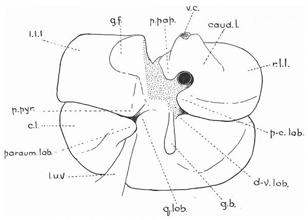
Fig. 2. — Outline of model of liver of 15-mm. pig embryo. Caudal surface. p.pap., papillary process; caud.l., caudate lobe; p.pyr., pyramidal process ; paraum. lob., paraumbilical lobule; p-c.lob., precaudate lobule; d-v.lob., dextro-vesical lobule; y.lob., ‘‘quadrate” lobule; v.c., vena cava. Other lettering as in fig. 1. The dotted area is not covered by peritoneum.
15-mm. (25 days’) Embryo
No feature of the liver of a 15-mm. embryo, and of the one next to be described, is more striking than the large proportion of it formed by the central lobe (fig. 2). That the whole organ has developed rapidly is indicated in many ways; but the rate of growth of the central lobe has greatly exceeded that of the lateral lobes. This being so, the central lobe has increased the lead, in regard to its volume, which it had in the younger embryos. There is, however, not the slightest indication of its subdivision into two parts.
The extra-hepatic portion of the right umbilical vein having disappeared, there is a single deep groove on the caudal surface of the central lobe, to the left of the gall-bladder, in which the left umbilical vein is lodged. The gall-bladder in the nineteen days’ embryo reaches slightly beyond the most ventral limit of the liver. In the next older embryo it just fails to reach this limit, and in the 15-mm. embryo its blunt, blind extremity lies some little distance from the liver margin. The same holds good for older embryos and also for the adult pig. This points to the conclusion that the liver and gall-bladder do not grow ventralwards at the same rate, and will perhaps account for the absence of a groove in the liver, such as Thompson (4) has described as being formed, in the human embryo, in preparation to receive the gall-bladder during its growth in a ventral direction.
The caudal or intestinal surface of the central lobe has become very uneven, partly on account of depressions for the reception of viscera, partly from the production of three incipient outgrowths, of some importance from their character and fate in older embryos. One of the projections is placed to the left of the dorsal end of the groove in which the left umbilical vein is accommodated (fig. 2, parawm. lob.). The second prominence is in the form of a rounded eminence between the umbilical vein and the gallbladder (fig. 2, g.lob.), and is connected with the first named by a bridge of liver-substance which crosses over the vein and so spans the valley along which it runs. The third projection is, as yet, very slight, and lies immediately to the right of the gall-bladder (fig. 2, d-v. lob.).
The two lateral lobes are approximately of the same size. The shape of the left lobe has not materially altered, though the processus pyramidalis is more prominent than in the 8-mm. embryo (fig. 2, p.pyr.). In the right lobe a fair amount of growth has taken place along the vena cava, and between this and the portal vein. In brief, the rudiment of a caval lobe (Ruge) has come into existence. Just ventral to, and to the right of the place where the portal vein enters the liver, there is a rounded projection which may be of moment (fig. 2, p-c. lob.), if, as is conjectured, it corresponds to the procaudate lobule present in the liver of some Primates (of. Buge (14)).
25-mm. (30 days’) Embryo
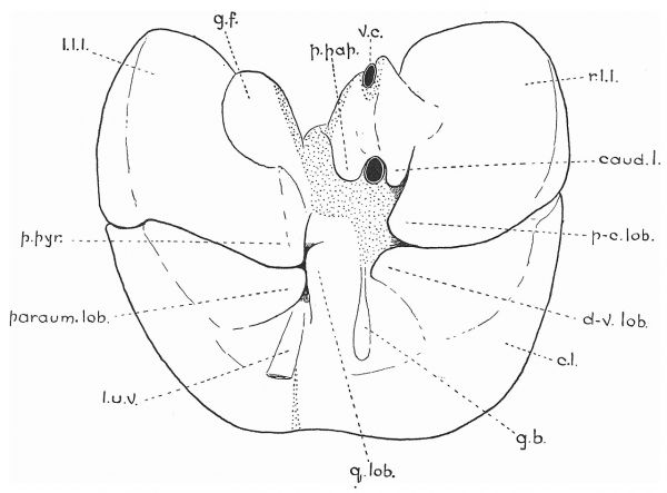
The central lobe still forms a large proportion of the entire liver; a feature best appreciated when the organ is viewed from the thoracic side. Not until now has there been the least sign of a division into right and left central lobes; and even now there is only a shallow groove along the bottom of which the falciform ligament is attached (fig. 3).
The depression produced by the umbilical vein is deeper than before ; as is also, in a minor degree, that for the gall-bladder. The three projections from the intestinal face of the central lobe previously mentioned are much more prominent than in the 15-mm. embryo. That to the left of the umbilical vein is particularly conspicuous, and that to the right of the gall-bladder has developed from a mere elevation into a veritable process (fig. 3, d-v. lob.). The prominence between the vein and the gall-bladder, though less obtrusive than the other two, is, nevertheless, a conspicuous feature.
Fig. 3. — Outline of model of liver of 25-mm. pig embryo. Caudal surface. Lettering as in preceding figures,
Of the two lateral lobes the left is now somewhat the larger, so that there is an advance towards the adult size-relationship. Of the form of the left lobe it will suffice to say that the gastric depression forms a smaller proportion of the caudal surface than formerly (fig. 3, gf), and that the processus pyramidalis has increased in size and has become blunt at its apex (fig. 3, p.py7.).
The caval lobe is now of fair size, and a groove, corresponding to the portal vein in position, produces a partial division into a processus papillaris and a caudate lobe (fig. 3, p.pap. and caud.l.). Ventral to the rudimentary caudate lobe is a projection of the right lateral lobe similar to the one already mentioned in the liver of the 15-mm. embryo (fig. 3, ip-c.lob.).
In this embryo, then, there are five projections grouped around the portal fissure. Two of them belong to the central lobe; one, the processus pyramidalis, to the left lobe; and two to the right lobe. Of the last mentioned, one—the caval lobe—is partly subdivided in consequence of its position astride the portal vein. A sixth projection, to the left of the umbilical vein, though not in close relationship to the portal fissure, is within a short distance of it. From the circumstance that they grow towards the portal fissure, and, by so doing, render it relatively less spacious and deeper, one is tempted to apply the name of “portal opercula” to these six projections. Before doing so, however, it would be necessary to discover if similar “opercula” develop and behave in a like manner in the embryonic liver of other mammals. The material at my disposal, though not demonstrating the contrary, is not sufficiently abundant to permit of any generalisation.
In order to facilitate description, it will be well to say here that there seems great probability that the various prominences mentioned above are homologous with certain lobules described by Ruge as occurring in the liver of the Primates. The homology of the projection already referred to as the processus pyramidalis appears beyond question; and the processus papillaris need not be further discussed. The prominence springing from the central lobe to the left of the umbilical vein seems to be comparable to the lobulus paraumbilicalis (figs. 2 and 3, parawm. lob.), the subsequent flattening which it undergoes, and its growth in a ventral direction, bringing it into a position quite similar to that figured by Ruge.
The projection between the gall-bladder and the umbilical vein may be referred to, for convenience only, as the “quadrate lobule” (figs. 2 and‘3, q.lob.). The eminence lying immediately ventral to the caudate lobe is most likely equivalent to the lobulus precaudatus (Ruge) of Primates (figs. 2 and 3, p-clob.). This is all the more probable since, in some embryos, its right lateral boundary is defined by a fissure occupying a position very like that of the fisswra precaudata (Ruge) of the monkey’s liver.
The prominence to the right of the gall-bladder does not seem to be represented in the material examined by Ruge. Its importance is not very great in older embryos, and, therefore, little attention need be paid to it at present. If it is necessary to give it a name for purposes of reference, it might be called the dextro-vesical lobule (figs. 2 and 3, d-v.lob.).
The livers of embryos larger than 25 mm. were not modelled, as they are sufficiently large to be capable of examination by the naked eye. It is searcely necessary to give a detailed description of each specimen, since all follow the same lines of development. A general survey will suffice. A 52-mm. embryo has a liver very similar to that of the 25-mm. embryo, the only differential points of moment being an increased prominence of the processus pyramidalis and a greater distinctness of the precaudate lobule. In larger embryos the central lobe retains its relatively large size, and the notch for the umbilical vein on its ventral border becomes deeper. In the oldest embryo examined a fissure extends from the notch for some distance into the lobe, thus producing the separation, present in the adult, into right and left central lobes. At no time, however, can the fissure be said to rank with those limiting the right and left lateral lobes. Independent of its comparative shallowness, it differs from the lateral fissures in its lateness of appearance and in the manner of its production.
The history of the processus pyramidalis in embryos larger than 52 mm. consists in its becoming less and less projecting and more and more flattened against the rest of the left lateral lobe, consequent upon the increase in the size of the stomach and.the correlated increase in the extent of the fossa, formed by the liver, in which it lies. But, in spite of the resultant flattening, it does not lose its independent character.
As has been related, in younger embryos the precaudate lobule gradually becomes more distinct and projecting; but from an 86-mm. embryo onwards it is compressed against the rest of the right lateral lobe. The compression begins in the shape of a groove for the duodenum, which gradually increases in width until the whole lobule is involved. In some embryos (122 mm., for example) the lobule is represented by a flattened area, ventral to the caudate lobe, continuous to the right, without line of demarcation, with the bulk of the right lateral lobe. In others, however, notably in that of 150 mm., a decided fissure forms its right boundary. From its variability in embryos, and from its inconstancy in the adult Primate liver, it must be concluded that the preecaudate lobule is not of so much importance as is the processus pyramidalis. Though, in the embryo, it forms an appendage to the right lateral lobe comparable to the pyramidal process of the left lobe, its close proximity to the portal vein precludes it from equalling the processus pyramidalis in magnitude and independence.
The process of the central lobe which has been considered above as equivalent to Ruge’s lobulus paraumbilicalis, shares in the general depression produced by the adjacent hollow viscera. At the same time, it grows over the umbilical vein, and extends towards the ventral border of the liver.
The “quadrate” and dextro-vesical lobules present little of interest in older embryos. They are simply represented by flattened areas, extending towards the portal fissure, one on each side of the cystic duct. Though their importance in the pig is not very great, they are evidently of much more moment in some mammals.
The fissure bounding the caudate lobe—which I have elsewhere (8) named the right dorso-lateral fissure—just reaches the margin of the liver in an 86-mm. embryo; and in an embryo measuring 122 mm. in length it crosses the margin. It clearly develops in the same manner as do the right and left lateral fissures; that is to say, it is produced in consequence of the growth in extent of the lobes between which it lies.
From the above it may be contended that the mammalian liver consists essentially of three lobes only. In the pig, and probably in all other ‘mammals, the main or central lobe is, at first, considerably greater than the sum of the other two lobes; and it is not until the adult form has been attained that its volume fails to constitute half of that of the liver as a whole. The central lobe, moreover, is really only one lobe, and not two, as described by Flower. In those mammals in which a right and a left central lobe can be recognised, the division is, so to speak, accidental, and the result of the presence of an umbilical vein. In some animals, the growth of hepatic tissue being abundant in its neighbourhood, the vein becomes completely surrounded by liver from the occurrence of a fusion between the tissue of the paraumbilical and “quadrate” lobules. In others, such a fusion is either absent or incomplete, and an “umbilical fissure” is the result. The “umbilical fissure,” therefore, cannot be allowed to rank as a true line of separation of the central lobe into two parts. Nor does it mark the true morphological line of division of the liver into right and left segments. Except in those animals in which the two umbilical veins fuse into one—and such a fusion is apparently of rare occurrence— the embryonic median plane lies to the right of the “umbilical fissure.” In mammals like the pig, in which both umbilical veins are involved in the liver, it is obvious that the true line of separation of right and left moieties must lie somewhere between the two veins. There seems little reason to doubt that it coincides in position with the gall-bladder and cystic duct, which also mark the original line of attachment of the mesogastrium ; and there seems good reason for assuming that the “quadrate lobe” really belongs to the left half of the liver.
That the contiguous viscera, solid as well as hollow, have a profound influence in moulding the liver, is undoubted. Toldt and Zuckerkand1 (28) have clearly demonstrated that the pressure of even so small an object as the gall-bladder may produce atrophy of liver tissue. But there is no convincing evidence to show that the adjacent organs have any influence whatever upon the production of the two lateral fissures by which the liver is divided into its three primary lobes. Doubtless the projections of liver-substance, to which attention has been directed, are produced in the first instance by growth along lines of least resistance. But, as has been said, with the exception of the papillary process and the caudate lobe, they early cease to be projections, and are flattened by the pressure of superposed organs; and at no time do they contribute to the essential morphology of the liver. The caval lobe should hardly be classed along with objects like the pyramidal process, since it develops along the vena cava and its mesentery, whereas the other projections of the primary lobes do not follow either a vessel or any other structure.
An interesting feature in the development of the fissures has been observed. It appears from certain embryos, notably the 15-mm. pig embryo and an 1l-mm. hedgehog embryo, that the hepatic tissue of the different lobes is separated by a thin layer of mesodermic tissue even before actual fissures make their appearance. Or, in other words, the fissures do not cut a primitively common mass into lobes, but develop along lines of mesoderm which form septa between lobes really independent from an earlier period (figs. 18 and 19). So far as can be gathered from an examination of the literature, this feature of development seems to have escaped notice. In what measure it affects the question of the causation of fissures can hardly be determined in the absence of more extensive investigations. It would be of the greatest interest to examine the early stages of development of a consolidated liver, with a view to the discovery of similar mesodermic septa.
Development of the Blood-Vessels of the Liver of the Pig
The development of the lobes having been sketched, the way is prepared for the consideration of the process of growth of the blood-vessels.
6-mm. Embryo
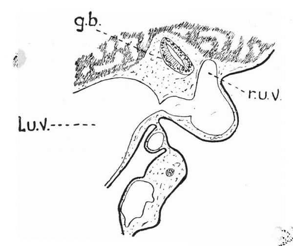
In this embryo there are two umbilical veins which, though the left is slightly the larger, have nearly the same calibre prior to their entrance into the liver. Some little distance before they reach the liver the two veins approach each other very closely, without, however, effecting an actual intercommunication. The intra-hepatic portion of the left vein has a much more considerable volume than has the corresponding part of the right vessel. At this stage, in the absence of a ductus venosus, the left vein terminates rather abruptly in the vicinity of the cesophageal notch of the liver. In the light of subsequent development, it is necessary to point out that a small vessel arises from the termination of the left umbilical vein and passes into the substance of the left lateral lobe (fig. 4, a). This will be further considered in connection with older specimens. Close to its termination, a vessel is connected with the right side of the left. umbilical vein and a branch leaves it to the left. the right is with the right omphalo-mesenteric vein, and the branch to
Fig, 4. — Semidiagrammatic outlines of hepatic vessels of 6-mm. pig embryo.
l.o-m.v., left omphalo-mesenteric vein; 7.0-m.v., right omphalo-mesenteric vein; l.h.v., left hepatic vein; m.h.v., middle hepatic vein; a, a small vein which ultimately becomes the ramus angularis. Other lettering as in preceding figures. The outlines of the lobes are indicated by dotted lines.
The connection on the left is doubtless the remains of the left vein of the same name (figs. 4 and 6, l.0-m.v.).
Within the liver the right umbilical vein follows a course parallel to that of the left vein. It ends by joining the omphalo-mesenteric vein about the point where this vessel divides into right and left branches (figs. 4 and 7, r.u.v.); or it would possibly be more correct to say that a short venous channel leaves the omphalo-mesenteric vein at its point of division and runs forwards (cranialwards) to join the termination of the umbilical vein (fig. 7).
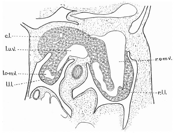
Fig. 5. — Camera-lucida drawing of section of liver, etc., of 6-mm. pig embyro. st., stomach. This and the three succeeding figures are of a series of sections proceeding from the dorsal to the ventral part of the liver.
The two omphalo-mesenteric veins are represented by a single vessel, which pursues the customary spiral course over the dorsal surface of the intestine in order to enter the liver. Immediately on its entrance it divides into two main branches. The larger one, to the right, follows the curvature of the surface of the right lateral lobe, in the form of a large, laterallyflattened channel, which finally leaves the liver without being subjected to any interruption whatever (figs. 4, 5, and 6, r.0-m.v.). It, beyond doubt, represents the right omphalo-mesenteric vein; but whether it is a reopened
Fig. 6. — Camera-lucida drawing of section of liver, etc., of 6-mm. pig embryo.
The section shows the continuity of the right omphalo-mesenteric vein (7.0-m.v.) in the right lateral lobe (r.l. channel, or one which has never been interrupted, lack of younger material leaves open to question.
The left branch of the omphalo-mesenteric vein, even in the oldest embryo examined, is not completely embedded in the liver, but runs in a groove more or less on the surface. It joins the left umbilical vein close to its termination, and opposite the vessel described above as a branch of the umbilical vein destined for the left lateral lobe. It seems probable that this is the vestige of the left omphalo-mesenteric vein, support being afforded to the supposition by the circumstance that it has a narrow, but unmistakable, connection with the rudimentary vena hepatica sinistra (fig. 4, l.o-m.v.). If this surmise be correct, the left omphalo-mesenteric vein pursues a course closely comparable to that of the right vein. That is, it follows a curved path conformable to the surface of the left lateral lobe.
Fig. 7. — Camera-lucida drawing of section of liver, ete., of 6-mm. pig embryo. d., duodenum ; d.a.p., dorsal anlage of pancreas. The connection between the right omphalo-mesenteric vein (7.0-m.v.) and the right umbilical vein (7.u.v.) is shown.
From what has just been said, it is evident that, as yet, there are no absolutely independent hepatic veins; but the proximal part of the left omphalo-mesenteric vein having become almost completely detached from the rest, there is a rudiment of the vena hepatica sinistra which, even at this early period, shows indications of a division into two branches—one for the left lateral lobe, the other for the central lobe (fig. 4, U.A.v.). A short and rather thick vessel leaves the central lobe to join the termination of the right omphalo-mesenteric vein. Subsequent development shows that it is the rudiment of the vena hepatica media as described later (fig. 4, m.h.v.).
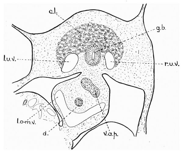
Fig. 8. — Camera-lucida drawing of section of liver, etc., of 6-mm. pig embryo.
v.a.p., ventral anlage of pancreas. The section illustrates the symmetrical disposition of the two umbilical veins, one on each side of the gall-bladder.
The nineteen days’ pig embryo, then, affords corroborative evidence in favour of the conclusions, arrived at by Brachet (21) and restated here as the result of the observation of the development of the hepatic lobes, that the liver is fundamentally composed of three lobes. The central of these is associated with the two umbilical veins; and the right and left lateral lobes are developed in connection with the right and left omphalo-mesenteric veins respectively. Furthermore, because the rudiments of the hepatic veins are produced from the omphalo-mesenteric veins, they primarily belong to the lateral lobes. Those vessels which ultimately drain the central lobe are of later development.
8-mm. Embryo
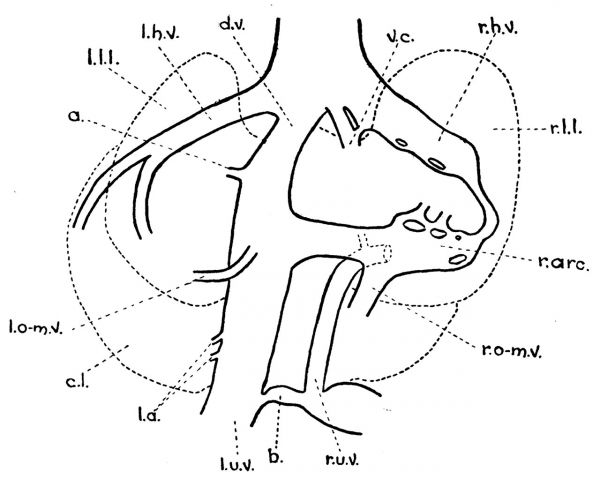
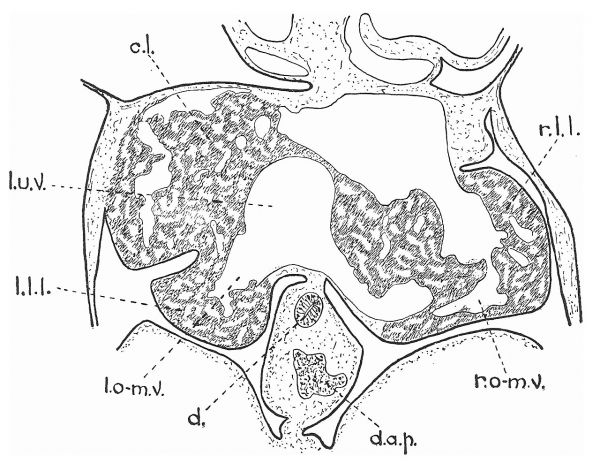
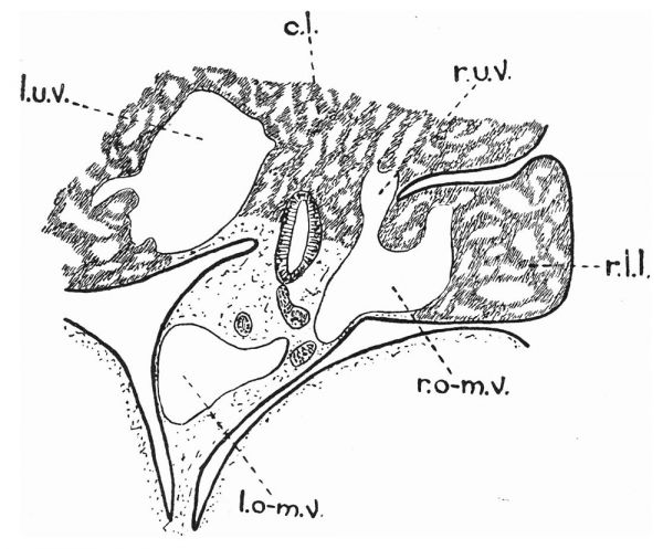
There are still two umbilical veins, but the duty of the intra-hepatic portion of the right has clearly been taken over in part by the left. Its extra-hepatie portion is of good size (figs. 9 and 14, rw.v.), but is mainly connected with the vessels of the body-wall. In the younger embryo the two veins approach each other very closely before entering the liver. In the 8-mm. embryo there is a considerable intercommunication between them at the place where the right vein is about to enter the liver (fig. 9,5; fig. 13). As a consequence, the intra-hepatic portion of the right vein is comparatively slender though easily recognised and traceable to its union, as in the younger specimen, with a vessel arising from the point of bifureation of the omphalo-mesenteric vein (fig. 12).
Fig. 9. — Semidiagrammatic outlines of hepatic vessels of 8-mm. pig embryo. d.v., ductus venosus; v.c., vena cava; r.arc., ramus arcuatus; l.a., left arborisation from the recessus umbilicalis (Rex); b, connection between the two umbilical veins. Other lettering as in preceding figures.
Fig. 10. — Camera-lucida drawing of section of liver, etc., of 8-mm. pig embryo.
2.¢., Vena cava; a@, a small, short vein which ultimately becomes the ramus angularis. Figs. 10 to 14, inclusive, are of sections taken in order from the dorsal to the ventral part of the embryo.
The communication between the two umbilical veins is of interest as indicating a similarity of development in the marsupials and in higher mammals. M‘Clure (29) has described the formation of two communications in an 8-mm. embryo of Didelphis marsupials: one at the umbilicus, whereby an umbilical sinus is produced ; the other on the ventral surface of the liver, by which another sinus is formed. After the formation of the second sinus, the two vessels pass “through the parenchyma of the liver in a channel common to both, which opens into the post-cava in common with the left hepatic vein.” In regard to 115-12 mm. embryos of Didelphis, M‘Clure says that there is one large umbilical vein forming the principal channel between the allantois and. the liver; “a vein which I regard as the left umbilical vein. This large vein lies in the ventral body-wall slightly to the left of the mid-ventral line, and to its right, is situated a much smaller vessel which is difficult to follow in consecutive sections, but which is probably the remains of the right umbilical vein. The two umbilical veins appear to anastomose in places with each other, so that one might almost regard the larger of the two veins as being formed, in places, through the fusion of the two umbilical veins.” Broom (30) gives a comparable account in his description of an 85 mm. Trichoswrus embryo, where he mentions a single vessel as carrying the blood from the allantois to an umbilical sinus, from which two large umbilical veins pass into the liver.
Fig. 11, — Camera-lucida drawing of section of liver, etc., of 8-mm. pig embryo.
The interruption of the right omphalo-mesenteric vein (r.0-m.v.) is only apparent, neighbouring section showing that it is continuous.
These observations on the vessels of the marsupial embryos tempt one to regard the much narrower communication in the pig embryo as of a like nature in a minor degree. Bonne (20) is of opinion that the single intrahepatic vein of the sheep is formed by the fusion of the two umbilical veins ; or, a8 one may say, the fusion is carried to a much further extent than in marsupials. There is, however, the bare possibility that the right vein may dwindle in the sheep in the same manner as in Didelphis, and that Bonne may have failed to detect it. This, naturally, is a mere supposition. Whatever may be the arrangement in the sheep, there is an anastomosis between the two veins in the pig, established about the time at which the intra-hepatic portion of the right vein becomes narrow prior to losing its continuity with the extra-hepatic part.
A ductus venosus has now been formed (fig. 9, d.v.). The establishment of a more direct route along which the blood may travel on its way to the heart has resulted in a very definite interruption in the continuity of the left omphalo-mesenteric vein, and the consequent production of an independent vena hepatica sinistra (figs. 9,10, and 15, lh.v.). A number of small branches leave the left side of the intra-hepatic course of the left umbilical vein, some of which become definitive vessels and form the left arborisation from the recessus umbilicalis of*Rex (fig. 9, J.a., and fig. 12). It should be noted that a small, short branch, arising from the umbilical vein at the point where it joins the ductus venosus (figs. 9 and 10, a), is comparable to a similar vessel in the nineteen days’ embryo.
Fig. 12, — Camera-lucida drawing of section of liver, etc., of 8-mm. pig embryo.
The section shows the connection between the right omphalo-mesenteric vein (7.0-m.v.) and the right umbilical vein (7.u.v.).
The right omphalo-mesenteric vein is similar to that of the previous embryo, except that it has become more involved in the formation of sinusoids (fig. 11, r.o-m.v.). In this embryo the branch to the left—joining the left umbilical vein—is relatively narrow; but this is probably of no significance. The right branch calls for no remark beyond that it is connected, by means of small channels, with the vena cava, along which a caudate lobe has begun to grow. The third branch of the omphalo-mesenteric vein, leaving the point of its bifurcation into right and left main branches, joins the termination of the right umbilical vein as it does in the nineteen days’ embryo. From the place of union a moderately large channel passes forwards (cranialwards) in the substance of the central lobe.
Fig. 13. — Camera-lucida drawing of section of liver, etc., of 8-mm. pig embryo.
This section shows the communication between the two umbilical’ veins (cf. fig. 9).
Fig. 14. — Camera-lucida drawing of section of liver, etc., of 8-mm. pig embryo,
The figure illustrates a section just ventral to that of the previous figure. Compare the calibre of the right umbilical vein (r.u.v.) in the two figures.
There are now independent vena hepatica sinistra and vena hepatica dextra formed by dorsal and ventral tributaries (fig. 15, Lh.v. and r.h.v.), The dorsal right vein is within the right lateral lobe, where it is still continuous, by means of a series of large channels, with the right omphalomesenteric vein. The ventral tributary of the right hepatic vein drains the right portion of the central lobe. In addition there is a third rudimentary hepatic vein taking origin in the middle part of the central lobe and constituting the forerunner of the vena hepatica media (fig. 15, m.h.v.).
Fig, 15. — Semidiagrammatic outlines of hepatic veins of 8-mm. pig embryo.
r.h.v., right hepatic vein ; m.h.v., middle hepatic vein; J.h.v., left hepatic vein.
The preponderance, in this embryo as in the younger one, of the right lateral lobe over its fellow, depends chiefly upon its great richness in venous channels. It is only when the vascularity in the two is more nearly balanced that the left lateral lobe begins to assume the relatively greater bulk.
15-mm. Embryo
The extra-hepatic portion of the right umbilical vein no longer communicates with vessels in the interior of the liver. It is mainly concerned in the vascular supply of the body-wall; but there is still a narrow, extra-hepatic connection between it and the left umbilical vein. The left vein is large, and, as before, contributes a number of moderately capacious branches to the left and anterior (cranial) parts of the central lobe in the shape of a left arborisation of Rex (figs. 16 and 20, l.a.). A few smaller vessels pass towards the right; that is to say, the right arborisation has begun to form (fig. 16, ”.a.).
Fig. 16. — Semidiagrammatic outlines of hepatic vessels of 15-mm. pig embryo.
r.ang., ramus angularis ; r.cys., ramus cysticus; r.a., right arborisation from the recessus umbilicalis (Rex). Other lettering as before.
Close to the point at which the left umbilical vein becomes continuous with the ductus venosus, a ramus angularis takes origin and passes into the left lateral lobe, where its branches lie dorsal to those of the dorsal left hepatic vein (figs. 16, 17, and 18, r.ang.). There seems good reason for supposing that the ramus angularis of the pig is not developed from the remains of the left omphalo-mesenteric vein. The vestige of the lastnamed vessel is clearly recognisable as arising ventral to the end of the umbilical vein, and traversing the left lateral lobe on about the same level as the smaller twigs of the dorsal left hepatic vein (fig. 19, l.o-m.v.). The ramus angularis, then, appears to be produced by the growth of the small branch which has been described in the smaller embryos as springing from the umbilical vein close to its termination (figs. 9 and 10, a). If this view be correct, the development of the ramus in the pig differs from that described by Mall as occurring in the human embryo.
Fig. 17. — Camera-lucida drawing of section of liver, etc., of 15-mm. pig embryo.
d.v., ductus venosus ; 7.arc., ramus arcuatus, developed from part of the right omphalo- ave mesenteric vein; 7.ang., ramus angularis. Figs. 17 to 20 are arranged so that tig. 17 “ry is of a dorsal and fig. 2u of a ventral section of the same embryo.
The portal (omphalo-mesenteric) vein divides, as in the younger embryos, into right and left main branches. The left joins the umbilical vein as before; and the right has become a ramus arcuatus no longer continuous with the dorsal right hepatic vein (figs. 16 and 17, r.ure.).
In the two younger embryos a third vessel has been mentioned as taking origin from the omphalo-mesenteric vein at its point of bifurcation and joining the intra-hepatic part of the right umbilical vein. In the 15-mm. embryo the vessel is of good size, and its homology with the ramus cysticus of Rex is beyond doubt. It possesses a ventral branch running downwards (ventralwards) in the central lobe, comparatively close to the surface, and just to the right of the cystic duct (figs. 16 and 20, rcys.). How much of this may have been formed from the right umbilical vein is difficult to say, but its position and general disposition are such that one can hardly avoid the supposition that some of it, at least, is the direct descendant of part of the umbilical vein. A short cranial branch leaves the ramus cysticus close to its origin, and immediately divides into right and left twigs which are placed in the central lobe, one on each side of the ventral right hepatic vein.
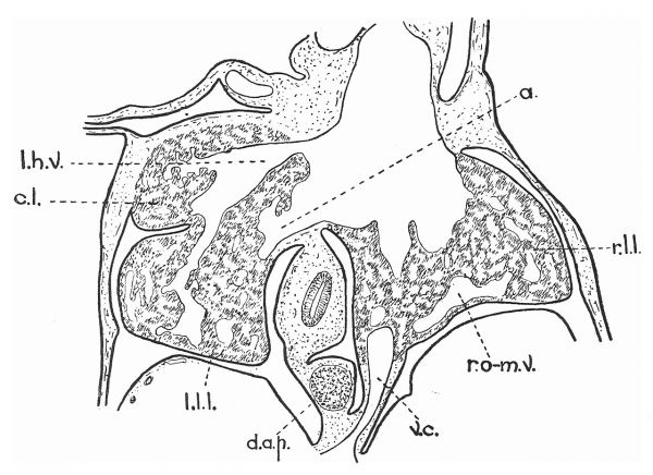
Fig. 18. — Combination camera-lucida drawing of three successive sections of liver, etc., of 15-mm. pig embryo. r.h.v, and l.h.v., right and left hepatic veins, developed from portions of the right and left omphalo mesenteric veins respectively ; r.ang., ramus angularis, developed from @ of figs. 4, 9, and 10. Figs. 17 and 18 show that the ramus lies dorsal to the left hepatic vein.
All the main hepatic veins are now clearly defined. The right vein divides into a dorsal and a ventral branch; the former (fig. 19, ~h.v.1) draining the right lateral lobe, the latter (fig. 19, r.h.v.2) proceeding from the right part of the central lobe. The left hepatic vein is similarly disposed on the left side of the liver (fig. 19, U.h.v.1 and Lh.v.2). A middle hepatic vein commences as right and left tributaries (fig. 20, m.h.v.1 and m.h.v, 2) and terminates very close to the end of the left hepatic vein.
25-mm. Embryo
The vessels of this embryo do not demand a detailed description, since, in the main, they resemble those of the 15-mm. embryo. Numerous fairly large vessels arise from the left and anterior (cranial) sides of the umbilical vein (the left arborisation from the recessus umbilicalis of the adult), and a few branches of notable size have been developed from the right side of the vein. It is obvious that, though a left arborisation is beginning to form as early as the twenty-second day, a corresponding right group of vessels is of somewhat later development.
Fig, 19. — Camera-lucida drawing of section of liver, etc., of 15-mm. pig embryo. The section shows the dorsal tributaries (r.h.v.1 and l.h.v.1) of the right and left hepatic veins draining the right and left lateral lobes, whereas their ventral tributaries (r.h.v. 2 and Lh.v. 2) drain the central lobe. A mesodermic septum produces a partial separation of right lateral and central lobes. :
The ramus angularis and ramus arcuatus are both large, but the latter is of somewhat greater volume than the former. The ramus cysticus at its origin is not much smaller than the other portal branches, and its disposition is very similar to that of the same vessel in the 15-mm. embryo.
As there has merely been an increase in the size and in the number of the branches of the hepatic veins, no call is made for special remark regarding them.
It is worthy of note that, so far as the embryos under consideration are concerned, neither ramus descendens nor ramus omentalis can be detected. The caudate lobe and the omental lobule contain no vessels larger than sinusoids; a circumstance probably to be associated with the fact that these portions of the embryonic liver of the pig are but poorly developed.
Fig, 20. — Camera-lucida drawing of section of liver, etc., of 15-mm. pig embryo.
The figure shows the two tributaries (m.h.v. 1 and m.h.v. 2) of the middle hepatic vein just about to join. The position of the ramus cysticus (r.cys.) should be compared with that Hi the right umbilical vein in some of the preceding figures (notably figs. 7, 8, and 12).
Development of the Lobes and Veins in Other Embryos
The rest of the available embryological material is somewhat scrappy, but there are some embryos of the mole, hedgehog, and ruminants which are not without interest. The livers of the adult mole and hedgehog form good representatives of the generalised mammalian organ, inasmuch as all the lobes are well developed and separated by deep fissures. The ox and the sheep, on the other hand, have comparatively compact livers, but with a well-marked caudate lobe and a fairly large processus papillaris.
Mole Embryos
So far as my material admits of conclusions, it appears that the lobes are formed at a comparatively early period in the mole. In an embryo 7 mm. in length they are all present; and in a 13-mm. embryo they have a form not unlike that of the adult lobes. The central lobe forms rather less than half of the total bulk of the liver, and is virtually undivided. The left umbilical vein is entirely surrounded by hepatic tissue, and there is consequently no umbilical fissure. The enclosure of the vein has obviously been caused by the coalescence and complete fusion of the two processes described in connection with the pig embryos as paraumbilical and “quadrate” lobules. The mole differs from the pig in having a cystic fissure cutting the ventral border of the central lobe on a level with the gall-bladder.
The liver of the 13-mm. mole embryo has two large lateral lobes, almost equal in size. A processus pyramidalis is not very obvious; but there is a conspicuous projection in the same position as the precaudate lobule of the pig. The most striking difference between the liver of the mole and that of the pig is the large size of the caudate lobe and the processus papillaris of the former.
The arrangement of the branches of the umbilical and portal veins is much the same in the mole and the pig. The ramus angularis, ramus arcuatus, and ramus cysticus have the same general disposition. The ramus cysticus of the mole runs forwards (towards the diaphragm) for a short distance and then divides into a vessel which continues the direction of the parent vein, and a ventral branch lying practically parallel to, and to the left of, the cystic duct. This corresponds to the vein which has so strong a resemblance to the intra-hepatie portion of the right umbilical vein in the pig. It is, perhaps, worth mentioning that the ramus arcuatus divides into two branches: one curves upwards dorsal to the vena hepatica dextra ; the other runs almost entirely towards the right. The latter is, to a certain extent, represented in the pig; but its volume in this animal is not nearly so great as in the mole.
The hepatic veins of the mole are somewhat different from the corresponding vessels of the pig. There are really only two veins—a right and a left—each having a dorsal and a ventral tributary, belonging respectively to a lateral lobe and to the adjacent part of the central lobe; in addition each vein receives a third vessel, apparently homologous with one of the tributaries of the vena hepatica media of the pig.
Since the mole has a large caudate lobe, a ramus descendens was carefully sought, and a small vessel, no doubt homologous with the ramus as described by Rex, was found leaving the portal vein close to the origin of the ramus arcuatus and ramus cysticus. This, however, does not form the only vessel supplying the lobe, for there are several small channels running into it from various contiguous parts of the portal vein. No definite ramus omentalis could be discovered.
According to the conclusion arrived at by Rex, the caudate lobe should be drained by the right hepatic vein; but in the mole embryos examined, the blood from the lobe finds it way, by several small venules, directly into the vena cava, much as it does in the pig.
Hedgehog Embryo
It is rather surprising to find, in a hedgehog embryo of 11 mm. in length, that the right omphalo-mesenteric vein is not yet interrupted ; or, if it has been interrupted in earlier stages, it has been reopened, and is represented by a very capacious channel uniting the ramus arcuatus and the right hepatic vein. The presence of a continuous passage is all the more striking because of the advanced stage at which the development of the lobes has arrived.
A ramus cysticus arises from the left main branch of the omphalomesenteric vein about midway between its origin and its union with the umbilical vein. As in the mole, a ramus descendens leaves the omphalomesenteric vein in common with the ramus arcuatus and supplies the large caudate lobe.
The absence of a ramus omentalis in the mole embryos has been remarked. In the hedgehog, a short vessel, rapidly terminating in sinusoids in the papillary process, was observed, and taken as being the beginning of a ramus omentalis. It leaves the left main branch of the omphalo-mesenterie vein almost exactly opposite the origin of the ramus cysticus.
In the arrangement of the hepatic veins, the hedgehog resembles the pig more closely than the mole. Instead of there being two separate middle hepatic veins, each terminating in common with the other hepatic vein of its own side, as in the mole, there is one middle vessel ending in company with the left hepatic vein, as in the pig. It should be remarked that a considerable amount of blood is drained from the right lateral lobe by a number of fairly large veins which open directly into the vena cava.
Calf Embryos
In the adult ox and sheep, the vestige of the umbilical vein is completely embedded in liver substance; but in the calf, a short time before birth, there is a decided “umbilical fissure.” That is, the paraumbilical and “quadrate” lobules have not completely fused over the vein. The rest of the liver of such a calf has practically all the adult features.
The large caudate lobe is supplied by a relatively wide ramus descendens, which, as in the mole and hedgehog, leaves the right main portal branch at nearly the same point as a good-sized ramus cysticus and a small ramus arcuatus. A ramus angularis is also easily demonstrable, but presents no peculiarities of moment.
The hepatic veins are three in number—right, middle, and left. The left and middle veins join the vena cava close together, and are not formed by large tributaries, but receive a number of small vessels at intervals. The right vein is different. It opens into the vena cava some distance caudal to the termination of the middle and left veins, and is formed by two tributaries, one draining the large caudate lobe, the other coming from the equivalent of the right lateral lobe.
Clearly the distribution of the hepatic veins of the pig, mole, hedgehog, and calf does not follow a common plan; nor does it entirely consist with the generalised statement of Rex. The most important differences are connected with the vessel which has been named the vena hepatica dextra in the above descriptions. In the ruminant it comports itself according to the method as enunciated by Rex. In the mole and pig, however, one of its tributaries drains the central lobe, this evidently being equivalent to the vena hepatica media accessoria described by Rex as taking origin in, and receiving vessels from, that portion of the central lobe which lies to the left of the stem of the ramus cysticus. If Rex be followed in this connection, the vena hepatica dextra of the pig and mole—and possibly also in man (ef. Mall)—is not formed by dorsal and ventral rami, and does not drain a district of the liver on the right equivalent to the area of drainage of the left vein. No doubt a more satisfying conclusion could have been arrived at had further embryological evidence been forthcoming, but the interpretation, as given above, appears to be satisfactory so far as information is available.
Only in the mole does the vena hepatica media depart from the arrangement present in the other embryos. In the pig, hedgehog, and calf it terminates in common with the left hepatic vein; whereas, in the mole, it is apparently represented by two veins, one joining the right and one the left hepatic vein. It must, however, be remembered that the vessels of only one mole embryo were modelled. It is possible that, had the others been similarly investigated, the disposition of the vein might have been found to resemble more closely that of the other animals.
The chief points of moment in the foregoing account may be summarised as follows :—
(1) The division of the liver as made by the human anatomist is convenient, but should not be held as resting upon an embryological or true morphological basis. The “umbilical fissure” in man, and mammals in general, is not a true fissure. Its appearance is late, and it may become obliterated in the adult. Its occurrence depends upon the presence of the left umbilical vein. If the right umbilical vein were to persist as well, two “fissures ” would be produced, and the error of considering one of them as the dividing line of the liver would be manifest.
There is much to be said for Cantlie’s (15) contention that a line drawn from the gall-bladder to the exit of the hepatic veins divides the liver into right and left halves.
(2) The mammalian liver consists essentially of three lobes — a central and two lateral. The usual division of the central lobe into two is correlated with the presence of an “umbilical fissure,” and therefore is not fundamental. The caudate and Spigelian “lobes” are merely appendages or processes of the right lateral lobe. In order to indicate their true morphology, it would be better to name them caudate and omental or pupillary processes.
In view of developmental evidence, it appears reasonable to ask that the following modifications should be made in Flower’s nomenclature :—
Flower. { Right lobule . . . Right central lobe. Central lobe \ Left lobule . . . . Left central lobe. Main part . . . . Right lateral lobe. Right lateral \ Processus caudatus . . Caudate lobe. lobe \ Processus omentalis or papillaris . . . . Spigelian lobe. Left lateral lobe . . . . . Left lateral lobe.
(3) Not only do the three fundamental lobes make their appearance independently, they also develop in connection with different embryonic veins. The central lobe is produced about the umbilical veins. The right and left lateral lobes grow along the course of the right and left omphalomesenteric veins.
The right and left hepatic veins are primarily connected with the right and left lateral lobes respectively. The hepatic veins of the central lobe are of later development.
(4) Whatever may be the cause or causes of the presence of fissures in the mammalian liver, it cannot be admitted that, as yet, a satisfactory connection has been proved between facts and factors. The question is complicated by the possibility that the fissures may be preceded by mesodermic septa which separate the lobes at an early period. The presence of such septa in the liver of the pig and hedgehog embryos points to a new field of inquiry.
References
(1) Routzsron, H. D., “Livers with Anomalies in their Lobulation,” Journ. Anat. and Phys., vol. xxvii., 1893, Proc. Anat. Soc. of Great Britain and Ireland, p. Xxxi.
(2) Parsons, F, G., “ Liver with Two Right and Two Left Lobes,” Journ. Anat. and Phys., vol. xxxviii., 1904, Proc. Anat. Soc. of Great Britain and Ireland, p. xxiii.
(3) THomson, A., “Some Variations in the Anatomy of the Human Liver,” Journ, Anat. and Phys., vol. xix., 1885, p. 303.
(4) THomson, A., “The Morphological Significance of Certain Fissures in the Human Liver,” Journ. Anat. and Phys., vol. xxxili., 1899, p. 546.
(5) Duvernoy, “Etudes sur le foie,” Ann. Sci. Nat., 2me série, t. iv., p. 269, 1835.
(6) Lessee, F. X., ‘Recherches anatomiques sur les Camélidés,” Arch. du Museum @hist. nat. de Lyon, t. viii., 1903, Mémoire No. 1,
(7) Kerru, A, “The Position and Manner of Fixation of the Liver of Primates, and the Part these Factors play in the Lobulation of the Liver,” Journ. Anat. and Phys., vol. xxxiii., 1899, Proc. Anat. Soc. of Great Britain and Ireland, p. xxi.
(8) Bradley, O. Cuarnock, “On the Abdominal Viscera of Cercocebus fuliginosus and Lagothrix humboldti,” Proc. Roy. Soc. Edin., vol. xxiv., 1903, p. 505.
(9) Ruex, G., “Der Verkiirzungprocess am Rumpfe von Halbaffen,” Morph. Jakrb., Bd. xviii., 1892, p. 185.
(10) Rex, H., “ Beitrige zur Morphologie der Sdugerleber,” Morph. Jahrb., Bd. xiv., 1888, p. 517.
(11) Rotueston, G., “On the Homologies of the Lobes of the Liver in Mammalia,” Brit. Assoc. Reports, 1861, Notices and Abstracts, p. 174.
(12) Owen, R., On the Anatomy of Vertebrates: vol. iii.. Mammals, 1868.
(13) Flower, W. H., “On the Comparative Anatomy of the Organs of Digestion of the Mammalia,” Med. Times and Gazette, 1872, vols. i. and ii. ‘On the Arrangement and Nomenclature of the Lobes of the Liver in Mammalia,” Nature, vol. vi., 1872, p, 346. Brit, Assoc. Reports, 1872, p. 150.
(14) Ruex, G., “Die aiisseren Formverhaltnisse der Leber bei den Primaten,” Morph. Jahrb., Bd. xxix., 1902; Bd. xxx., 1903; and Bd. xxxv., 1906.
(15) Cantuis, J., “On a New Arrangement of the Right and Left Lobes of the Liver,” Journ. Anat. and Phys., vol. xxxii., 1898, Proc. Anat. Soc. of Great Britain and Ireland, p. iv. ‘On the Lobes of the Liver,” Brit. Med. Journ., 1899, vol. ii. p. 833.
(16) His, W., Anatomie menschlicher Embryonen, Leipzig, 1880-1885.
(17) Hocusterrer, F., “ Beitrége zur Entwicklungsgeschichte des Venensystems der Amniotem,” iii. Siuger, Morph. Jahrb., Bd. xx., 1893, p. 542.
(18) Minot, C. S., “On a hitherto Unrecognised Form of Blood Circulation without Capillaries in the Organs of Vertebrata,” Proc. Boston Soc. Nat. Hist., vol. xxix., 1900, p. 185.
(19) Lewis FT. The development of the vena cava inferior. (1902) Amer. J Anat. 1(3): 229-244.
(20) Bonng, C., ‘Recherches sur le développement des veines du foie chez le lapin et le mouton,” Journ. de l’ Anat. et de la Phys., t. xl., 1904, p. 223.
(21) Bracugt, A., “ Recherches sur le développement du diaphragme et du foie chez le lapin,” Journ. de l’ Anat. et de la Phys., t. xxxi., 1895, p. 511. 42 Morphology and Development of the Mammalian Liver
(22) Mall FP. A study of the structural unit of the liver. (1906) Amer. J Anat. 5:227-308.
(23) Giraupet, E., “ Morphogenése du systéme circulatoire du foie,” Rev. de Méd., Ann. xxvii., 1907, p. 70. .
(24) Hint, E. C., “On the Schultze Clearing Method as used in the Anatomical Laboratory of the Johns Hopkins University,” Johns Hopkins Hosp. Bull., April 1906, p. 111.
(25) Kerpet, F., Normentafel zur Entwicklungsgeschichte des Schweines (Sus scrofa domesticus), Jena, 1897.
(26) THompson, P., and Taytor, G., “ Specimens of Liver showing the Processus Pyramidalis,” Journ. Anat, and Phys, vol. xxxix., 1905, Proc. Anat. Soc. of Great Britain and Ireland, p. xxi.
(27) Lewis FT. The gross anatomy of a 12 mm pig. (1903) Amer. J Anat. 2: 221-225.
(28) Totpr and ZucKERKANDL, Sttzungsber. d. Wiener Akademie, Bd. Ixxii., 1876. (Quoted by Mall.)
(29) M‘Cuurs, C. F. W., ‘‘ A Contribution to the Anatomy and Development of the Venous System of Didelphis marsupialis (L.)”; part. ii. Development, Amer. Journ. Anat., vol. v., 1906, p. 163.
(30) Broom, R., ‘On the Arterial Arches and Great Veins in the Foetal Marsupial,” Journ. Anat. Phys., vol. xxxii., 1898, p. 477.
Cite this page: Hill, M.A. (2024, April 26) Embryology Paper - A contribution to the morphology and development of the mammalian liver. Retrieved from https://embryology.med.unsw.edu.au/embryology/index.php/Paper_-_A_contribution_to_the_morphology_and_development_of_the_mammalian_liver
- © Dr Mark Hill 2024, UNSW Embryology ISBN: 978 0 7334 2609 4 - UNSW CRICOS Provider Code No. 00098G


