Paper - A Human Embryo Twenty-Six Days Old
| Embryology - 1 May 2024 |
|---|
| Google Translate - select your language from the list shown below (this will open a new external page) |
|
العربية | català | 中文 | 中國傳統的 | français | Deutsche | עִברִית | हिंदी | bahasa Indonesia | italiano | 日本語 | 한국어 | မြန်မာ | Pilipino | Polskie | português | ਪੰਜਾਬੀ ਦੇ | Română | русский | Español | Swahili | Svensk | ไทย | Türkçe | اردو | ייִדיש | Tiếng Việt These external translations are automated and may not be accurate. (More? About Translations) |
Mall FP. A human embryo twenty-six days old. (1891) J Morphol. 5: 459-480.
| Historic Disclaimer - information about historic embryology pages |
|---|
| Pages where the terms "Historic" (textbooks, papers, people, recommendations) appear on this site, and sections within pages where this disclaimer appears, indicate that the content and scientific understanding are specific to the time of publication. This means that while some scientific descriptions are still accurate, the terminology and interpretation of the developmental mechanisms reflect the understanding at the time of original publication and those of the preceding periods, these terms, interpretations and recommendations may not reflect our current scientific understanding. (More? Embryology History | Historic Embryology Papers) |
A Human Embryo Twenty-Six Days Old
Introduction
SEVERAL years ago Dr. C. 0. Miller of the Johns Hopkins University gave me a very young human embryo which was so well preserved and so perfect in all respects that it justified a very careful study. He very kindly has procured for me the following history.
- ‘‘The woman, twenty-nine years old, had been married nine years, and had always menstruated regularly every twenty-eight days, the period each time lasting three days. She had given birth to four healthy children, the last having been born January I, 1888. Her last menstrual period began on October 6, 1888, and ended on the 9th. Her next menstrual period should have begun on November 3, but on account of its falling out, she concluded that she was pregnant, and, on November 20,began taking large doses of ergot, which she had repeatedly taken to produce abortion in earlier pregnancies, but with no result, Several days later she applied to a professional abortionist who used instruments, after which she had a continuous metrorrhagia, and called for me to attend her. On November 27, just fifty-two days after the beginning of the last menstrual period, the unbroken ovum came away. It was kept in a cool place for three hours, and then without opening placed in eighty per cent alcohol.”
When the specimen came into my hands it was found covered with villi two or three millimetres in length, without which it measured 22 mm. in diameter. Upon opening it I found that the embryo had been hardened without any irregular shrinkage. A year later it was shown by staining a portion of the membranes that the cells were preserved excellently; and the embryo was then stained with alum carmine, imbedded in paraffin, and cut into sections at right angles to the branchial arches 15 microns thick.
Age
The nape-breech length measured 7 mm. and the vertex- breech 6 mm. The study of mammalian embryos, as well as a series of human embryos, tells us that this embryo cannot be over a month old. From the results of post-mortem examinations of women shortly before the beginning of menstruation, Bischoff, Williams, Dalton, Leopold, and others place ovulation two or three days before the beginning of menstruation. Especially on account of the study of several cases in which the earliest possible cohabitation took place a week or two after the last menstrual period, embryologists and gynecologists reckon the duration of pregnancy from the beginning of the first period which has fallen out.
So in order to estimate more accurately the age of this embryo, we must subtract twenty-eight from the time which has elapsed since the beginning of the last period (fifty-two days), and add two for the time between ovulation and menstruation. The shape and size of this embryo correspond with that described by others as the fourth week, and twenty-six days is in all probability its age.
External Form
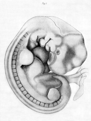
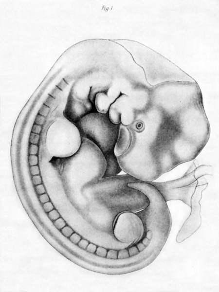
The embryo is flexed upon itself, forming almost a circle (Pl. XXIX., Fig. I). The head shows the outline of the brain within, and also a marked elevation over the region of the Gasserian ganglion. The nasal pit is a large shallow depression, being well exposed on both sides. The lense is small, and is surrounded by a groove which is continued between the superior maxillary process and nasal pit.
Three branchial arches are visible on the right side, and four on the left. The ventral end of the first is bulbous, while from its dorsal end the superior maxillary process arises. The second is also bulbous on its ventral end, the major portion of the trunk hanging over the third arch. This is the embryonic operculum which will finally close the sinus praecervicalis. The third arch lies more towards the median line, that is, it is within the sinus praecervicalis. The fourth arch is visible only on the left side; it lies deep in the sinus praecervicalis, and is almost covered by the third arch.
The clefts are irregular in shape, as shown in the figure; and the first, second, and third show marked depressions at their dorsal ends, which indicate the blending of the ectoderm with the seventh, ninth, and tenth nerves.
On the dorsal side of the branchial region, alike on both sides of the head, there is a marked depression which lies immediately over the otic vesicle. The vesicle, however, is fully separated from the ectoderm.
The protovertebrae are more marked on the right than the left side ; twenty-seven on the right, and twenty-four on the left. On the right only the last seven cervical, all the dorsal and lumbar, and five sacral are visible; while on the left two occipital, all the cervical and dorsal, and but two lumbar are seen. The sections, however, show that the muscle plates are the same on both sides.
The extremities are well marked, the anterior being somewhat larger than the posterior. The anterior on the right side is flat, and bent directly towards the median line ; while on the left it hangs away from the mouth. The posterior on the right is bent towards the head, and on the left side it is simply an oval mound.
Upon the body proper there are three marked elevations: two for the heart, and one for the liver. In general, these elevations are the same on both sides.
The umbilical cord is large, and lies on the left side of the body, as described by Waldeyerl and by Janoiik.2 In most embryos described it lies on the right side. The cord is short, and midway between the embryo and its attachment to the chorion it shows a decided enlargement. The umbilical vesicle is large, measuring 7 mm. in length and 5 in diameter.
Method of Study
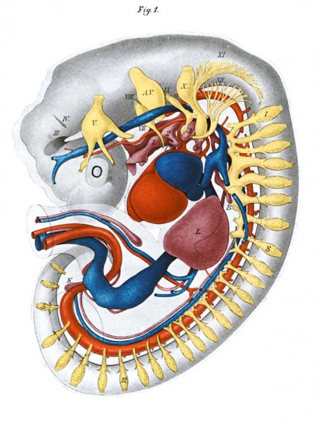
The embryo was stained with alum carmine, and cut into serial sections I 5 microns thick. Although thinner sections undoubtedly would have been better to study the histology, for my purpose I desired sections in no way distorted. On account of greater ease in studying the sections, I cut the embryo at right angles to the nape-breech length, at the same time striking the branchial arches nearly at right angles. In all there were 351 perfect sections, none lost, and only two or three slightly distorted. The sections were enlarged 662 diameters, which at the same time increased their thickness to I mm. Every second section was now drawn on wax plates 2 mm. thick, and the external outline of the section cut out. Before working the interior of the enlarged sections, they were carefully piled, in order to obtain the form of the exterior of the embryo. Next, I made a plaster mould of the pile of plates, in order to keep them in position. The plates were constantly kept within the half-mould, in order to keep the model from becoming distorted. This latter procedure proved of great value, as the plates were cut into pieces in order to model the various organs. All the wax representing the tissue between the organs and the exterior of the body on the left side was removed, exposing the organs as shown in the figure on Plate XXX. Then the organs were freed from the opposite side, and the pieces of wax blended so as to isolate each organ by itself.
The body cavity was modelled as a corrosion preparation, by drawing its outline on a second set of plates, and removing all the wax representing the body cavity, then piling the plates again, and finally casting the whole with Wood's metal. The metal was next smoothed and imbedded in plaster of paris, from which it was removed by boiling. The plaster mould was now cast with solder, and the wax broken off. By this method a metal cast of the whole ccelom was obtained.
The shrinkage while imbedding in paraffin was slightly over ten per cent, so our model, which represents the section of the embryo enlarged 662/8 times, is but sixty times larger than the specimen while in alcohol. All the measurements I give have been reduced to correspond with those of the alcoholic specimen.
Central Nervous System
When the neural tube is straightened it measures from end to end 17mm. with a diameter of 1.5 mm. for the fore-brain, and 0.5 mm. at the point between the posterior extremities. Its general shape is shown, Plate XXX., which corresponds quite well with His’s BY. On the exterior it is plainly shown that the cerebrum and optic vesicle are attached to the fore-brain.
The cerebral hemispheres are represented on both sides by oval projections from the fore-brain, extending somewhat over the surrounding tissue on the mid-brain side, and under the eye. Viewed from the median side the pit represents a stage midway between His’s BY. and K.0.2. On the median line between the two hemispheres there is a fold of medullary tube-wall which extends more than a third across the cerebrum, and has a tendency to cut it in half. It measures 0.4 mm. in perpendicular, and projects 0.16mm. into the fore-brain vesicle. Compared with His’s figures, this is undoubtedly the epithelial covering of the choroid plexus.
Towards the mouth from the cerebrum there is the opening into the optic vesicle. It is triangular in shape, with the base on the oral side, and the apex pointing towards the inter-brain. The opening within the stem of the optic vesicle is round, and ends as a circle about the secondary ocular vesicle. Viewed from the outside, the secondary ocular vesicle is a round pit, in which lies the lense. There is no slit on the oral side of the stem (Plate XXX., Fig. I).
The inter-brain shows a marked constriction in its middle, both on the outside and also within its lumen. This undoubtedly was caused by a shrinkage when the specimen was hardened. As the mid-brain and hind-brain are approached, the walls gradually become thicker and thicker, until the after-brain is reached, when the ventral side alone increases, while the dorsal walls become very thin. From the origin of the facial nerve to the origin of the pneumogastric, the walls of the dorsal half of the neural tube are very thin. From now on, the thickness of the walls of the tube, with the exception of the extreme dorsal and ventral sides,l is much the same in transverse section, the thickness gradually diminishing as the tail is approached.
Cranial Nerves
The olfactory pit is sharply defined and is composed of five or six layers of cells. Throughout its extent cell divisions are present, most numerous, however, at its concave oral side. In this region there are marked pyramidal cells with their base towards the outside of the body and their apices pointing towards the brain. These cells undoubtedly mark the beginning of the olfactory nerve as pointed out by His. In Amblystoma and Necturus these cells are much more pronounced and can be traced in the various stages from the olfactory pit to the brain.
There is as yet no indication of a permanent optic nerve. The primary optic vesicle still communicates with the fore-brain while the distal or rod-and-cone layer of the secondary vesicle shows peculiar changes. The layer is about twice as thick as the proximal or pigment layer, and in both these are nuclear figures. The location of these cells, which are dividing, is on the margin which corresponds with the layer about the central canal in the spinal cord. The pigment layer is about five cells thick, while the rod-and-cone layer is about eight.
The rod-and-cone layer is composed of two distinct zones, - a distal or hyaline and a proximal or cellular. The hyaline zone lies next to the lense and seems to be composed of cilia, all being directed towards a centre lying within the lense. The granular zone is composed in great part of round cells between which are many bipolar and unipolar cells. The unipolar cells are more numerous than the bipolar and project, with their pole, towards the position which is later to be rods and cones. The bipolar send one pole in the same direction and the other into the hyaline layer.
At a point in the stem of the optic vesicle nearest the mouth there enters a vessel which is undoubtedly the arteria centralis retinz. As this perforates the hyaline layer, the “cilia” become shorter, but are in no way directed towards this artery, ie.thefuturedirectionoftheopticnerve. Nodoubtthislayer is identical with the peripheral veil as described by His.1
It is extremely difficult to locate the origin of the third nerve. At the floor of the mid-brain there is a suspicious spot composed of several dozen cells which are somewhat separated from the remaining cells and lie partly within the terminal veil (Pl. XXX., Fig. 1, 111.). No nerve fibres extend from the brain into the surrounding tissue.
The trochlear nerve is well marked as a small group of cells in the ventral wall of the isthmus between the mid-brain and hind-brain. The cells lie just under the terminal veil, and each sends a single short pole towards the dorsal part of the brain. They extend but half-way around the tube.
The Gasserian ganglion with its three branches marks the trifacial. Upon its ophthalmic branch a small group of cells indicates the ciliary ganglion. From the Gasserian ganglion numerous fibres enter the hind-brain as its sensory root. The motor root arises more ventral from a large group of cells and passes as a large bundle of fibres into the inferior maxillary branch of the nerve.
The sixth nerve is represented as a small group of cells, dorsal, but somewhat aboral from the first branchial cleft. None of the cells send prolongations from the brain to form a distinct nerve, but all of the unipolar motor cells are pointed in one direction.
It is extremely difficult to isolate the facial nerve from the auditory nerve ganglion. Following it from the second branchial arch it passes through the heart of the acoustico-facial ganglion, and after entering the neural tube passes towards the ventral side of the same, making an arch around the ganglion of the sixth to take its origin near the median line.
The acoustic ganglion extends from the facial to the auditory vesicle, to which it is adherent, and then with the facial nerve sends twigs into the after-brain. The auditory vesicle is olive-shaped, is placed at right angles to the after-brain, and from its dorsal end there is a marked prolongation, the beginning of the aquaeductus vestibuli. The walls are of quite even thickness throughout, and the lumen is of the same general shape as the vesicle.
Between the auditory vesicle and the after-brain lies the upper ganglion of the glosso-pharyngeal which receives fibres from a group of cells lying in the floor of the after-brain, and sends a bundle of fibres more dorsally into the same. On the ventral side there is a bundle of nerves which communicates with a second ganglion, the ganglion petrosum. The ganglion petrosum is in direct continuity with a slight invagination of ectoderm at the dorsal part of the second branchial groove, sends a branch into the third arch, and communicates with the ganglion jugulare of the vagus.
The vagus is composed of two enormous ganglia, as shown in P1.XXX. The two ganglia are united by a band of cells, and from the ganglion nodosum a branch passes to the aboral and lateral side of the fourth branchial arch. At the central end of the nerve numerous branches pass into the after-brain. The ganglion jugulare receives at once twigs from the accessory nerve, which soon arrange themselves into a bundle to become fully separated before the ganglion nodosum is reached.
The accessory nerve arises as a row of bundles between the vagus and first cervical, and emerges from the after-brain midway between its dorsal and ventral walls. As the twigs approach the first cervical nerve, the origin becomes more ventral, and are continuous with the ventral root of this nerve. Although the rudimentary ganglion, first described by Froriep as the ganglion of the accessory, has been verified by His for the human embryo, I cannot find any trace of it in this specimen. I have had no difficultyin finding it in dog and cat embryos, and therefore must conclude that it is wanting in this embryo. This is what we should expect to find from time to time, especially in an organ which is in process of degeneration.
The hypoglossal nerve arises as a group of fibres parallel to but more ventral than the accessory. The bundles are arranged to correspond with the myotomes of the head, and on the aboral side arise in common with the accessory and the first cervical nerve.
The Spinal Nerves
The spinal nerves are all distinctly marked by a large dorsal ganglion which sends small bundles of fibres into the cord, and a ventral root which arises from the motor cells in the anterior horn of the same. The ganglia are largest in the cervical region, and gradually diminish in size as the tail is approached. The eight cervical nerves are united by anastomoses, which are destined to form the cervical and brachial plexuses respectively. The distal ends of the upper eight dorsal nerves are divided into two branches. Beyond these the remaining dorsal, lumbar, and sacral nerves end as a single branch. In all, twenty-nine spinal nerves can be identified ; i.e. eight cervical, twelve dorsal, five lumbar, and four sacral. Beyond this there is a group of myotomes, which towards the tip of the tail run into one another. In this region the dorsal ganglia are-not fully separated from the spinal cord ; in fact, the whole seems yet to be blended with the ectoderm.
Sympathetic Nerves
Onodi has shown quite conclusively that the sympathetic nerves arise from the spinal. In our embryo, although there are as yet no sympathetic ganglia, there are marked branches from the first six dorsal nerves extending directly towards the chorda. These branches, without their ganglia, are spoken of by His for a human embryo 7 mm. long, in his last communication.a It will be seen from the reconstruction that all the sympathetic nerves arise from the oral branches of the spinal nerves. Three are in front of the celiac axis, and three behind. From the study of other mammalian embryos I find that at about this stage the sympathetic nerves become encircled about the celiac axis; and as this vessel moves aboralwards, the successive branches are entangled, and in this way form the splanchnic nerve. In the adult the celiac axis is back of the twelfth dorsal vertebra, and the solar plexus encircles it. This plexus communicates by means of the great splanchnic nerve with the fifth to the tenth dorsal nerves. Under favorable conditions it may be traced to the third, second, or even the first dorsal.2 Now in the various stages of development, the celiac axis is successively opposite the various dorsal vertebrae; and as it moves backwards it carries with it these sympathetic twigs from the spinal nerves, which all unite to form the splanchnic.
Skeleton
The bulk of the framework in this specimen is composed of multipolar cells. Although these play a very minor part in adult higher animals, they are no doubt in embryos almost the sole element which holds the tissues together. Besides these there is the chorda dorsalis, which begins in the entoderm at the base of the third branchial arch, and extends to the tip of the tail. As the chorda passes between the two vertebral arteries, it is surrounded by various conipact groups of cells which mark the bodies of the future vertebrae. On the oral side of the first cervical nerve the group marks the occipital bone; it is the most conspicuous mass, extends from one side of the body to the other, and sends two processes oralwards and lateral to the two vertebral arteries. Next in distinctness is the first cervical vertebra, while the following cervicals gradually diminish ; the first three dorsal are faintly outlined.
The Muscle Plates
The occipital cartilage is bound to the first cervical by means of a muscle plate on either side. On the oral side of the occipital there are'three muscle plates lying over the three bundles of nerve roots of the hypoglossal nerve. The plate nearest the occipital is bound to the cartilage on one end, while the remaining two have no cartilage between them. The plates gradually become larger and larger as the dorsal region is approached, and beyond this become smaller and smaller, until the tip of the tail is reached. In all, there are thirty-eight in number.
Especially well marked in the dorsal region are hollow outgrowths from the plates into the body wall. The rest of the plate is solid, but from the aboral and ventral corner these epithelioid prolongations extend beyond the beginning of the body cavity. On the median side of each plate there is a hyaline border which marks the beginning of muscle fibres. Between this border and the nerve stem lies a group of cells quite marked at the point the nerve ends, and gradually diminishing in number as the dorsal ganglion is approached. Many of these cells, as they enter the hyaline border of the myotome, become unipolar with the pole pointing towards the nerve root.
Heart
The heart, shown in profile in the figures, is 1.6mm. wide, 1.5 high, and I deep. The ventricles are contracted and empty while the auricle is distended with blood. The right auricle is larger than the left, and into it empties the sinus reuniens, which is guarded by a well-marked valve. The left auricle is smaller, and partly separated by a septum, which extends towards, and half-way to, the auriculo-ventricular opening. Between this s e p turn and the auriculo-ventricular opening is a free communication between the two auricles, -the embryonic foramen ovale. Theventricle is also partly divided into two compartments which communicate with each other, and also with the auricle above. A partial septum divides the auriculo-ventricular opening into two channels, making the left auricle and ventricle, and right auricle and ventricle communicating freely with each other. There is no free direct communication between an auricle and an opposite ventricle. The whole aorta arises from the right ventricle, The arrangement is such that the flow of blood may be from the right auricle to the right ventricle, and bulbus aortz, or right auricle, foramen ovale, left auricle, left ventricle, right ventricle, and then bulbus aortz. At its origin the bulbus is, upon transverse section, hour-glass shaped, and nearly separated into two tubes.
The walls of the auricles are much thinner than those of the ventricles. In the ventricle there are many bridges extending across the lumen, making the walls sponge-like in appearance. Between the auricle and ventricle there is a marked constriction in the walls which extends about one-tenth the distance to the auricular-ventricular opening.
Arteries
After the bulbus aortae leaves the ventricle it is fully surrounded by the cavity of the pericardium until it breaks up into the aoftic arches. In this region the walls are quite thick, being composed in great part of round cells. As the bulbus passes into the aortic arches, these cells are continued into the indifferent mesodermal cells of the body.
There are three completed aortic arches lying within the third, fourth, and the tissue aboralwards from the fourth branchial arches. They are the third, fourth, and fifth aortic arches. Their general direction and shape is shown on Plate XXX. The two sets of arches unite to form the two aortae, which again unite, between the sixth and seventh cervical nerves, to form a single aorta. In His’s embryo B the division is about at the same point;l while in embryo A it is at the fourth dorsal.a As the aorta passes backward, it gradually becomes larger and larger, so that its diameter in the lumbar region is several times that in the cervical.~Here it very abruptly breaks up into two large branches which pass into the cord as the umbilical arteries.
Although there are many blood corpuscles scattered throughout the tissue of the embryo, I can make out definitely only a few arteries. The artery arising from the third aortic arch, and passing along the dorsal side of the branchial cavity up to the eye, is undoubtedly the internal carotid. Slightly beyond the eye it breakc up into numerous small branches, the most prominent passing towards the mid-brain, and undoubtedly represents the posterior cerebral. In His’s diagram4this same twig passes between the inter- and mid-brains, and this throws it in front of the third nerve. In the neighborhood of the eye no branches could be found which arise from the carotid, but a large branch passes through the retina. This indicates that the ophthalmic is present, but cannot be followed in the sections.
From the fifth aortic arch, on either side, there is a branch which passes to the lung, and breaks up into a network of capillaries about the pulmonary buds. This is the pulmonary artery.
On the dorsal side of the aorta there are, on either side, twenty-one segmental arteries, the first being in front of the first cervical nerve, and the last behind the twelfth dorsal. The second segmental communicates on either side with a large branch - the vertebral. This branch extends as far forward as the otic vesicle, and gives off two distinct twigs, one extending in front of the vagus nerve, and the other just in front of the first cervical ; they probably represent the anterior and posterior cerebellar arteries.
The origin of the vertebral is much more anterior than in the adult. His,' and recently, Hochstetter, a have shown that the vertebral is in many respects a segmental artery, and the condition of things in this embryo confirms this view. The second cervical segmental artery ends in a T, as shown in the plate. Undoubtedly the cross-piece of the T is to transfer the origin of the vertebral back to the second; and so on. Following the segmental branches backward, I find that between the fifth and sixth cervical nerves, a branch passes from the segmental into the anterior extremity. Although this twig arises (as far as the relation of the subclavian artery to the brachial plexus is concerned) one segment too far forward, I think it must represent the future subclavian.s From this point on down to the first lumbar, the segmental arteries gradually diminish in size. Below the first lumbar nerve no segmental branches arise from the aorta.
Between the lateral and ventral sides of the aorta there are fourteen pairs of segmental branches, which pass to the Wolffian body. Each pair arises on the ventral side just opposite the origin of the branches which pass towards the spinal cord. The anterior branches, which are quite small, arise between the seventh and eighth cervical nerves, and pass directly to the anterior end of the Wolffian body. Back of this pair the branches are of about equal size, the last being in front of the first lumbar nerve.
On the ventral median line of the aorta there are two distinct branches, -coeliac axis and the omphalo-mesenteric. At the fourth lumbar several small branches are given off from the ventral side of the aorta, and break up into a capillary network which extends throughout the mesentery.
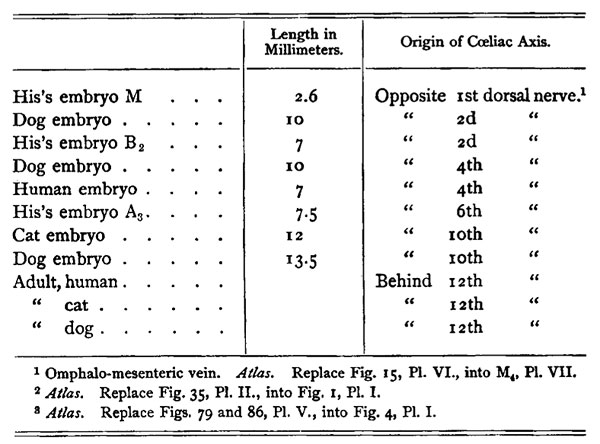
Although the branch to the stomach and liver has already all its relations to these organs, as the celiac axis in the adult, its origin is far too far forward. Other embryos, however, demonstrate that the origin of this vessel is constantly shifting as the following table shows -
It is only left for us to conclude that the stomach, liver, and pancreas receive their artery while they lie dorsal to the heart, and as their organs move backward the origin of the coeliac axis is gradually shifted in the same direction.
In these cases the omphalo-mesenteric artery is also shifted with the coeliac axis. In our embryo this vessel has a double origin, which indicates that this movement may be brought about by an anastomosis forming and then occlusion of the old origin. There is no twig from the omphalo-mesenteric to the mesentery.
The twigs which arise from the lumbar aorta at once break up into a capillary network which extends to and encircles the intestine, finally communicating with a vein which empties into the omphalo-mesenteric vein.
Veins
The veins which I have followed out are the jugular, cardinal, subclavian, omphalo-mesenteric, and umbilical. Their general course is shown in the reconstruction. It will be noticed that the cardinal extends along the whole length of the Wolffian body, receiving the blood from this organ. The omphalo-mesenteric receives a vein from the mesentery, -the inferior mesenteric. The jugular and cardinal unite to form the ductus Cuvieri, which on the right side passes directly into the sinus reuniens, while on the left it flows across the dorsal side of the heart before it empties into the same.
On either side there are several large veins which arise in the anterior extremity, soon unite to form a single branch and empty into the ductus Cuvieri. On the left side these veins form a sinus around the united branch from the fourth and fifth cervical nerves, and then communicate with the cardinal vein as well as with the ductus Cuvieri.
It is the left omphalo-mesenteric vein which remains in the specimen. The vein passes around the dorsal side of the alimentary canal, and about in the middle of the liver unites with the umbilical vein. The united veins now become greatly narrowed and then again enlarge to form the sinus reuniens. From the distal side of the constriction the veins pass to the substance of the liver, while on the proximal side these efferent branches enter, forming a portal system.
Coelom
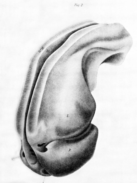
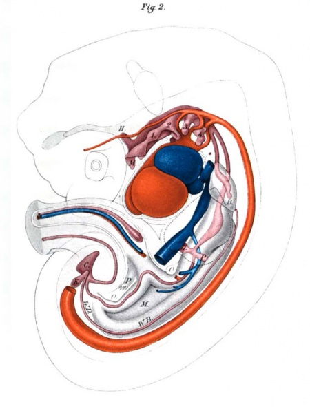
A cast of the ccelomic cavity is shown in Fig. 2, P1. XXIX. The picture was taken from the inverted model viewed from the dorsal and left sides. The slit along the dorsal side marks the mesentery while the grooves on either side of this indicate the position of the Wolffian bodies. The bulbous end of this model represents the pericardial cavity.
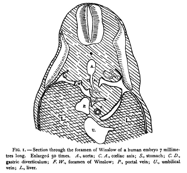
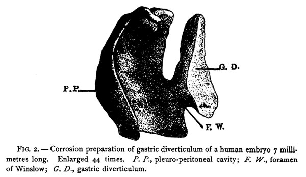
The pericardial cavity surrounds the whole heart, as shown in the sagittal section in Fig. 2, PI. XXX. The cavity is perforated only where the large veins enter, and where the artery leaves the heart. The cavity completely surrounds the bulbus aortae to its origin in the ventricle, the ventral side of this cavity being directly continuous with the ventral pericardial cavity. On the dorsal side the cavity is broken through for the transmission of the veins to the heart. Between the bulbus and the entrance of these veins the cavity extends across this median line as three distinct openings. On the dorsal side of the heart on either side of the lungs the pericardial cavity communicates with the pleuro-peritoneal cavity by means of two openings, each of which has a long diameter of 0.5 mm and a short diameter of 0 .1 mm. The cavity now encircles the lungs, leaving, however, a dorsal and a ventral mesentery. More aboralwards a slit passes about the liver, and one about the stomach, as shown in Fig. I . On the left side these two slits are quite smooth, and again run together on the aboral side of the liver. On the right, however, instead of a slit we have a pocket, or diverticulum, the cast of which is given in Fig. 2. The asymmetry of the two sides corresponds with the shifting of the stomach, and later the diverticulum forms the lesser peritoneal cavity.' Farther back the cavities on either side become symmetrical again, and then communicate with each other on the ventral side of the alimentary canal, as shown in Fig. 2, Pl. XXX.
The cavity next encircles the omphalo-mesentric vessels of the cord extending to the outside of the body. Behind the cord there is again a large communication from one side to the other. On the ventral side of the intestines, from the junction of the cord with the mesentery, a large papilliform projection hangs into this cavity (P). Still farther back the cavity encircles the Wolffian bodies, and finally ends by projecting deep into the tissues of the pelvis.
Alimentary Canal
A lateral view of the cast of the branchial region is shown on Pl. XXX. The entoderm is directly continuous with the ectoderm, and the cast is carried to the full exterior of the body. On the dorsal median line the pocket which forms the hypophysis, exfending between the mid-brain and after-brain, is shown. The first branchial pocket is bulbous and projects laterally, and then passes as a marked groove on the ventral side of the branchial cavity obliquely away from the mouth and towards the median line. It is destined to form the Eustachian tube. The second pocket is equally as well marked but is hook-shaped, with the point reaching half-way to the median line. The third pocket points with its free extremity toward the second, and is yet in free communication with the branchial cavity. It is destined to become the thymus, The fourth pocket is irregular in shape, as shown in the figure, It is in general parallel with the alimentary canal and rests wholly between the fourth and fifth aortic arches.
The thyroid gland is as a small nodule lying in the median line on the ventral side of the second branchial arch. Throughout their whole extent the branchial pockets at no point communicate with the exterior of the body. In no section is there a rupture of the membrane of His.
Immediately behind the branchial region the embryonic pharynx gives off the larynx, which farther back divides into the two bronchi. The intestinal tube now becomes dilated to form the stomach, below which arise the sprouts to form the pancreas and liver. The pancreas is composed of a small group of cells which lie in the mesogastrium, contains a lumen and communicates directly with the duodenum.
On the ventral side, however, this sprout is irregular, one branch boring into the liver, and the other ending as a sphere in the septum transversum. The liver is composed of two lobes, the right being about I mm. in diameter and half a millimetre thick. Its lateral border is symmetrical, and on its median side it is convex. It communicates along the septum transversum to the left lobe, which is very regular in shape.
Behind the liver the intestine makes an irregular curve towards the umbilical cord and to the left, and finally ends in the cloaca.
The cloaca is pyramidal in shape, with the apex pointing towards the tip of the tail. The highest point of the apex is blended with the ectoderm, and no doubt is about to break through. The intestine enters on the oral side of the base (speaking of the cloaca as a pyramid), and the allantois arises somewhat more aboralwards. The allantois is a small tube which is markedly dilated as it enters the cord, and then again becomes more constricted. On either side of the allantois and on the dorsal side of the intestine the Wolffian duct enters. Shortly before the duct enters the cloaca it gives off a blind tube, the beginning of the kidney.
The Wolffian body is a very large, somewhat lobulated body extending on either side of the intestine from the cloaca to the sixth dorsal nerve. Its general outline is given in Fig. 2 , PI. XXX. Upon transverse section, its free surface is small and forms a complete semicircle in the cervical region, larger but more flat in the dorsal, and again somewhat smaller but semicircular in the lumbar region. The Wolffian duct extends throughout the body, extending just below the free surface and lying on the dorsal side in the lumbar region and on the ventral side in the cervical region. The glomeruli extend from the sixth cervical nerve to the fifth lumbar, and there are about two or three glomeruli to a segment. In the cervical region the tubule connecting the glomerulus with the Wolffian duct is slightly bent, in the dorsal region greatly convoluted, and in the lumbar region it is straight.
CLARK UNIVERSITY, WORCESTER, MASS.,
May 14,1891.
Plates
Plate 1
DESCRIPTION OF PLATE XXIX.
|
|
| FIG.1 . External view of the embryo before it was sectioned. Enlarged 20 times. | FIG.2. Corrosion preparation of the pericardial and pleuro-peritoneal cavities. Enlarged 44 times. P. pericardial space; A. opening for aorta; K opening for vein; L. space over liver; M slit for mesentery; W.B. space for Wolffian body. |
Plate 2
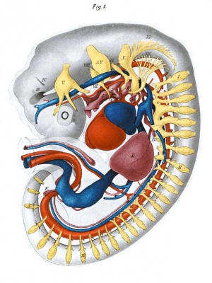
DESCRIPTION OF PLATE XXX.
|
|
| Fig. 1. Reconstruction viewed from the left side. Enlarged 15 times. HI., IV., V., etc. cranial nerves; A. V.auditory vesicle; I. 2, 3, and 4 branchial pockets; T. thyroid gland; B. bronchus; L. liver; K.kidney; yellow, nerves; red, arteries; blue, veins. The dotted lines mark the extremities. | Fig. 2. The same as Fig. I. Deeper view. H. hypophysis; M. mouth, mesentery; I, 2, 3, 4, branchial pockets; B. bronchus; P. pancreas; L. liver; W.B. Wolffian body; W.D. Wolffian duct; K.kidney; C. cloaca; 0. openings by which the pleuro-peritoneal cavities communicate; P. papilliform projection into the lower opening. |
| Historic Disclaimer - information about historic embryology pages |
|---|
| Pages where the terms "Historic" (textbooks, papers, people, recommendations) appear on this site, and sections within pages where this disclaimer appears, indicate that the content and scientific understanding are specific to the time of publication. This means that while some scientific descriptions are still accurate, the terminology and interpretation of the developmental mechanisms reflect the understanding at the time of original publication and those of the preceding periods, these terms, interpretations and recommendations may not reflect our current scientific understanding. (More? Embryology History | Historic Embryology Papers) |
Glossary Links
- Glossary: A | B | C | D | E | F | G | H | I | J | K | L | M | N | O | P | Q | R | S | T | U | V | W | X | Y | Z | Numbers | Symbols | Term Link
Cite this page: Hill, M.A. (2024, May 1) Embryology Paper - A Human Embryo Twenty-Six Days Old. Retrieved from https://embryology.med.unsw.edu.au/embryology/index.php/Paper_-_A_Human_Embryo_Twenty-Six_Days_Old
- © Dr Mark Hill 2024, UNSW Embryology ISBN: 978 0 7334 2609 4 - UNSW CRICOS Provider Code No. 00098G

