Neural - Pons Development
| Embryology - 9 Jul 2026 |
|---|
| Google Translate - select your language from the list shown below (this will open a new external page) |
|
العربية | català | 中文 | 中國傳統的 | français | Deutsche | עִברִית | हिंदी | bahasa Indonesia | italiano | 日本語 | 한국어 | မြန်မာ | Pilipino | Polskie | português | ਪੰਜਾਬੀ ਦੇ | Română | русский | Español | Swahili | Svensk | ไทย | Türkçe | اردو | ייִדיש | Tiếng Việt These external translations are automated and may not be accurate. (More? About Translations) |
Introduction


(Latin, pons = "bridge") A brain stem region within the central nervous system, anatomically lying above the medulla before the nervous system becomes the spinal cord.
Historically, the pons was described as originating from rhombomere r1 to r6, molecular studies have shown that the basilar pons is located only within rhombomeres r3 and r4.[1]
Neural development is one of the earliest systems to begin and the last to be completed after birth. This development generates the most complex structure within the embryo and the long time period of development means in utero insult during pregnancy may have consequences to development of the nervous system. The early central nervous system begins as a simple neural plate that folds to form a groove then tube, open initially at each end. Failure of these opening to close contributes a major class of neural abnormalities (neural tube defects). Within the neural tube stem cells generate the 2 major classes of cells that make the majority of the nervous system : neurons and glia. Both these classes of cells differentiate into many different types generated with highly specialized functions and shapes. This section covers the establishment of neural populations, the inductive influences of surrounding tissues and the sequential generation of neurons establishing the layered structure seen in the brain and spinal cord.
Some Recent Findings

|
|

| More recent papers |
|---|
 This table allows an automated computer search of the external PubMed database using the listed "Search term" text link.
More? References | Discussion Page | Journal Searches | 2019 References | 2020 References Search term: Pons Development | Pons Embryology |
| Older papers |
|---|
| These papers originally appeared in the Some Recent Findings table, but as that list grew in length have now been shuffled down to this collapsible table.
See also the Discussion Page for other references listed by year and References on this current page.
|
Development Overview
Neuralation begins at the trilaminar embryo with formation of the notochord and somites, both of which underly the ectoderm and do not contribute to the nervous system, but are involved with patterning its initial formation. The central portion of the ectoderm then forms the neural plate that folds to form the neural tube, that will eventually form the entire central nervous system.
- Early developmental sequence: Epiblast - Ectoderm - Neural Plate - Neural groove and Neural Crest - Neural Tube and Neural Crest
| Neural Tube | Primary Vesicles | Secondary Vesicles | Adult Structures |
|---|---|---|---|
| week 3 | week 4 | week 5 | adult |
| prosencephalon (forebrain) | telencephalon | Rhinencephalon, Amygdala, hippocampus, cerebrum (cortex), hypothalamus, pituitary | Basal Ganglia, lateral ventricles | |
| diencephalon | epithalamus, thalamus, Subthalamus, pineal, posterior commissure, pretectum, third ventricle | ||
| mesencephalon (midbrain) | mesencephalon | tectum, Cerebral peduncle, cerebral aqueduct, pons | |
| rhombencephalon (hindbrain) | metencephalon | cerebellum | |
| myelencephalon | medulla oblongata, isthmus | ||
| spinal cord, pyramidal decussation, central canal | |||
Early Brain Vesicles
Primary Vesicles

Secondary Vesicles

Fetal Pons
- 130 -140 mm CRL - first myelinated fibers in each motor root of the trigeminal, abducent, and facial nerves.[4]
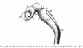
Adult Pons MRI

|
A T1-weighted sagittal MR image from a control subject, showing the midline structures of the posterior cranial fossa and the brainstem and the cerebellum.
|
References
- ↑ 1.0 1.1 Watson C, Bartholomaeus C & Puelles L. (2019). Time for Radical Changes in Brain Stem Nomenclature-Applying the Lessons From Developmental Gene Patterns. Front Neuroanat , 13, 10. PMID: 30809133 DOI.
- ↑ Kratochwil CF, Maheshwari U & Rijli FM. (2017). The Long Journey of Pontine Nuclei Neurons: From Rhombic Lip to Cortico-Ponto-Cerebellar Circuitry. Front Neural Circuits , 11, 33. PMID: 28567005 DOI.
- ↑ Tate MC, Lindquist RA, Nguyen T, Sanai N, Barkovich AJ, Huang EJ, Rowitch DH & Alvarez-Buylla A. (2015). Postnatal growth of the human pons: a morphometric and immunohistochemical analysis. J. Comp. Neurol. , 523, 449-62. PMID: 25307966 DOI.
- ↑ 4.0 4.1 Hatta T, Satow F, Hatta J, Hashimoto R, Udagawa J, Matsumoto A & Otani H. (2007). Development of the pons in human fetuses. Congenit Anom (Kyoto) , 47, 63-7. PMID: 17504389 DOI. Cite error: Invalid
<ref>tag; name 'PMID17504389' defined multiple times with different content - ↑ Fumagalli M, Ramenghi LA, Righini A, Groppo M, Bassi L, De Carli A, Parazzini C, Triulzi F & Mosca F. (2009). Cerebellar haemorrhages and pons development in extremely low birth weight infants. Front Biosci (Elite Ed) , 1, 537-41. PMID: 19482668
- ↑ Achiron R, Kivilevitch Z, Lipitz S, Gamzu R, Almog B & Zalel Y. (2004). Development of the human fetal pons: in utero ultrasonographic study. Ultrasound Obstet Gynecol , 24, 506-10. PMID: 15459939 DOI.
- ↑ Sekula RF, Jannetta PJ, Casey KF, Marchan EM, Sekula LK & McCrady CS. (2005). Dimensions of the posterior fossa in patients symptomatic for Chiari I malformation but without cerebellar tonsillar descent. Cerebrospinal Fluid Res , 2, 11. PMID: 16359556 DOI.
Reviews
Angeles Fernández-Gil M, Palacios-Bote R, Leo-Barahona M & Mora-Encinas JP. (2010). Anatomy of the brainstem: a gaze into the stem of life. Semin. Ultrasound CT MR , 31, 196-219. PMID: 20483389 DOI.
Articles
Gesemann M, Litwack ED, Yee KT, Christen U & O'Leary DD. (2001). Identification of candidate genes for controlling development of the basilar pons by differential display PCR. Mol. Cell. Neurosci. , 18, 1-12. PMID: 11461149 DOI.
Ozawa H & Takashima S. (1998). Immunocytochemical development of transferrin and ferritin immunoreactivity in the human pons and cerebellum. J. Child Neurol. , 13, 59-63. PMID: 9512304 DOI.
Search PubMed
Search Pubmed: Pons Embryology | Pons Development
Additional Images
Historic Images
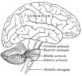
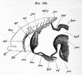
Fig. 677. Brain (lateral view)
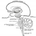
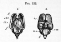
Fig. 678. Brain - Schematic representation of the chief ganglionic categories (I to V).
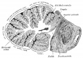
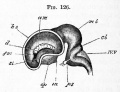
Cerebellum
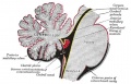
Cerebellum, Pons and Fourth Ventricle


External Links
External Links Notice - The dynamic nature of the internet may mean that some of these listed links may no longer function. If the link no longer works search the web with the link text or name. Links to any external commercial sites are provided for information purposes only and should never be considered an endorsement. UNSW Embryology is provided as an educational resource with no clinical information or commercial affiliation.
Glossary Links
- Glossary: A | B | C | D | E | F | G | H | I | J | K | L | M | N | O | P | Q | R | S | T | U | V | W | X | Y | Z | Numbers | Symbols | Term Link
Cite this page: Hill, M.A. (2026, July 9) Embryology Neural - Pons Development. Retrieved from https://embryology.med.unsw.edu.au/embryology/index.php/Neural_-_Pons_Development
- © Dr Mark Hill 2026, UNSW Embryology ISBN: 978 0 7334 2609 4 - UNSW CRICOS Provider Code No. 00098G