Introduction
Neural development is one of the earliest systems to begin and the last to be completed after birth. This development generates the most complex structure within the embryo and the long time period of development means in utero insult during pregnancy may have consequences to development of the nervous system. The early central nervous system begins as a simple neural plate that folds to form a groove then tube, open initially at each end. Failure of these opening to close contributes a major class of neural abnormalities (neural tube defects).
The midbrain/hindbrain boundary (MHB, isthmic organizer) is a secondary organizer region lying at the junction of the Mesencephalon and Metencephalon.
Some Recent Findings
- Mouse Fgf8-Cre-LacZ lineage analysis defines the territory of the postnatal mammalian isthmus[1] "The isthmus is recognized as the most rostral segment of the hindbrain in non-mammalian vertebrates. In mammalian embryos, transient Fgf8 expression defines the developing isthmic region, lying between the midbrain and the first rhombomere, but there has been uncertainty about the existence of a distinct isthmic segment in postnatal mammals. We attempted to find if the region of early embryonic Fgf8 expression (which is considered to involve the entire extent of the prospective isthmus initially) might help to identify the boundaries of the isthmus in postnatal animals. By creating an Fgf8-Cre-LacZ lineage in mice, we were able to show that Fgf8-Cre reporter expression in postnatal mice is present in the same nuclei that characterize the isthmic region in birds. The 'signature' isthmic structures in birds include the trochlear nucleus, the dorsal raphe nucleus, the microcellular tegmental nuclei, the pedunculotegmental nucleus, the vermis of the cerebellum, rostral parts of the parabrachial complex and locus coeruleus, and the caudal parts of the substantia nigra and VTA. We found that all of these structures were labeled with the Fgf8-Cre reporter in the mouse brain, and we conclude that the isthmus is a distinct segment of the mammalian brain lying caudal to the midbrain and rostral to rhombomere 1 of the hindbrain."
- Isthmus organizer for mesencephalon and metencephalon[2] " Brain vesicles formation is the first sign of regionalization. Classical transplantation using quail and chick embryos revealed that the mesencephalon-metencephalon boundary (isthmus) functions as an organizer of the mesencephalon and metencephalon. Fgf8 is accepted as a main organizing molecule of the isthmus. Strong Fgf8 signal activates the Ras-ERK signaling pathway to differentiate the cerebellum. In this review, the historical background of the means of identifying the isthmus organizer and the molecular mechanisms of signal transduction for tectum and cerebellum differentiation is reviewed."
- Role of Lmx1b and Wnt1 in mesencephalon and metencephalon development[3] "The isthmus is the organizing center for the tectum and cerebellum. Fgf8 and Wnt1 are secreted molecules expressed around the isthmus. The function of Fgf8 has been well analyzed, and now accepted as the most important organizing signal. Involvement of Wnt1 in the isthmic organizing activity was suggested by analysis of Wnt1 knockout mice. But its role in isthmic organizing activity is still obscure. Recently, it has been shown that Lmx1b is expressed in the isthmic region and that it may occupy higher hierarchical position in the gene expression cascade in the isthmus. We have carried out misexpression experiment of Lmx1b and Wnt1, and considered their role in the isthmic organizing activity. Lmx1b or Wnt1 misexpression caused expansion of the tectum and cerebellum. Fgf8 was repressed in a cells that misexpress Lmx1b, but Fgf8 expression was induced around Lmx1b-misexpressing cells. As Lmx1b induced Wnt1 and Wnt1 induced Fgf8 expression in turn, Wnt1 may be involved in non cell-autonomous induction of Fgf8 expression by Lmx1b. Wnt1 could not induce Lmx1b expression so that Lmx1b may be put at the higher hierarchical position than Wnt1 in gene expression cascade in the isthmus. We have examined the relationship among isthmus related genes, and discuss the mechanism of the formation and maintenance of isthmic organizing activity."
- Early mesencephalon/metencephalon patterning and development of the cerebellum[4] "Fate mapping studies in chick have shown that at early stages the cerebellum derives from cells in the mesencephalon and metencephalon (mes-met). Transplantation studies in chick have implicated the mes-met junction (isthmus) as a source of secreted factors that organize development of the entire mes-met, perhaps by stimulating proliferation and specifying positional values across the region. Fgf-8 has been implicated as a major factor involved in the isthmus organizing activity. Gene expression studies indicate that the anterior and posterior expression domains of the homeobox genes Otx-2 and Gbx-2, respectively, are the earliest indication of a division of the brain."
|
| More recent papers
|
|
This table allows an automated computer search of the external PubMed database using the listed "Search term" text link.
- This search now requires a manual link as the original PubMed extension has been disabled.
- The displayed list of references do not reflect any editorial selection of material based on content or relevance.
- References also appear on this list based upon the date of the actual page viewing.
References listed on the rest of the content page and the associated discussion page (listed under the publication year sub-headings) do include some editorial selection based upon both relevance and availability.
More? References | Discussion Page | Journal Searches | 2019 References | 2020 References
Search term: Metencephalon Embryology
<pubmed limit=5>Metencephalon Embryology</pubmed>
|
Development Overview
Neuralation begins at the trilaminar embryo with formation of the notochord and somites, both of which underly the ectoderm and do not contribute to the nervous system, but are involved with patterning its initial formation. The central portion of the ectoderm then forms the neural plate that folds to form the neural tube, that will eventually form the entire central nervous system.
- Early developmental sequence: Epiblast - Ectoderm - Neural Plate - Neural groove and Neural Crest - Neural Tube and Neural Crest
Neural Tube Development
| Neural Tube
|
Primary Vesicles
|
Secondary Vesicles
|
Adult Structures
|
| week 3
|
week 4
|
week 5
|
adult
|
neural plate
neural groove
neural tube
Brain
|
prosencephalon (forebrain)
|
telencephalon
|
Rhinencephalon, Amygdala, hippocampus, cerebrum (cortex), hypothalamus, pituitary | Basal Ganglia, lateral ventricles
|
| diencephalon
|
epithalamus, thalamus, Subthalamus, pineal, posterior commissure, pretectum, third ventricle
|
| mesencephalon (midbrain)
|
mesencephalon
|
tectum, Cerebral peduncle, cerebral aqueduct, pons
|
| rhombencephalon (hindbrain)
|
metencephalon
|
cerebellum
|
| myelencephalon
|
medulla oblongata, isthmus
|
| spinal cord, pyramidal decussation, central canal
|
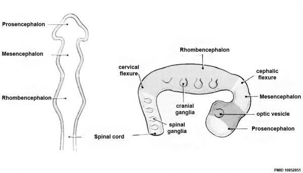
Early Brain Vesicles
Primary Vesicles
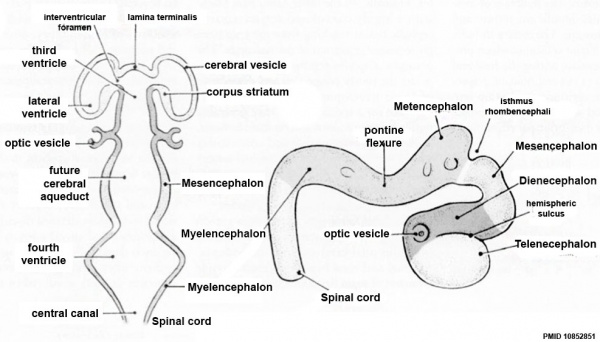
Secondary Vesicles
References
- ↑ <pubmed>28510270</pubmed>
- ↑ <pubmed>18494704</pubmed>
- ↑ <pubmed>12399317</pubmed>
- ↑ <pubmed>9509514</pubmed>
Reviews
<pubmed>19206138</pubmed>
Articles
Search PubMed
Search Pubmed: Metencephalon Embryology | Metencephalon Development |
Glossary Links
- Glossary: A | B | C | D | E | F | G | H | I | J | K | L | M | N | O | P | Q | R | S | T | U | V | W | X | Y | Z | Numbers | Symbols | Term Link
Cite this page: Hill, M.A. (2026, July 12) Embryology Neural - Metencephalon Development. Retrieved from https://embryology.med.unsw.edu.au/embryology/index.php/Neural_-_Metencephalon_Development
- What Links Here?
- © Dr Mark Hill 2026, UNSW Embryology ISBN: 978 0 7334 2609 4 - UNSW CRICOS Provider Code No. 00098G


