Lymph Node Development
| Embryology - 27 Jul 2026 |
|---|
| Google Translate - select your language from the list shown below (this will open a new external page) |
|
العربية | català | 中文 | 中國傳統的 | français | Deutsche | עִברִית | हिंदी | bahasa Indonesia | italiano | 日本語 | 한국어 | မြန်မာ | Pilipino | Polskie | português | ਪੰਜਾਬੀ ਦੇ | Română | русский | Español | Swahili | Svensk | ไทย | Türkçe | اردو | ייִדיש | Tiếng Việt These external translations are automated and may not be accurate. (More? About Translations) |
Introduction


Lymphatic vasculature drains lymph fluid from the organ tissue space and returns it to the blood vasculature for recirculation. The lymph node lies on the path of lymph vessels and these structures monitor and carry out immune surveillance of this fluid for antigens and pathogens, trapping them within the lymph nodes and generating immune responses.
In early node development, vein endothelial cells form a spherical body (lymph sac) that is surrounded and then invaded by mesenchymal cells that contribute the lymph node stroma. The current mouse model shows lymphoid tissue inducer cells initially regulating the activation of mesenchymal lymphoid tissue organizer cells.[1]
The reticular cells that form the stroma of the node were also initially seen as purely structural and supportive, more recent studies have shown that the reticular network that they form is also part of the immune process.
Some Recent Findings
|
| More recent papers |
|---|
 This table allows an automated computer search of the external PubMed database using the listed "Search term" text link.
More? References | Discussion Page | Journal Searches | 2019 References | 2020 References Search term: Lymph Nodes Embryology | Lymph Nodes Development | Lymph Nodes | tissue inducer cell | mesenchymal lymphoid tissue organizer cell |
| Older papers |
|---|
| These papers originally appeared in the Some Recent Findings table, but as that list grew in length have now been shuffled down to this collapsible table.
See also the Discussion Page for other references listed by year and References on this current page.
|
Early Development
- lymph sacs (primitive lymph sacs) form from endothelial cells.
- bud from the veins during early development
- then form buds that branch and form the lymphatic network.
- lymphoid tissue inducer cells - (LTi) first hematopoietic cells to enter and induce lymphoid tissue development.
Molecular
Prox1
- expressed by lymphatic endothelial cells.
Reticular Cells
Mesoderm derived fibroblastic reticular cells (FRCs, RCs) form the stromal supporting network of cells and extracellular matrix in secondary lymphoid organs (lymph nodes, spleen and thymus). These specialized myofibroblasts form the structural "sponge" within lymphoid tissue, through with T cells, B cells, dendritic cells (DCs), plasma cells and macrophages interact and move.[8]
- synthesise reticular fibres (type III collagen) that form the main extracellular matrix component
- differentiate as distinct subpopulations distributed in the cortical and medullar regions
- T cell zone fibroblastic reticular cells (TRCs)
- bind dendritic cells that initiate immunity by presenting antigens to T lymphocytes also initiate remodelling of lymph node RCs
- may regulate cellular traffic and activation of specific lymphocyte populations
Model of Reticular Network Formation[9]

| Accumulation of lymphocytes induces alteration of mesenchymal progenitors. | Lymphocytes and differentiated FRCs gradually degrade preexisting matrix, and FRCs weave RF meshwork from the newly produced components. | Mature RN forms and antigen presentation occur on the stromal reticulum. |

Lymph Node and Reticular Cell Classification[10]
Molecular
- express myofibroblast markers - desmin, vimentin, CD90, CD73, CD103, α-smooth muscle actin (αSMA) and ERTR7 antigen.
- express - podoplanin (PDPN), platelet-derived growth factor receptor-α (PDGFRA) and genes from antigen presentation and cytokine response pathways.
- lack of expression of CD45 and CD31.
Dendritic Cells
Dendritic cells (DCs, antigen-presenting cells, APCs) present antigens and induce a primary immune response in resting naïve T lymphocytes. These cells originate from the same common progenitor as monocytes.[11] Recently it has been shown that recirculation of antigen-bearing dendritic cells in remote lymphoid organs can prime T cells in other locations.[12]
In 2011 Ralph M. Steinman received half the Nobel Prize half of the award to to Ralph M. Steinman for his discovery of the dendritic cell and its role in adaptive immunity.
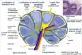
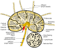
Adult Lymph Node
|

Legend
|
Lymph Node Cartoon Gallery
Detailed structure
Cell movement
Detailed structure
Simple structure
Wiki image
internal structure
lymph node cells


- Links: Immunobiology - Figure 1.8. Organization of a lymph node | MBoC Figure 24-16. A simplified drawing of a human lymph node

|
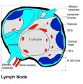
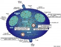
Schematic representation of the organization of a lymph node.[13]
|

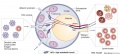
Mesenteric Lymph Node

Gastrointestinal tract intestine immune overview showing mesenteric lymph nodes.[14]
Histology
- Lymph Node Histology: Subcapsular Sinus | Follicle | Germinal Centre | Medullary Cords and Sinuses | High Endothelial Venules | Macrophages | Node cartoons
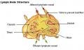
Adult Lymph Node Structure
- Capsule - dense connective tissue
- Trabeculae - dense connective tissue
- Reticular Tissue - Reticular cells and fibers, supporting meshwork
- Macrophages - process antigen, difficult to distinguish from the reticular cells.


Lymph
- enters the node through afferent vessels
- filters through the sinuses
- leaves through efferent vessels
Subcapsular sinus = marginal sinus

Continuation of trabecular sinus

Adult Lymphocytes
Lymphocyte Electron Micrographs
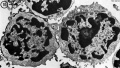
T and B Lymphocyte
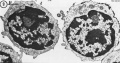
T and B Lymphocyte
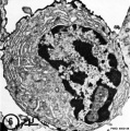
Plasma cell (B)
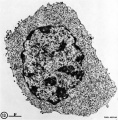
Cytotoxic (T)


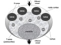
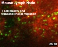
Cell Trafficking into and out of Lymph Nodes
Lymphocyte Traffic in and out of the Lymph Node[15]
The following data is from an in vivo mouse study[16] of live adult mouse lymphocytes (T and B cells) imaged within a lymph node.
Both lymphocyte types:
- Spend 8 to 24 h in the lymph node interstitium.
- Transit across a lymphatic endothelium to exit.
- Enter a network of medullary sinuses.
- Drain from sinuses into efferent lymphatic vessels.
Lymphocyte Migration Speeds
T cells - 10–12 μm/min in the follicle diffuse cortex, peak velocities up to 30 μm/min. (move more slowly in the medullary region near the hilus of the lymph node than in the paracortex)
B cells - 6 μm/min in the follicle diffuse cortex, peak velocities up to 20 μm/min.
Both cortical T cells and follicular B cells move in random directions following "guide cells".
Lymphocyte Guide Cells
FDC - Follicular Dendritic Cells, may guide B cells in the follicle.
FRC - Fibroblastic Reticular Cells, may guide T cells in the follicle.
Subcapsular Space
The subcapsular region is not only the initial entry site for lymph flow into the node, but has also been shown to have important immune functions associated with cells located in this space.
- Macrophages - clear lymph-borne viruses and present them to antiviral B cells.[17] This has also been recently demonstrated in the mouse model.[2]
- Memory B cells - have been shown to be reactivated in subcapsular proliferative foci of lymph nodes.[18]
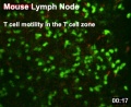
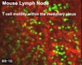
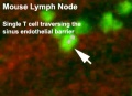
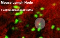
Lymphocyte Movies
| Adult Mouse Lymphocyte Motility | |||||||||||||||
|---|---|---|---|---|---|---|---|---|---|---|---|---|---|---|---|
|
|
|
| ||||||||||||
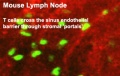
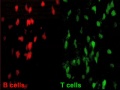
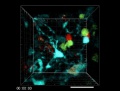
| Transendothelial migration |
T cell zone | Medullary sinus | Sinus endothelial barrier | ||||||||||||
|
|
|
| ||||||||||||
| Bi-directional traffic | Cross the sinus endothelial barrier |
T and B cell motility | T and B cell coupling | ||||||||||||
| Mouse Immune Movies: Transendothelial migration | T cell zone | Medullary sinus | Sinus endothelial barrier | Bi-directional traffic | cross the sinus endothelial barrier | T and B cell motility | T and B cell coupling | T cell Elimination | Immune System Development | Mouse Development | |||||||||||||||
References
- ↑ Onder L & Ludewig B. (2018). A Fresh View on Lymph Node Organogenesis. Trends Immunol. , 39, 775-787. PMID: 30150089 DOI.
- ↑ 2.0 2.1 Moran I, Grootveld AK, Nguyen A & Phan TG. (2019). Subcapsular Sinus Macrophages: The Seat of Innate and Adaptive Memory in Murine Lymph Nodes. Trends Immunol. , 40, 35-48. PMID: 30502023 DOI.
- ↑ Khaksary-Mahabady M, Khazaeel K, Pourmahdi Borujeni M & Yazdanjoo B. (2018). Morphometric development of sheep (Ovis aries) lymph nodes in fetal period. Vet Res Forum , 9, 121-128. PMID: 30065800 DOI.
- ↑ Huang SS, Li YW, Wu JL, Johnson FE & Huang JS. (2018). Development of the LYVE-1 gene with an acidic-amino-acid-rich (AAAR) domain in evolution is associated with acquisition of lymph nodes and efficient adaptive immunity. J. Cell. Physiol. , 233, 2681-2692. PMID: 28833090 DOI.
- ↑ Gentek R & Bajénoff M. (2017). Lymph Node Stroma Dynamics and Approaches for Their Visualization. Trends Immunol. , 38, 236-247. PMID: 28214099 DOI.
- ↑ van de Pavert SA & Mebius RE. (2014). Development of secondary lymphoid organs in relation to lymphatic vasculature. Adv Anat Embryol Cell Biol , 214, 81-91. PMID: 24276888 DOI.
- ↑ Vondenhoff MF, van de Pavert SA, Dillard ME, Greuter M, Goverse G, Oliver G & Mebius RE. (2009). Lymph sacs are not required for the initiation of lymph node formation. Development , 136, 29-34. PMID: 19060331 DOI.
- ↑ Fletcher AL, Acton SE & Knoblich K. (2015). Lymph node fibroblastic reticular cells in health and disease. Nat. Rev. Immunol. , 15, 350-61. PMID: 25998961 DOI.
- ↑ Fletcher AL, Acton SE & Knoblich K. (2015). Lymph node fibroblastic reticular cells in health and disease. Nat. Rev. Immunol. , 15, 350-61. PMID: 25998961 DOI.
- ↑ Alvarenga HG & Marti L. (2014). Multifunctional roles of reticular fibroblastic cells: more than meets the eye?. J Immunol Res , 2014, 402038. PMID: 24829927 DOI.
- ↑ Liu K & Nussenzweig MC. (2010). Origin and development of dendritic cells. Immunol. Rev. , 234, 45-54. PMID: 20193011 DOI.
- ↑ Hervouet C, Luci C, Bekri S, Juhel T, Bihl F, Braud VM, Czerkinsky C & Anjuère F. (2014). Antigen-bearing dendritic cells from the sublingual mucosa recirculate to distant systemic lymphoid organs to prime mucosal CD8 T cells. Mucosal Immunol , 7, 280-91. PMID: 23801305 DOI.
- ↑ Mueller SN & Germain RN. (2009). Stromal cell contributions to the homeostasis and functionality of the immune system. Nat. Rev. Immunol. , 9, 618-29. PMID: 19644499 DOI.
- ↑ Macpherson AJ & Smith K. (2006). Mesenteric lymph nodes at the center of immune anatomy. J. Exp. Med. , 203, 497-500. PMID: 16533891 DOI.
- ↑ Miyasaka M & Tanaka T. (2004). Lymphocyte trafficking across high endothelial venules: dogmas and enigmas. Nat. Rev. Immunol. , 4, 360-70. PMID: 15122201 DOI.
- ↑ Wei SH, Rosen H, Matheu MP, Sanna MG, Wang SK, Jo E, Wong CH, Parker I & Cahalan MD. (2005). Sphingosine 1-phosphate type 1 receptor agonism inhibits transendothelial migration of medullary T cells to lymphatic sinuses. Nat. Immunol. , 6, 1228-35. PMID: 16273098 DOI.
- ↑ Junt T, Moseman EA, Iannacone M, Massberg S, Lang PA, Boes M, Fink K, Henrickson SE, Shayakhmetov DM, Di Paolo NC, van Rooijen N, Mempel TR, Whelan SP & von Andrian UH. (2007). Subcapsular sinus macrophages in lymph nodes clear lymph-borne viruses and present them to antiviral B cells. Nature , 450, 110-4. PMID: 17934446 DOI.
- ↑ Moran I, Nguyen A, Khoo WH, Butt D, Bourne K, Young C, Hermes JR, Biro M, Gracie G, Ma CS, Munier CML, Luciani F, Zaunders J, Parker A, Kelleher AD, Tangye SG, Croucher PI, Brink R, Read MN & Phan TG. (2018). Memory B cells are reactivated in subcapsular proliferative foci of lymph nodes. Nat Commun , 9, 3372. PMID: 30135429 DOI.
Textbook
Immunobiology 5th edition The Immune System in Health and Disease Charles A Janeway, Jr, Paul Travers, Mark Walport, and Mark J Shlomchik.
Part I. An Introduction to Immunobiology and Innate Immunity
- Chapter 1. Basic Concepts in Immunology
- The components of the immune system
- Figure 1.3 All the cellular elements of blood, including the lymphocytes of the adaptive immune system, arise from hematopoietic stem cells in the bone marrow
- Figure 1.4 Myeloid cells in innate and adaptive immunity
- Figure 1.5 Lymphocytes are mostly small and inactive cells
- Figure 1.6 Natural killer (NK) cells
- Figure 1.7 The distribution of lymphoid tissues in the body
- Figure 1.8 Organization of a lymph node
- Figure 1.9 Organization of the lymphoid tissues of the spleen
- Summary to Chapter 1
- The components of the immune system
Part III. The Development of Mature Lymphocyte Receptor Repertoires
- Chapter 7. The Development and Survival of Lymphocytes
Reviews
Onder L & Ludewig B. (2018). A Fresh View on Lymph Node Organogenesis. Trends Immunol. , 39, 775-787. PMID: 30150089 DOI.
Kuper CF, van Bilsen J, Cnossen H, Houben G, Garthoff J & Wolterbeek A. (2016). Development of immune organs and functioning in humans and test animals: Implications for immune intervention studies. Reprod. Toxicol. , 64, 180-90. PMID: 27282947 DOI.
Hartiala P & Saarikko AM. (2016). Lymphangiogenesis and Lymphangiogenic Growth Factors. J Reconstr Microsurg , 32, 10-5. PMID: 25665098 DOI.
Articles
Bailey RP & Weiss L. (1975). Light and electron microscopic studies of postcapillary venules in developing human fetal lymph nodes. Am. J. Anat. , 143, 43-58. PMID: 165702 DOI.
Bailey RP & Weiss L. (1975). Ontogeny of human fetal lymph nodes. Am. J. Anat. , 142, 15-27. PMID: 1167215 DOI.
Search Pubmed
Search Pubmed: Lymph Node Development | Lymphocyte Development
External Links
External Links Notice - The dynamic nature of the internet may mean that some of these listed links may no longer function. If the link no longer works search the web with the link text or name. Links to any external commercial sites are provided for information purposes only and should never be considered an endorsement. UNSW Embryology is provided as an educational resource with no clinical information or commercial affiliation.
- The Jackson Laboratory JAX Mice Strains - Lymphoid Tissue Defects
- Cambridge Immunology Network Dr Louise Gaynor - Immunogenetics of uterine Natural Killer
Terms
- adenoid - (Greek " +-oeides = in form of) in the form of a gland, glandular; the pharyngeal tonsil.
- afferent lymph - vessel carrying lymph towards a node containing antigen-presenting cells, antigen, effector and memory T cells, and regulatory T cells.
- acquired immune deficiency syndrome - (AIDS) note this is now better described as "advanced HIV disease", decrease in the number of CD4 T cells. (More? Immunobiology - AIDS)
- anastomose - joining of two tubes or structures together.
- Antibody mediated immunity - the immune function of plasma cells (active B lymphocytes) secreting antibody which binds antigen.
- antibodies - mammals have five classes (IgA, IgD, IgE, IgG, and IgM)
- antigen - any substance that is recognised by the immune system and stimulates antibody production.
- appendix - is a gut-associated lymphoid tissue (GALT) located at the beginning of the colon. The anatomy is as a finger-like structure that arises from the cecum. The length (2.5-13 cm) is longer in both infants and children and also has more abundant lymphatic tissue in early life. The wall structure is similar to the small intestine (though with no villi), nor plicae circularis. Lymph nodules surround the lumen of the gastrointestinal tract and extend from the mucosa into the submucosa.
- B cell - (B-cell, B lymphocyte) historically named after a structure called the bursa of Fabricius in birds, a source of antibody-producing lymphocytes. These immune cells develop in the bone marrow. (More? Electron micrographs of nonactivate and activated lymphocytes)
- blood - liquid connective tissue containing cells of the lymphatic system see Cardiovascular terms
- B lymphocyte - (B cell, B-cell)
- BALT - (Bronchus Associated Lymphoid Tissue) immune tissue associated with the respiratory tract.
- band cell - (band neutrophil or stab cell) immature neutrophil seen in bone marrow smear, a cell undergoing granulopoiesis, derived from a metamyelocyte, and leading to a mature granulocyte. Also occasionally seen in circulating blood.
- bone marrow sinusoid - endothelial cells and no supporting cells vascular space supplied by arteriole and capillary vessels, interconnected by inter-sinusoidal capillaries, spanning throughout the bone marrow. Radially distributed around the draining central sinus (about 100 µm in diameter). Bone marrow sinusoids are unique and are not comparable with regular veins.
- cecum - (caecum, Latin, caecus = "blind") within the gastrointestinal tract a pouch that connects the ileum with the ascending colon of the large intestine.
- cell - has a specific cell biology definition, but is often used instead of "lymphocyte" when describing B and T cells.
- cell-mediated immunity - the immune function of T lymphocytes. (More? Immunobiology - T Cell-Mediated Immunity)
- central tolerance - in thymus mediated by cortical epithelial cells, medullary epithelial cells and thymic dendritic cells, involves deletion of self reactive thymocytes (T cell) (see [https://www.ncbi.nlm.nih.gov/pubmed/30476234 PMID30476234).
- "clockface" - a term used to describe the appearance of plasma cell nuclei due to the clumping of the chromatin at the nucleus periphery. More clearly seen in tissue plasma cells that the bone marrow smear, where they are sometimes confused with the basophilic erythroblasts. Image - plasma cell
- CD - (cluster of differentiation) identifies immunological surface markers on cells. Positive (+) generally means that the substance is expressed/identified, while negative (-) means that it is missing/not identified.
- CD4+ - (T helper cells) refers to T lymphocytes that express CD4 (cluster of differentiation 4, a glycoprotein of the immunoglobulin superfamily) on their surface, associated with helper/inducer function. These cells can be infected by human immunodeficiency virus (HIV).
- CD4/CD8 ratio - clinical measurement of different immune cell types (ratios between 1.5 to 2.5 are considered normal). Viral infections such as HIV, cytomegalovirus, Epstein-Barr virus, and influenza virus, associated with an inversion of the ratio.
- CD8+ - (cytotoxic T cells) refers to T lymphocytes that express CD8 (glycoprotein of the immunoglobulin superfamily) on their surface, associated with cytotoxic/suppressor activity.
- "clockface" - a term used to describe the appearance of plasma cell nuclei due to the clumping of the chromatin at the nucleus periphery. More clearly seen in tissue plasma cells that the bone marrow smear, where they are sometimes confused with the basophilic erythroblasts.
- cords of Billroth - spleen cellular columns located in red pulp. surrounded by splenic sinusoids. Cords contain reticular cells, macrophages, lymphocytes, plasma cells and erythrocytes.
- cortex - outer layer, used in association with medulla (innner layer or core) a general description that can be applied to describing an organ with a layered structure.
- cortical Thymic Epithelial Cell - (cTEC, types I - IV) support and antigen presenting cells located in the cortex regions of the thymus required for positive and negative selection of maturing T cells. See also medullary epithelial cell.
- crypt - (tonsil crypt) tonsil squamous epithelium infold, with intraepithelial passages containing non-epithelial cells. Functions include: intimate contact between immune response effector cells, facilitate transport of antigens, synthesise secretory components, and contain a pool of immunoglobulins. PMID 7559106
- dendritic cell - (DC, antigen-presenting cell, APC) cells that present antigens and induce a primary immune response in resting naïve T lymphocytes. Originate from the same common progenitor as monocytes (PMID 20193011). In 2011 Ralph M. Steinman received half the Nobel Prize half of the award to to Ralph M. Steinman for his discovery of the dendritic cell and its role in adaptive immunity.
- Effector cells - the immune functioning (active) B and T lymphocytes.
- Efferent lymph - vessel carrying lymph away from a node.
- fibroblastic reticular cell - (FRC) specialized myofibroblasts that form the structural mesenchymal network "sponge" within lymphoid tissue that regulate immune cell migration, activation, and survival. Immune T cells, B cells, dendritic cells (DCs), plasma cells and macrophages move and interact.
- follicular dendritic cell - (FDC) in B cell follicles of secondary lymphoid organs, cells interspersed within the stromal cell network function: Primary - help B cells to cluster. Secondary - in GC long-term retention of intact antigen and support B cell survival.
- GALT - Gut Associated Lymphatic Tissue consisting of Peyer’s patches, isolated lymphoid follicles and mesenteric lymph nodes.
- germinal centre - (GC) centre of B cell follicles of secondary lymphoid organs, where antigen-activated B-cell clones expand and undergo immunoglobulin gene hypermutation and selection.
- haemopoiesis (hemopoiesis) formation of blood cells.
- Hassall's corpuscles - (Hassall's body, thymic corpuscle) Epithelial reticular cells located in the thymic medulla. Named after Arthur Hill Hassall (1817-1894) a British physician and chemist.
- high endothelial venule - (HEV) the specialised post-capillary venous region that enables blood lymphocytes and pre-dendritic cells to enter a lymph node. The endothelial cells express ligands that bind lymphocytes, aiding their adhesion and subsequent transmigration into the lymph node. With inflammation, monocytes and NK cells can also enter here.
- humoral immune response - production of antibody by plasma cells derived from B lymphocytes (B cells).
- IEL - Intraepithelial Lymphocyte are T lymphocytes located in the gastrointestinal tract epithelium. Natural IELs (previously ‘type b’ IELs) acquire activated phenotype during development in the thymus in the presence of self antigens. Induced IELs (previously ‘type a’ IELs) progeny of conventional T cells activated post-thymically in response to peripheral antigens.
- IgA - the main class of antibody released at mucosal surfaces and in secretions (saliva, tears, milk, and respiratory and intestinal secretions) and the most abundantly produced antibody (70%). PMID 22566964
- IgD - the immunoglobulin B cell starts to produce as a cell-surface molecule after leaving the bone marrow.
- IgE - bind Fc receptors (surface of mast cells in tissues and basophils in the blood) release of potent pro inflammatory molecules mediators of allergic reactions.
- IgG - the major class of immunoglobulin in the blood.
- IgM - the first class of antibody made by a developing B cell, which may switch to making other classes of antibody.
- immunodeficiency - when one or more components of the immune system is defective. (More? Immunobiology - immunodeficiency)
- immunoglobulin - (antibody, Ab) protein produced by plasma cells.
- immunosenescence - in ageing and disease, refers to a weaker immune responses producing a progressive deterioration and increased susceptibility to infectious diseases, neoplasia, and autoimmune diseases.
- innate lymphoid cells - (ILCs) subset of lymphocytes that lack antigen-specific receptors, are located in peripheral tissues and abundant at barrier surfaces, decrease in number with age. PMID 29924974
- intraepithelial lymphocyte (IEL) immune cells residing in the gastrointestinal tract epithelium. image - Intraepithelial lymphocyte differentiation
- involution - in the thymus refers to the replacement, mainly in the cortex, of cells by adipose tissue. (More? PubMed- thymus involution) | Cancer Medicine - Thymomas and Thymic Tumors)
- Kupffer cells - stellate macrophage cells located in the liver sinusoids, named after Karl Wilhelm von Kupffer (1829 - 1902) a German anatomist who originally identified these cells. (More? Liver Development)
- lacteal - term used to describe the lymphatic vessels of the small intestine.
- lamina propria - a layer of loose connective tissue found underneath an epithelium, together with the epithelium described as mucosa.
- Langerhans cell - (LC, dendritic cell) Antigen-presenting immune cell found mainly in the basal/suprabasal layers of adult skin and mucosa. Cells lie in the basal/suprabasal layers of stratified epidermal and mucosal tissues. First in the innate antiviral immune defines and can migrate to lymph nodes and induce a T cell–mediated adaptive immune response. (More? Integumentary | Immune System Development)
- Leukocyte - (Greek, lukos = clear, white) white blood cell.
- lingual - related to the tongue, as in lingual tonsil, forms part of Waldeyer’s ring.
- lymph node - connective tissue encapsulated lymphoid organ (1mm - 2cm in size), positioned in the pathway of lymph vessels. (More? Lymph Node Development)
- lymphangion - the functional unit of a lymph vessel that lies between two semilunar (half moon-shaped) valves.
- lymphangiogenesis - formation of new lymph vessels from pre-existing lymphatic structures. During embryogenesis and in adult tissues as reaction to inflammation or injury.
- M cell - (microfold cell) found in the follicle-associated epithelium of the Peyer's patch. Function to transport gut lumen organisms and particles to immune cells across the epithelial barrier.
- macrophage - a large highly motile white blood cell which engulfs foreign material (bacteria etc) and both degenerating cells and cell fragments. Differentiates from a monocyte and found in many different tissues and locations. Current theory suggests tissue macrophage is also derived from resident stem cell population in many tissues. More? Immunobiology - Defects in phagocytic cells are associated with persistence of bacterial infection)
- MALT - Mucosa Associated Lymphoid Tissue.
- medulla - inner layer or core, used in association with cortex (outer layer) a general description that can be applied to describing an organ with a layered structure.
- medullary Thymic Epithelial Cell - (mTEC, types I-VII) support and antigen presenting cells located in the medullary regions of the thymus, required for central tolerance (negative selection) of maturing T cells (PMID 11375064). See also cortical thymic epithelial cell.
- Memory Cell - effector T cell (lymphocyte)
- mesenteric lymph nodes - Part of GALT as well as being involved in gut-draining. image - mesenteric lymph nodes
- Mononuclear Phagocytic System - (MPS, Lymphoreticular System, Reticuloendothelial System, RES) Consists of circulating monocytes in the peripheral blood and non-circulating (fixed) tissue macrophages (MΦ) located in tissues and organs.
- NAVL - (naval) mnemonic to remember the neurovascular bundle components Nerve Artery Vein Lymph found travelling together within organs and tissues.
- negative selection - T cells bearing autoreactive T cell antigen receptors (TCRs) are eliminated during their development in the thymus, protects against autoimmunity.
- normoblast - seen in bone marrow smear, a developing erythroblast (red blood cell) that still retains a nucleus.
- nude mice - (nu/nu) mice which are congenitally hairless and athymic, therefore they do not reject tissue and tumor grafts.
- PALS - acronym for PeriArterial Lymphoid Sheath in the spleen white pulp.
- parenchyma - (Greek = enkeim "to pour in") cells forming the functional cells of an organ or tissue. These cells carry out the function of the organ at a cellular level, and are not the structural cells, connective tissue, extracellular matrix (stromal).
- periarterial lymphoid sheath - (PALS) in the spleen the white pulp that surrounds the central arteries. (T-lymphocytes,macrophages and plasma cells)
- pharyngeal pouch III - origin of endodermal component of the thymus (also formed from neural crest). Pharyngeal arches
- Plasma Cell - active B cell (lymphocyte) which is secreting antibody. Located in either bone marrow or peripheral lymphoid tissues, these cells have and increased cytoplasmic volume (due to increase rough endoplasmic reticulum) in comparison to the inactive (non-secreting) lymphocyte.
- primary follicle - follicle that does not contain germinal centre, secondary follicles do germinal centre.
- red pulp - spleen region, organized as cell cords (splenic cords, cords of Billroth) and vascular sinuses.
- regulatory T cells - (Tregs) maintain self tolerance and suppress pathological immune responses by control of immune response to non-self antigens.
- reticular fibres - reticular cells secrete this extracellular matrix protein, composed of type III collagen.
- right lymphatic duct - drains most of the right upper quadrant. See also thoracic duct.
- secondary follicle - contain germinal centre, primary follicle does not contain germinal centre.
- sentinel lymph node - the hypothetical first lymph node or group of nodes reached by metastasizing cancer cells from a primary tumour.
- sinus - a larger vessel or space usually curved that may contain air, blood, or lymph. e.g. splenic medullary sinus, lymph node medullary sinus, sub-capsular sinus, trabecular sinus.
- sinusoid - a tiny vessel with a tortuous path and many connections to similar vessels. e.g. hepatic and bone marrow sinusoids.
- splenic capillary sheaths - in spleen around capillary endothelium and consist of three main cell types: CD271+ stromal capillary sheath cells, CD68+CD163− macrophages and recirculating B-lymphocytes. Sheaths may; 1. allow interaction among sheath macrophages and B-lymphocytes, 2. attract recirculating B-lymphocytes from the open circulation of the red pulp to start migration into white pulp follicles. 30356180
- splenic sinusoids - enlarged splenic spaces located in red pulp and surrounding cords of Billroth.
- sphingosine-1-phosphate - (S1P) sphingolipid secreted into the extracellular space establishing a gradient acting through G protein-coupled receptors to attract lymphocytes out of lymphoid organs (lymph node, thymus, spleen) into the circulation.
- stroma - (Greek = "a cover, table-cloth, bedding") tissue forming the framework/support of an organ or tissue. That is the structural cells which form connective tissue and secrete extracellular matrix, rather than the functional cells (parenchymal). All organs can therefore be functionally divided into these 2 components, stromal/parenchymal.
- Subcapsular sinus (=marginal sinus) space lying under the connective tissue capsule which receives lymph from afferent lymphatic vessels.
- T cell - (T-cell, T lymphocyte) named after thymus, where they develop, the active cell is responsible for cell-mediated immunity (killer T cells and helper T cells). Cells express T-cell receptor on surface and directly kill virally or bacterially infected cells. These cells can themselves be infected by HIV. (More? Electron micrographs of nonactivate and activated lymphocytes)
- TEC - (Thymic Epithelial Cell) thymus support and antigen presenting cells further divided anatomically and functionally into medullary TEC (mTEC, types I-VII, for central tolerance) and cortical epithelial cell (cTEC, types I-IV, positive and negative selection) populations (see PMID 28800929 PMID 30308217).
- T cell activation - (T lymphocyte activation)The activation process begins with T-cells searching for and encountering antigen-bearing dendritic cells within lymph nodes.
- thoracic duct - (TD) largest and main lymphatic vessel, drains the lower body including the extremities and abdomen. Intra-thoracic tributaries include: intercostal, mediastinal, and bronchomediastinal trunks.
- Thymic corpuscle - (Hassall's corpuscle) a mass of concentric epithelioreticular cells found in the thymus. The number present and size tend to increase with thymus age. (see classical description of Hammar, J. A. 1903 Zur Histogenese und Involution der Thymusdriise. Anat. Anz., 27: 1909 Fiinfzig Jahre Thymusforschung. Ergebn. Anat. Entwickl-gesch. 19: 1-274.)
- thymic epitheliocytes - reticular cells located in the thymus cortex that ensheathe the cortical capillaries, creating and maintain the microenvironment necessary for the development of T-lymphocytes in the cortex.
- T helper cells - (helper T-cells) (Th cells, CD4+) refers to T lymphocytes that when mature express CD4 (glycoprotein of the immunoglobulin superfamily) on their surface.
- T lymphocyte - (T cell, T-cell) regulate cell-mediated immunity.
- thymus - an immune/endocrine (thymic hormone, thymosins) organ involved in the maturation (differentiation) of T lymphocytes (T-cells).
- tonsils - lymph nodules embedded in the mucus membranes located at the back of the mouth and top of the throat. The overlying epithelium helps identify the location.
- tonsillitis - a common bacterial infection of the palatine tonsils, occurring mostly in children and young adults and can also become recurrent tonsillitis.
- vermiform appendix - see appendix, anatomical region containing gut-associated lymphoid tissue located within the gastrointestinal tract at the beginning of the colon. The anatomy is as a finger-like structure that arises from the cecum. The length (2.5-13 cm) is longer in both infants and children and also has more abundant lymphatic tissue in early life. The wall structure is similar to the small intestine (though with no villi), nor plicae circularis. Lymph nodules surround the lumen of the gastrointestinal tract and extend from the mucosa into the submucosa.
- VDJ recombination - (variable, diversity and joining gene segments) genetic recombination event that occurs in immune cell maturation in primary lymphoid organs, B cells ((bone marrow) and T cells (thymus).
- Waldeyer’s ring - ring of lymphoid tissue in the pharyngeal wall: palatine tonsils, nasopharyngeal tonsil (adenoid) and lingual tonsil. First described in 1884 by von Waldeyer-Hartz.
- white pulp - (Malpighian follicles, Malpighian bodies of the spleen, white nodules, splenic lymphoid nodules) spleen lymphoid region, organized as lymphoid sheaths with both T-cell and B-cell compartments, around the branching arterial vessels (resembles lymph node structure).
| Other Terms Lists |
|---|
| Terms Lists: ART | Birth | Bone | Cardiovascular | Cell Division | Endocrine | Gastrointestinal | Genital | Genetic | Head | Hearing | Heart | Immune | Integumentary | Neonatal | Neural | Oocyte | Palate | Placenta | Radiation | Renal | Respiratory | Spermatozoa | Statistics | Tooth | Ultrasound | Vision | Historic | Drugs | Glossary |
Glossary Links
- Glossary: A | B | C | D | E | F | G | H | I | J | K | L | M | N | O | P | Q | R | S | T | U | V | W | X | Y | Z | Numbers | Symbols | Term Link
Cite this page: Hill, M.A. (2026, July 27) Embryology Lymph Node Development. Retrieved from https://embryology.med.unsw.edu.au/embryology/index.php/Lymph_Node_Development
- © Dr Mark Hill 2026, UNSW Embryology ISBN: 978 0 7334 2609 4 - UNSW CRICOS Provider Code No. 00098G