Embryology History - Arthur Meyer
| Embryology - 30 Jul 2026 |
|---|
| Google Translate - select your language from the list shown below (this will open a new external page) |
|
العربية | català | 中文 | 中國傳統的 | français | Deutsche | עִברִית | हिंदी | bahasa Indonesia | italiano | 日本語 | 한국어 | မြန်မာ | Pilipino | Polskie | português | ਪੰਜਾਬੀ ਦੇ | Română | русский | Español | Swahili | Svensk | ไทย | Türkçe | اردو | ייִדיש | Tiếng Việt These external translations are automated and may not be accurate. (More? About Translations) |
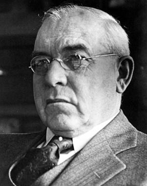
Arthur William Meyer (1873 – 1966)
Department of Medicine Stanford University.
Stanford University first full-time appointee to the faculty of the School of Medicine (1909), Professor of Anatomy and executive head of the Department until his retirement in 1938.
- "Arthur William Meyer was born August 18, 1873, near Cedarburg, Wisconsin, attended local schools, and graduated from the University of Wisconsin in 1898. After a period of teaching, he entered the Johns Hopkins University Medical School, from which he received the M.D. degree in 1905. He taught for two years at Johns Hopkins and for one year each at the University of Minnesota and Northwestern University before coming to Stanford. He was the author of "An Analysis of the Exercitationes de Generatione Animalium of William Harvey," 1936, of the "Rise of Embryology," 1939, and of many articles dealing with anatomy and embryology. He was a member of and an active participant in the activities of a number of scientific societies. The Stanford Anatomy Museum, thanks to his efforts, possesses one of the most nearly complete collections of human bones, both normal and pathological, available for study."
(excerpt from Memorial Resolution Stanford Historical Society)
As well as an Anatomist and Embryologist, Arthur Meyer was also a keen medical historian for embryology. Firstly, in a 1932 series of brief essays, then in 1936 a series on the Hunters and finally in 1939 in a textbook "The Rise of Embryology".
Meyer AW. 1932 - Essays on the History of Embryology: Part I | Part II | Part III | Part IV | Part V | Part VI | Part VII | Part VIII | Part IX | Part X | Part XI | Arthur Meyer | Historic Embryology Papers
| Embryologists: William Hunter | Wilhelm Roux | Caspar Wolff | Wilhelm His | Oscar Hertwig | Julius Kollmann | Hans Spemann | Francis Balfour | Charles Minot | Ambrosius Hubrecht | Charles Bardeen | Franz Keibel | Franklin Mall | Florence Sabin | George Streeter | George Corner | James Hill | Jan Florian | Thomas Bryce | Thomas Morgan | Ernest Frazer | Francisco Orts-Llorca | José Doménech Mateu | Frederic Lewis | Arthur Meyer | Robert Meyer | Erich Blechschmidt | Klaus Hinrichsen | Hideo Nishimura | Arthur Hertig | John Rock | Viktor Hamburger | Mary Lyon | Nicole Le Douarin | Robert Winston | Fabiola Müller | Ronan O'Rahilly | Robert Edwards | John Gurdon | Shinya Yamanaka | Embryology History | Category:People | ||
|
The Rise of Embryology
By Arthur William Meyer. Stanford University Press, 1939. xv —|— 367 pages, 97 figures. Price, $6.00.
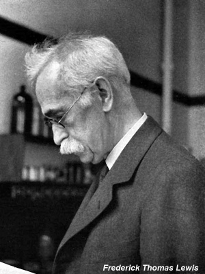
Book Review - by Frederic Lewis
A nation-wide interest in medical history is a recent development in America. Corner’s Anatomical Texts of the Earlier Middle Ages, Sarton’s History of Science, Breasted ’s Smith Papyrus, McMurrich’s Leonardo da Vinci, Fulton’s Readings in the History of Physiology, Boyden’s Talmudic Anatomy, are products of the last 15 years. Professor A. W. Meyer, Whose “Analysis of Harvey’s De Generatione” has already won him a place in this distinguished coterie, dedicates his new volume “in due humility to the nascent but transcendent cause of the history of science.”
“The Rise of Embryology” is concerned almost. wholly with events preceding the epochal discovery of von Baer—that “accidental” emergence of an egg from the (-}raafia.n follicle of a dog, reported in 1827. Chapter I sketches the ignorance of primitive peoples in regard to sex, with photographs of Australian aboriginees and African pygmies. Out of such depravity the science of embryology arose. “Early historic ideas of generation” follow. Here Dr. Meyer might have mentioned that Breasted considered possible as early as date as 3000 B.C. for the Egyptian reference to the “throbbing weak place of an infant ’s crown,” in the ancient description of meninges exposed in a compound fracture of the skull. The functions of ovary and testis, as Dr. Meyer reports, were sufficiently understood in China so that castration is mentioned as early as 1100 B0. In ancient China sows were spayed, and boars and cocks mutilated, for endocrine and other effects. In India, “before the sixth century B.C.,” viviparous and oviparous animals were distinguished from a third class produced from germs, yet such distinctions, if unknown to Adam, would have been told him by his boys.
With long prehistory, Greek medicine and science dawned, with a multiplicity of doctors, surgeons, naturalists and philosophers. Limiting the story to ten pages, Dr. Meyer can do little more than introduce the great names — Hippocrates, Aristotle, and Galen — with casual reference to certain others. Chapter II. concludes with embryology systematized by Galen. It comprises four stages of development, viz. (1) the formless geniture; (2) the embryo, of certain consistency, with heart, brain and liver still indistinct; (3) the fetus, with its major organs clearly present and some indications of the others; and (4) the infant with all its parts, growing until term.
Thus introduced, Dr. Meyer proceeds with a series of thirteen essays, or remaining chapters, on such fundamental topics as spontaneous generation, the search for the mammalian ovum, the problem of malformation, and the growth of morphology. Each essay begins with early speculations — with Plato, Aristotle, or a passage from Holy Writ. Not all the pioneers were on the right track. Strait is the gate and narrow is the way which leadeth unto truth, as unto life; and Dr. Meyer “steeped in the literature not only of generation but of anatomy before and during Harvey’s time,” tells of those who lost the trail quite as effectively as the climb of those who found it.
As to “Spontaneous Generation” (Ch. III), who knows whether it occurs? Only by forgetting that “we ourselves remain uncertain regarding the nature and origin of ultrascopic viruses” can we conclude that “Pasteur ’s experiments afforded a complete explanation,” and “finally disposed of the theory of spontaneous generation.” But Paracelsus held that all things were or could be generated by the help of putrefaction. He gave directions for making a true and living infant in a retort. On the title page of his Opera, in the vignette which Dr. Meyer shows enlarged, does not Minerva wink? She is shown above a three—line inscription, SCIENTIA IMMUTABILIS (Science unchanged), in which lurk more appropriate suggestions than Scientia immutabilis.
Allied to spontaneous generation, the doctrine of epigenesis (Ch. IV) teaches that the embryo develops from some amorphous generative substance in which the future organs are not specifically represented. The Psalmist (Ps. 139, v. 16) and Aristotle “espoused this fruitful idea,” later defended by Wolf, and “finally established” by Pander and von Baer, who respectively “sounded the doom” and “tolled the death knell” of the opposing doctrine of preformation. Preformation (Oh. V) means that the future organism is potentially and causally present in the germ — the future organs are already there. “No part of the animal body is created after another and all are created and appear at the same time,” wrote Haller, mentioning the second teeth and the stag ’s antlers. Malpighi, Swaminerdam, Réaumur, Bonnet, and Spallanzani were preforrnationists; and as Dr. Meyer further notes, “prominent biologists of the present day have held that the embryo is aft.er all, in a certain sense, preformed in the ovum.”
After the brief chapters (VI and VII) on pangenesis and panspermia, in which an opportunity to exploit the diverting views of Father Kircher on anthropomorphous orchids has been declined, Meyer writes fully and ably on the discovery of the ovum and spermatozoon (Chs. VIII and IX). He revives the excitement of the search and the fantastic suggestions of amateur microscopists who drew what they imagined and took each other seriously. The only modern pictures in the book are those here introduced, after Stieve, representing human spermatozoa, one showing a head with parallels of latitude and meridians of longitude, and microsomes at their points of intersection.
With the recognition of spermatozoa and ova, the problem of fertilization is seen in a new light, and the advance to Hert-wig’s superb demonstration, though well beyond the period set, is briefly noted (Ch. X). The short chapters on hybrids (Ch. XI, The Role of the “Mule”), and on monstrosities including maternal impressions (Ch. XII), revert to ancient lore.
As a digression or intermission (Ch. XIII) Dr. Meyer turns to the early history of the microscope, the invention of the microtome, and the introduction of embedding and staining methods, on which the advances about to be reported depend, as well as many of those already considered. In the preceding essays Dr. Meyer has dealt with the “leading older beliefs regarding reproduction and genesis, quoting at some length the words of the early scholars to reveal the intellectual atmosphere of their time, and carrying these ideas to the modern viewpoint” (Professor Torrey ’s comments). It remains to sketch the basic studies of organogenesis, carried out chiefly on the chick, which form the rather vast subject of chapter XIV.
Aldrovandi (1522-1605) was “the first biologist since Aristotle to open the eggs of hens regularly during their incubation” and record what is found therein. He publishes a large clear picture, drawn by a Dr. Ulm, of the ovary, pedunculated yolks, uterus and the shellcovered egg in situ—a worthy predecessor of figure 1 in Duval’s “Atlas d’Embryologie.” It is an error, therefore, to adopt Dr. Needham ’s statement concerning Aldrovandi that “unfortunately there is only one picture of embryological interest, namely, a chick in the act of hatching.” More precisely the figure to which Dr. Needham refers is to show the position of the chick in the egg just prior to hatching. Presumably it is reversed in printing: possibly it is one of the abnormal presentations, some of which make hatching impossible. Between the unlaid egg and the chick ready to hatch, there are no figures. Aldrovandi quotes what peasant girls say (a/imzt mulierculae) about the shedding of the protuberance of the beak in newly hatched chicks — a structure which he is the first to record. From the same source he learned that the chalazae are the sperm of the cock; and he believed it, contrary to Aristotle. “It seems very strange,” we are told, “that he should have decided that the thoracic organs are formed from the seed of the cock.” But the reader should not infer that Aldrovandi segregated the thoracic organs as having a special origin. He is said also “to have stated that the heart is formed on the yolk.” Like the rest of the chick, in his opinion, the heart formed in the fertilized white (aipparebat /in albumine), and the yolk is nutriment.
Prompted by Aldrovandi, with whom he was studying at Bologna, Coiter, in May of 1564, ordered two set.t.ing hens, with tvventy-three eggs apiece, and Wrote his better version of the daily disclosures. He could draw (skeletons in other papers), yet did not Venture to picture the chick.
Coiter is mentioned; and a paragraph follows about Spigelius, too brief for clarity. It assumes that the reader knows that Spigelius ljielieved that an allantoic sac occurs in man—though none exists. \Vitl1 the authorities at odds, Spigelius was in a quandary. “Yet I think it’s there,” he said, for there is just much need of it as in the embryos “of other quadrupeds.” To have the urine spread hither and thither between chorion and amnion does not seem consonant with nature. “Another reason is that my fellow-countryman, Andreas Vesalius, has pictured it in the human fetus, like the intestine from which sausages are made” (I Fabrica, Lib. V, cap. 17). Dr. Meyer adds, “It also is surprising that Spigelius seems to have offered no explanation for the presence of so peculiar a thing as the vernix caseosa, which he described.” His explanation is in Caput VIII of “De formato foetu. ” But Fabricius well and adequately presented, and for Harvey there is Dr. Meyer’s separate book, indispensable on that topic.
“Descartes deserves no place in the history of embryology.” In that field “it would be unnecessary to mention the renowned Frenchman . . . except . . . that he attempted to explain the development of all animals on a mechanical basis.” Dr. Meyer of course recognizes the fruitful mechanist conception, but finds not a whit of tangible evidence to support it in Descartes’ “Formation du Foetus.”
In contrast, Malpighi, with no words about the animal machine, described what takes place in the hen’s egg, sending his let.ters of revelation to the Royal Society. “The greatest of the microscopists,” — “not only one of medicine ’s greatest names, but one of its most attractive personalities” (Garrison). Dr. Meyer devotes two pages to Malpighi, supplemented by casual references scattered through the book. “The space allotted to an author does not necessarily correspond with his importance,” Dr. Meyer has warned in his Preface. Debraw, on bees, perhaps because less well-known, receives five pages. The discovery and crude portrayal of the origin of the brain and cord as a furrow in the skin would seem to need more than the statement, “he noticed the elevation of the medullary folds.” As to the heart, Singer is quoted that some of Malpighi’s figures “though inadequately described, are yet so good that they would fit a modern text-book Without much alteration.” The fascinating figures are, in fact, easily reproduced, and the Way in which Hunter ’s drawings interpret them has been briefly presented in Dr. Cushing’s “Bicentenary of the Birth of John Hunter” (N. E. J. of Med, 1929, vol. 200, p. 820).
Hunter, with eight pages, is more sympathetically and substantially treated, and the book closes, touching upon Pander, von Baer, Purkinje, Schvvann, and Koelliker. The brief final chapter (XV), on Early Experimental Embryology, is sufficient to show that Roux, in 1859, was not “the creator of experimental embryology” — nor was Claude Bernard, as Dr. Meyer might have added, “the founder of experimental medicine.” The experimental method is as old as mankind. It proceeds from “the dark unfathomed retrospect.”
“For what is the present after all but a growth out of the past‘? (As a projectile form’d, impell’d, passing a certain line, still keeps on, So the present, utterly form ’d, impell’d by the past.) ”
In postscript, some unusual features and errata may be noted. The volume is attractively printed in Bodoni type, spaced so as to be easily read. The running heads are off center, indented a half-inch from the outer margins of the pages, where in fact they are more readily seen on turning the leaves; but the page numbers must then be placed at the bottom of the page. The ninety-seven figures (half-tones and line cuts) are printed as plates, on both sides of glazed unnumbered leaves, scattered through the book either singly or in groups of six or eight pages of pictures. Very few of them are referred to in the text, for they t.ell their own story. The running head, “The Rise of Embryology,” with black rules, is placed above all the pages of figures. often in the margin of a portrait, in a way not altogether agreeable; and the plates are all brought to a larger size than the letter-press, often opposite. Thus there are several innovations.
The plates range widely in quality. Some are excellent, others badly made and badly printed, occasionally too pale, but more often overinked. Such, for example, are Redi’s cherry flies (fig. 13); Frannhofer’s drum microscope (fig. 66) ; and the execrable bedraggled fowl (fig. 77) which blacken Ledermiiller’s poor copy of l\Ialpighi’s pictures, the final chick, out on the farm, being his own addition. Figure 91, Pander’s beautiful plate, is upside down and ascribed to von Baer.
The portraits range from the two ancient busts so familiarly called Hippocrates and Aristotle that we mistake them for authentic likenesses (compare Sarton, in Isis, 1939, XXX, p. 226), down to the bewigged Redi, from the Opere, 1809, “through the courtesy of Professor Evans.” Dr. Meyer considers the latter a better likeness than the “magnificent” folio portrait by Tempesti, issued with Redi.’s Sonetti of 1702. The Redi and four plates from Stirling’s “Apostles” have been needlessly enlarged, so that their texture is coarsened. The nineteen portraits include but one unfamiliar face — that of Abbé Nollet (1700-1770). He made taffety breeches for some male frogs in a patient study of their mating.
There are some typographical errors, such as primae concepta (p. 101) for primi conceptus; testis muliebre (104) for testis muliebris; testes muliebre (pp. 100, 104, 105) for testes muliebres; osteogenia foetum (297) for osteogenia foetuum; animalculi (146) for a11imalcula; epididymes (136, in a quotation) for epididymides. One is reminded of the impassioned appeal to an audience of Boston doctors, made by one of their number: “‘We must educate,” he exclaimed, “we must educate the gcmos medicus.”’ 1 In the Haighto-I1 quotation (p. 123) inadequate has been substituted for adequate; in the first line of p. 214, height should read eighth.
“Restriction and selection of material were unavoidable,’ yet the valuable bibliography extends over 19 pages. It contains no reference to Aldrovandi, nor to a book especially relevant to chapter I - M. F. Ashley-Montague, “Coming into Being among the Australian Aboriginees: An examination of all the evidence bearing upon the procreative beliefs of the native tribes of Australia,” London, 1937. The barnacle story in chapter 111 is told without reference to HeronAllen’s book, “Barnacles in Nature and in Myth,” Oxford, 1928, on which the author “wasted most pleasantly a year” (but his work was soon out of print).
In company with Needham’s ‘ ‘ History of Embryology’ ’ and Singer ’s “Evolution of Anatomy,” Dr. Meyer ’s “Rise of Embryology” should be in every biological library and laboratory, for labora.tory studies Without such orientation can hardly yield members of a learned profession.
1 Correctly, 9'6’)?/££8 m-ed-i-cum. Many of us, perhaps, can join Professor Stieve in his remark (Folia Anat. Jap., 1937, XV, p. 152): Wiihrend der neun Jahrc, in denen ich meine Hosen auf den Schulbiinken eines humanistischen Gymnasiums blank scheucrte, habe ich mich ausfiihrlich genug init allen solchen Dingen beschfaiftigt.
Harvard Medical School
- Bodoni type - serif typefaces first designed by Giambattista Bodoni (1740–1813)
References
- Meyer AW. On the structure of the human umbilical vesicle. (1904) J. Anat. 3(2):155-166.
- Meyer AW. Retrogressive changes in the fetal vessels and the suspensory ligament of the liver. (1914) Amer. J Anat. 477-521.
- Meyer AW. The Debt of Human Embryology to the Practitioner. (1913) Cal State J Med. Dec;11(12):479-81. PMID 18736116
- Meyer AW. Retrogressive changes in the fetal vessels and the suspensory ligament of the liver. (1914) Amer. J Anat. 477-521.
- Meyer AW. On the nature, occurrence, and identity of the plasma cells of Hofbauer. (1918) J Morphol. 32(1): 327-350.
- Meyer AW. and Wynne HMN. Some aspects of ovarian pregnancy - with report of a case. (1919) Johns Hopkins Hospital Bulletin 30:
- Mall FP. and Meyer AW. Studies on abortuses: a survey of pathologic ova in the Carnegie Embryological Collection. (1921) Contrib. Embryol., Carnegie Inst. Wash. Publ. 275, 12: 1-364.
- Meyer, AW. The rise of embryology. Stanford University Press, (1939). xv + 367 pages, 97 figures.
- Meyer, AW. The elusive human allantois in older literature. In Science, Medicine and History. (1953) Oxford University Press, Oxford, pp. 510-520.
Search PubMed: Meyer AW
Essays on the History of Embryology
- Meyer AW. Essays on the History of Embryology: Part I. Cal West Med. 1931 Dec;35(6):447-51. PMID 18741978
- Meyer AW. Essays on the History of Embryology: Part II. Cal West Med. 1932 Jan;36(1):40-4. PMID 18742009
- Meyer AW. Essays on the History of Embryology: Part III. Cal West Med. 1932 Feb;36(2):105-9. PMID 18742030
- Meyer AW. Essays on the History of Embryology: Part IV. Cal West Med. 1932 Mar;36(3):176-80. PMID 18742069
- Meyer AW. Essays on the History of Embryology: Part V. Cal West Med. 1932 Apr;36(4):241-4. PMID 18742100
- Meyer AW. Essays on the History of Embryology: Part VI. Cal West Med. 1932 May;36(5):341-3. PMID 18742132
- Meyer AW. Essays on the History of Embryology: Part VII. Cal West Med. 1932 Jun;36(6):394-7. PMID 18742154
- Meyer AW. Essays on the History of Embryology: Part VIII. Cal West Med. 1932 Jul;37(1):41-4. PMID 18742183
- Meyer AW. Essays on the History of Embryology: Part IX. Cal West Med. 1932 Aug;37(2):111-5. PMID 18742212
- Meyer AW. Essays on the History of Embryology: Part X. Cal West Med. 1932 Sep;37(3):184-7. PMID 18742252
- Meyer AW. Essays on the History of Embryology: Part XI. Cal West Med. 1932 Oct;37(4):243-6. PMID 18742279
Mr. John Hunter on Generation
- Meyer AW. Mr. John Hunter on Generation : Part I. Cal West Med. 1935 Aug;43(2):145-50. PMID 18743345
- Meyer AW. Mr. John Hunter on Generation: Part II. Cal West Med. 1935 Sep;43(3):222-6. PMID 18743376
- Meyer AW. Mr. John Hunter on Generation: Part III. Cal West Med. 1935 Oct;43(4):283-8. PMID 18743402
- Meyer AW. Mr. John Hunter on Generation : Part IV. Cal West Med. 1935 Nov;43(5):358-63. PMID 18743437
The Hunters in Embryology
- Meyer AW. The Hunters in Embryology: Part I. Cal West Med. 1936 Nov;45(5):420-2. PMID 18743862
- Meyer AW. The Hunters in Embryology: Part II. Cal West Med. 1936 Dec;45(6):492-5. PMID 18743892
- Meyer AW. The Hunters in Embryology: Part III. Cal West Med. 1937 Jan;46(1):38-40. PMID 18743916
- Links: William Hunter
Cite this page: Hill, M.A. (2026, July 30) Embryology Embryology History - Arthur Meyer. Retrieved from https://embryology.med.unsw.edu.au/embryology/index.php/Embryology_History_-_Arthur_Meyer
- © Dr Mark Hill 2026, UNSW Embryology ISBN: 978 0 7334 2609 4 - UNSW CRICOS Provider Code No. 00098G