Bat Development
| Embryology - 10 Jul 2026 |
|---|
| Google Translate - select your language from the list shown below (this will open a new external page) |
|
العربية | català | 中文 | 中國傳統的 | français | Deutsche | עִברִית | हिंदी | bahasa Indonesia | italiano | 日本語 | 한국어 | မြန်မာ | Pilipino | Polskie | português | ਪੰਜਾਬੀ ਦੇ | Română | русский | Español | Swahili | Svensk | ไทย | Türkçe | اردو | ייִדיש | Tiếng Việt These external translations are automated and may not be accurate. (More? About Translations) |
Introduction
The bat (chiroptera) family consists of about 1,000 species throughout the world today (90 in Australia) and is not a common model of mammalian embryonic development.
The taxon chiroptera can also be further divided into the Megachiroptera (flying foxes) and Microchiroptera suborders. Echolocation sounds have been shown to differ in Microchiroptera (vocal cords) and Megachiroptera (tongue clicks).
| Bat: Hendra Virus | Category:Bat | ||
| Bat Images: Craniofacial Development Carollia perspicillata Stage 10-13 | Stage 12-17 | Stage 18-23 | Miniopterus schreibersii fuliginosus Stage 13-17 | Limb Stage 13-17 | Limb Growth Stage 13-17 | Stage 18-23 | Hipposideros pratti Stage 11-22 | ||
|
| Animal Development: axolotl | bat | cat | chicken | cow | dog | dolphin | echidna | fly | frog | goat | grasshopper | guinea pig | hamster | horse | kangaroo | koala | lizard | medaka | mouse | opossum | pig | platypus | rabbit | rat | salamander | sea squirt | sea urchin | sheep | worm | zebrafish | life cycles | development timetable | development models | K12 |
Some Recent Findings
|
| More recent papers |
|---|
 This table allows an automated computer search of the external PubMed database using the listed "Search term" text link.
More? References | Discussion Page | Journal Searches | 2019 References | 2020 References Search term: Fruit Bat Embryology | Bat Embryology | Bat Development |
| Older papers |
|---|
Template:Older paper
|
Taxon

Chiroptera
Genbank common name: bats
Taxonomy Id: 9397 Rank: order
Genetic code: Translation table 1 (Standard)
Mitochondrial genetic code: Translation table 2 (Vertebrate Mitochondrial)
Lineage( abbreviated ): Eukaryota; Fungi/Metazoa group; Metazoa; Eumetazoa; Bilateria; Coelomata; Deuterostomia; Chordata; Craniata; Vertebrata; Gnathostomata; Teleostomi; Euteleostomi; Sarcopterygii; Tetrapoda; Amniota; Mammalia; Theria; Eutheria; Laurasiatheria
Species Comparison
Carollia perspicillata
Myotis thysanodes and M. lucifugus
|
<html5media height="220" width="270">File:Bat embryo stage 19.mp4</html5media> Bat embryo (stage 19) |
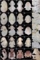
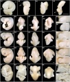
Embryonic Stages - Carollia perspicillata
stage 10 to 13
stage 12 to 17
stage 18 to 24


|
Stage |
Key features |
Somites |
Age |
Uterus diameter |
Crown-rump length |
Mass |
|
12 |
Forelimb buds form; tail bud forms; caudal neuropore closes; 3 pharyngeal arches. |
21-29 |
40 |
5.75 |
3.4 |
4.3 |
|
14 |
Retinal pigment; nasal pits; end of somitogenesis; propatagium and plagiopatagium primordia; hindlimb AER. |
36-40 |
44 |
6.95 |
5.35 |
24.6 |
|
15 |
Hand plate and footplate form; lens vesicle; auditory hillocks; premaxillary centers. |
46 |
8.65 |
7.45 |
56 | |
|
16 |
Nose-leaf primordium; pinna and tragus form; forelimb digital condensations, uropatagium primordium. |
50 |
12.06 |
8.66 |
110 | |
|
17 |
Tongue protruding; cervical flexure straightens; hindlimb interdigit tissue receding; eyes begin to close. |
54 |
13.45 |
9.15 |
114 | |
|
18 |
Free thumb; head and body smoother, rounder; eyes half-closed; postaxial flexure at wrist; calcar. |
60 |
16.32 |
12.35 |
278 | |
|
20 |
Distal forelimbs overlap over face; head larger; eyelids cover pigmented retina; claw primordia form. |
70 |
20.0 |
16.35 |
617 | |
|
22 |
Prominent, triangular nose-leaf; eyelids reopening; wing membranes corrugated; claws pigmented, hooked. |
80 |
23.03 |
20.02 |
1527 | |
|
24 |
Fetal period commences; eyes completely open; face and nose-leaf pigmenting. |
90 |
23.53 |
21.13 |
2097 | |
| Values are mean n= 2-6, +/- standard deviation, original table contains more detailed data. | ||||||
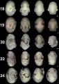
Embryonic Stages
Miniopterus schreibersii fuliginosus Stages 13-17
Embryonic stages 18-23
H. pratti Embryonic stages 11-22
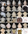
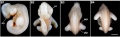
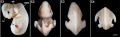
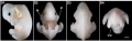
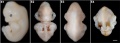
Craniofacial development


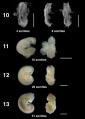
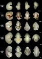
Miniopterus schreibersii fuliginosus
Stages 13-17
Stage 13
Stage 14
Stage 15
Stage 16
Stage 17
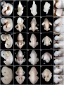
Limb Development


Images of the bat embryo Miniopterus schreibersii fuliginosus at embryonic Stages 13-17.[7]

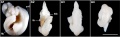
Bat - adult and fetal limbs[6]
A - Left limbs of adult Myotis ricketti. DI, DII, DIII, DIV and DV represent digits I-V of the forelimb
B, C - Left limbs of Miniopterus schreibersii fuliginosus in the Fetal Stage as an example of samples used for the Myotis ricketti libraries. Libraries Hand DI and Hand DII-V are constructed from forelimb digit I and digits II-V, respectively. Library Foot is constructed from hindlimb digits I-V.
Bar = 1 cm in A; bar = 1 mm in B and C.
Neural Development

The short-tailed fruit bat Carollia perspicillata Stage 14 embryo nervous system as identified by neurofilament antibody (brown) staining. Neurofilament is an intermediate filament protein, forming part of the neuronal cytoskeleton.
Historic Images
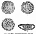
Fig. 55. Four stages in the development of the bat
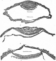
Fig. 59. Sections of blastodermic vesicle of bat


Abnormalities

Hendra Virus
- Hendra virus is a paramyxoviridae (ssRNA negative-strand virus) that mainly infects large fruit bats (flying foxes) which can be passed on to horses.
- The infection has occasionally been passed onto people who have been in close contact with an infected horse.
- There is evidence of fetal and placental infection in flying fox[8] and animal models.[9]
- There is currently insufficient information to determine whether there are developmental effects in humans.
- Links: NSW Public Health Sheet 2011 | Viralzone - Paramyxoviridae | Genome | Abnormal Development - Viral Infection
Rabies Virus
Rabies is a fatal encephalitis that can infect humans and is caused by lyssaviruses. Lyssavirus circulation has emerged in Southeast Asian bats.[10]
References
- ↑ 1.0 1.1 Cretekos CJ, Weatherbee SD, Chen CH, Badwaik NK, Niswander L, Behringer RR & Rasweiler JJ. (2005). Embryonic staging system for the short-tailed fruit bat, Carollia perspicillata, a model organism for the mammalian order Chiroptera, based upon timed pregnancies in captive-bred animals. Dev. Dyn. , 233, 721-38. PMID: 15861401 DOI.
- ↑ Nogueira MR, Ventura A, da Veiga CCP, Monteiro LR, Pinheiro NL & Peracchi AL. (2017). Dicephalic Parapagus Conjoined Twins in a Large Fruit-eating Bat, Genus Artibeus (Chiroptera, Phyllostomidae). Anat Histol Embryol , 46, 319-324. PMID: 28621033 DOI.
- ↑ Rasweiler JJ, Badwaik NK & Mechineni KV. (2011). Ovulation, fertilization, and early embryonic development in the menstruating fruit bat, Carollia perspicillata. Anat Rec (Hoboken) , 294, 506-19. PMID: 21337714 DOI.
- ↑ Martínez-Cerdeño V, Camacho J, Ariza J, Rogers H, Horton-Sparks K, Kreutz A, Behringer R, Rasweiler JJ & Noctor SC. (2018). The Bat as a New Model of Cortical Development. Cereb. Cortex , 28, 3880-3893. PMID: 29136119 DOI.
- ↑ Chen J, Rossiter SJ, Flanders JR, Sun Y, Hua P, Miller-Butterworth C, Liu X, Rajan KE & Zhang S. (2010). Contrasting genetic structure in two co-distributed species of old world fruit bat. PLoS ONE , 5, e13903. PMID: 21085717 DOI.
- ↑ 6.0 6.1 Wang Z, Dong D, Ru B, Young RL, Han N, Guo T & Zhang S. (2010). Digital gene expression tag profiling of bat digits provides robust candidates contributing to wing formation. BMC Genomics , 11, 619. PMID: 21054883 DOI.
- ↑ Wang Z, Han N, Racey PA, Ru B & He G. (2010). A comparative study of prenatal development in Miniopterus schreibersii fuliginosus, Hipposideros armiger and H. pratti. BMC Dev. Biol. , 10, 10. PMID: 20092640 DOI.
- ↑ Plowright RK, Field HE, Smith C, Divljan A, Palmer C, Tabor G, Daszak P & Foley JE. (2008). Reproduction and nutritional stress are risk factors for Hendra virus infection in little red flying foxes (Pteropus scapulatus). Proc. Biol. Sci. , 275, 861-9. PMID: 18198149 DOI.
- ↑ Williamson MM, Hooper PT, Selleck PW, Westbury HA & Slocombe RF. (2000). Experimental hendra virus infectionin pregnant guinea-pigs and fruit Bats (Pteropus poliocephalus). J. Comp. Pathol. , 122, 201-7. PMID: 10684689 DOI.
- ↑ <pubmed>21738801</pubmed>| PLoS Negl Trop Dis.
Reviews
Adams RA. (2008). Morphogenesis in bat wings: linking development, evolution and ecology. Cells Tissues Organs (Print) , 187, 13-23. PMID: 18163246 DOI.
Sears KE. (2008). Molecular determinants of bat wing development. Cells Tissues Organs (Print) , 187, 6-12. PMID: 18160799 DOI.
Bernard RT & Cumming GS. (1997). African bats: evolution of reproductive patterns and delays. Q Rev Biol , 72, 253-74. PMID: 9293029
Rasweiler JJ. (1993). Pregnancy in chiroptera. J. Exp. Zool. , 266, 495-513. PMID: 8371094 DOI.
Articles
Martínez-Cerdeño V, Camacho J, Ariza J, Rogers H, Horton-Sparks K, Kreutz A, Behringer R, Rasweiler JJ & Noctor SC. (2018). The Bat as a New Model of Cortical Development. Cereb. Cortex , 28, 3880-3893. PMID: 29136119 DOI.
Sears KE. (2008). Molecular determinants of bat wing development. Cells Tissues Organs (Print) , 187, 6-12. PMID: 18160799 DOI.
Chen CH, Cretekos CJ, Rasweiler JJ & Behringer RR. (2005). Hoxd13 expression in the developing limbs of the short-tailed fruit bat, Carollia perspicillata. Evol. Dev. , 7, 130-41. PMID: 15733311 DOI.
Rasweiler JJ & Badwaik NK. (1996). Improved procedures for maintaining and breeding the short-tailed fruit bat (Carollia perspicillata) in a laboratory setting. Lab. Anim. , 30, 171-81. PMID: 8783180 DOI.
Search Pubmed: bat development | chiroptera development
Additional Images
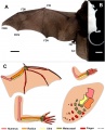
Image - Limb comparisons
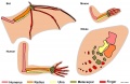
Image - Limb comparison cartoon
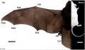
Image - Bat and Mouse limbs


Historic Images
| Historic Disclaimer - information about historic embryology pages |
|---|
|
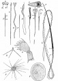
Fig. 5. Various types of spermatozoa
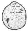
Fig. 9. Mature Ovum of Bat
Fig. 55. Four stages in the development of the bat
Fig. 59. Sections of blastodermic vesicle of bat
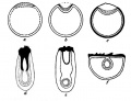
Fig. 5. Diagram to illustrate the condition of Entypy of the the Germinal Area


External Links
External Links Notice - The dynamic nature of the internet may mean that some of these listed links may no longer function. If the link no longer works search the web with the link text or name. Links to any external commercial sites are provided for information purposes only and should never be considered an endorsement. UNSW Embryology is provided as an educational resource with no clinical information or commercial affiliation.
- University of Michigan, Museum of Zoology Animal Diversity - Order Chiroptera
- The University of Texas, Department of Molecular Genetics Richard R. Behringer Lab
- Developmental Dynamics Poster of Bat Development PDF
- The University Queensland, Vision Touch and Hearing Research Centre Prof Jack Pettigrew |
Megabat | Molecular Phylogeny of Bats in Disarray
| Animal Development: axolotl | bat | cat | chicken | cow | dog | dolphin | echidna | fly | frog | goat | grasshopper | guinea pig | hamster | horse | kangaroo | koala | lizard | medaka | mouse | opossum | pig | platypus | rabbit | rat | salamander | sea squirt | sea urchin | sheep | worm | zebrafish | life cycles | development timetable | development models | K12 |
Glossary Links
- Glossary: A | B | C | D | E | F | G | H | I | J | K | L | M | N | O | P | Q | R | S | T | U | V | W | X | Y | Z | Numbers | Symbols | Term Link
Cite this page: Hill, M.A. (2026, July 10) Embryology Bat Development. Retrieved from https://embryology.med.unsw.edu.au/embryology/index.php/Bat_Development
- © Dr Mark Hill 2026, UNSW Embryology ISBN: 978 0 7334 2609 4 - UNSW CRICOS Provider Code No. 00098G