BGDB Gastrointestinal - Abnormalities

From Embryology
BGDsmall.jpg
Practical 1: Trilaminar Embryo | Early Embryo | Late Embryo | Fetal | Postnatal | Abnormalities | Lecture | Quiz


Cleft Lip and Cleft Palate

Cleft palate.jpgBilateral cleft palate.jpg

The concepts of cleft palate and cleft lip and palate will be covered in detail in the BGDB Face and Ear Development lecture/practical.

Atresia and Stenosis

The gastrointestinal tract can be considered as a simple tube or pipe, anything which blocks the tube (at different levels) can have different effects.

Gastrointestinal tract duplication sites based upon 78 clinical studies.[1]

There are two types of abnormalities that impact upon the continuity of the gastrointestinal tract lumen.

Atresia - interuption of the lumen (esophageal atresia, duodenal atresia, extrahepatic biliary atresia, anorectal atresia)

Stenosis - narrowing of the lumen (duodenal stenosis, pyloric stenosis)

Duplication - incomplete recanalization resulting in parallel lumens, this is really a specialized form of stenosis.

Classification of Oesophageal atresia.jpg Classification of Oesophageal Atresia
  • A Esophageal atresia (EA) without tracheoesophageal fistula (TEF)
  • B Proximal TEF with distal EA
  • C Distal TEF with proximal EA
  • D Proximal and distal TEF
  • E TEF without EA or “H”-type TEF

fistula - abnormal connection between either hollow or tubular organs.

Persistent Vitelline Duct

Meckel's diverticulum 01.jpg Endoderm cartoon.jpg


Links: Meckel's Diverticulum | Meckel's Image 1 | Meckel's Image 2 | Meckel's Image 3

Abnormal Gut Rotation

Intestinal malrotation.jpg Presents clinically in symptomatic malrotation as:

Neonates - bilious vomiting and bloody stools.


Newborn - bilious vomiting and failure to thrive.


Infants - recurrent abdominal pain, intestinal obstruction, malabsorption/diarrhea, peritonitis/septic shock, solid food intolerance, common bile duct obstruction, abdominal distention, and failure to thrive.


Ladd's Bands - are a series of bands crossing the duodenum which can cause duodenal obstruction.


Links: Midgut Volvulus | Situs Inversus | Gastrointestinal Tract - Abnormalities

Organ Abnormalities

Extrahepatic Biliary Atresia, Accessory Pancreatic Tissue, Anular Pancreas, Accessory Spleen

Motility Disorders

Aganglionic colon (Hirschprung's disease) - abnormalities of neural crest migration.


Links: Aganglionic colon | Enteric Nervous System | Neural Crest Development

Herniation Abnormalities

  • omphalocele - herniation of the bowel, liver and other organs into the intact umbilical cord, the tissues being covered by membranes unless the latter are ruptured
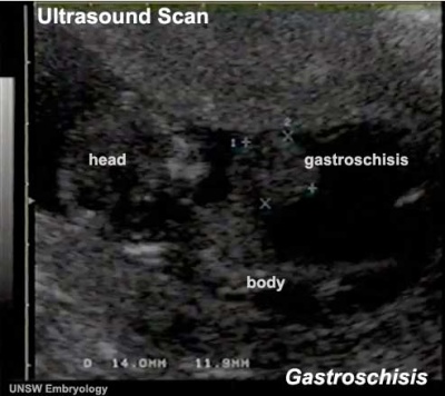
  • gastroschisis - intact umbilical cord and evisceration of the bowel through a defect in the abdominal wall, generally to the right of the cord, with no membrane covering

Omphalocele

Omphalocele ruptured during birth exposing liver and small intestine.[2]

Abdominal wall herniation abnormality occurs in both omphalocele and gastroschisis. Omphalocele involves "covered by membranes" and a lack of normal return of the bowel to the abdominal cavity and has a different position relative to the umbilical cord. The origin differs, as this is a failure of midgut loops to return to the body cavity after initial herniation into the umbilical cord during week 6 - 10.

Gastroschisis

Gastroschisis 01.jpg

Gastroschisis is a developmental abnormality occurs due to an abdominal wall defect, that allows the evisceration of the intestine.


Clefting

Bilateral cleft palate

Cleft lip and palate can affect postnatal nutrition, due to the inability of the infant to form a liquid seal on the breast during feeding.


Note - this topic will be covered in detail in the BGDB Practical - Face and Ear Development practical.


Links: Palate Development | Cleft Palate Foundation - feeding your baby

Cleft Lip

An abnormality of face development leading to an opening in the upper lip. Due to failure during the embryonic period of maxillary process fusion with the frontonasal prominence. Clefting of the lip and or palate occurs with 300+ different abnormalities. Depending on many factors, this cleft may extend further into the oral cavity leading to a cleft palate. In most cases clefting of the lip and palate can be repaired by surgery.

Cleft Palate

An abnormality of face development leading to an opening in the palate, the roof of the oral cavity between the mouth and the nose. If it occurs alone, due to failure during the early fetal period of palatal shelves. Clefting of the lip and or palate occurs with 300+ different abnormalities. In most cases clefting of the lip and palate can be repaired by surgery.

Polyhydramnios

Fetal polyhydramnios MRI-01.jpg Polyhydramnios (hydramnios, amniotic fluid disorder) refers to abnormally high amniotic fluid levels.


Fetal Causes
Decreased Fluid Swallowing or Absorption Increased Fluid Production
  • Lung disorders
  • Multiple pregnancy
  • Hydrops fetalis
Maternal Causes - Poorly controlled diabetes.

Meconium Peritonitis

A condition caused by intra-uterine intestinal perforation leading to a sterile inflammatory reaction of the peritoneum.

Metabolic Disorders

Guthrie test icon.jpg

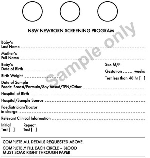
Inborn Errors of Metabolism (IEMs) are caused by mutations in the specific genes that lead to impaired proteins or enzymes production and are usually detected by the neonatal Guthrie test.

  • phenylketonuria (PKU) - 1 in 10,000 live births (about 10 babies per year). PKU causes high blood levels of phenylalanine and severe intellectual disability. A diet low in phenylalanine, started in the first two to three weeks results in normal development.
  • galactosaemia - 1 in 40,000 births (about 1-3 cases per year), incidence rate is different for other groups. Babies cannot process galactose, a component of lactose, (enzyme galactose-1-phosphate uridyl transferase) metabolizes galactose in milk sugar. Life-threatening liver failure and infections can occur. A galactose-free diet instituted in the first week is life saving.
  • Rarer metabolic disorders - Some fatty acid, organic acid and other amino acid defects can now be detected using Tandem Mass Spectrometry. These much rarer metabolic disorders affect about 15 – 18 babies per year. Early detection is important as diet and medications can treat most of these disorders. Without appropriate management they can cause severe disability or death.


Links: Guthrie test | neonatal diagnosis | milk | MedlinePlus - Galactosemia


Gastrointestinal Tract Terms  
  • allantois - An extraembryonic membrane, endoderm in origin extension from the early hindgut, then cloaca into the connecting stalk of placental animals, connected to the superior end of developing bladder. In reptiles and birds, acts as a reservoir for wastes and mediates gas exchange. In mammals is associated/incorporated with connecting stalk/placental cord fetal-maternal interface.
  • amnion - An extra-embryonic membrane, ectoderm and extraembryonic mesoderm in origin, also forms the innermost fetal membrane, that produces amniotic fluid. This fluid-filled sac initially lies above the trilaminar embryonic disc and with embryoic disc folding this sac is drawn ventrally to enclose (cover) the entire embryo, then fetus. The presence of this membrane led to the description of reptiles, bird, and mammals as amniotes.
  • amniotic fluid - The fluid that fills amniotic cavity totally encloses and cushions the embryo. Amniotic fluid enters both the gastrointestinal and respiratory tract following rupture of the buccopharyngeal membrane. The late fetus swallows amniotic fluid.
  • atresia - is an abnormal interruption of the tube lumen, the abnormality naming is based upon the anatomical location.
  • buccal - (Latin, bucca = cheek) A term used to relate to the mouth (oral cavity).
  • bile salts - Liver synthesized compounds derived from cholesterol that function postnatally in the small intestine to solubilize and absorb lipids, vitamins, and proteins. These compounds act as water-soluble amphipathic detergents. liver
  • buccopharyngeal membrane - (oral membrane) (Latin, bucca = cheek) A membrane which forms the external upper membrane limit (cranial end) of the early gastrointestinal tract. This membrane develops during gastrulation by ectoderm and endoderm without a middle (intervening) layer of mesoderm. The membrane lies at the floor of the ventral depression (stomodeum) where the oral cavity will open and will breakdown to form the initial "oral opening" of the gastrointestinal tract. The equivilent membrane at the lower end of the gastrointestinal tract is the cloacal membrane.
  • celiac artery - (celiac trunk) main blood supply to the foregut, excluding the pharynx, lower respiratory tract, and most of the oesophagus.
  • cholangiocytes - epithelial cells that line the intra- and extrahepatic ducts of the biliary tree. These cells modify the hepatocyte-derived bile, and are regulated by hormones, peptides, nucleotides, neurotransmitters, and other molecules. liver
  • cloaca - (cloacal cavity) The term describing the common cavity into which the intestinal, genital, and urinary tracts open in vertebrates. Located at the caudal end of the embryo it is located on the surface by the cloacal membrane. In many species this common cavity is later divided into a ventral urogenital region (urogenital sinus) and a dorsal gastrointestinal (rectal) region.
  • cloacal membrane - Forms the external lower membrane limit (caudal end) of the early gastrointestinal tract (GIT). This membrane is formed during gastrulation by ectoderm and endoderm without a middle (intervening) layer of mesoderm. The membrane breaks down to form the initial "anal opening" of the gastrointestinal tract.
  • coelomic cavity - (coelom) Term used to describe a space. There are extra-embryonic and intra-embryonic coeloms that form during vertebrate development. The single intra-embryonic coelom forms the 3 major body cavities: pleural cavity, pericardial cavity and peritoneal cavity.
  • crypt of Lieberkühn - (intestinal gland, intestinal crypt) intestinal villi epithelia extend down into the lamina propria where they form crypts that are the source of epithelial stem cells and immune function.
  • duplication - is an abnormal incomplete tube recanalization resulting in parallel lumens, this is really a specialized form of stenosis. (More? Image - small intestine duplication)
  • esophageal - (oesophageal)
  • foregut - first embryonic division of gastrointestinal tract extending from the oral (buccopharyngeal) membrane and contributing oesophagus, stomach, duodenum (to bile duct opening), liver, biliary apparatus (hepatic ducts, gallbladder, and bile duct), and pancreas. The forgut blood supply is the celiac artery (trunk) excluding the pharynx, lower respiratory tract, and most of the oesophagus.
  • galactosemia - Metabolic abnormality where the simple sugar galactose (half of lactose, the sugar in milk) cannot be metabolised. People with galactosemia cannot tolerate any form of milk (human or animal). Detected by the Guthrie test.
  • gastric transposition - clinical term for postnatal surgery treatment for esophageal atresia involving esophageal replacement. Typically performed on neonates between day 1 to 4. (More? gastrointestinal abnormalities | PMID 28658159
  • gastrointestinal divisions - refers to the 3 embryonic divisions contributing the gastrointestinal tract: foregut, Midgut and hindgut.
  • gastrula - (Greek, gastrula = little stomach) A stage of an animal embryo in which the three germ layers (endoderm/mesoderm/ectoderm) have just formed. All of these germ layers have contributions to the gastrointestinal tract.
  • gastrulation - The process of differentiation forming a gastrula. Term means literally means "to form a gut" but is more in development, as this process converts the bilaminar embryo (epiblast/hypoblast) into the trilaminar embryo (endoderm/mesoderm/ectoderm) establishing the 3 germ layers that will form all the future tissues of the entire embryo. This process also establishes the the initial body axes. (More? gastrulation)
  • Guthrie test - (heel prick) A neonatal blood screening test developed by Dr Robert Guthrie (1916-95) for determining a range of metabolic disorders and infections in the neonate. (More? Guthrie test)
  • heterotaxia - (Greek heteros = different; taxis = arrangement) is the right/left transposition of thoracic and/or abdominal organs.
  • hindgut - final embryonic division of gastrointestinal tract extending to the cloacal membrane and contributing part of the transverse colon (left half to one third), descending colon, sigmoid colon, rectum, part of anal canal (superior), urinary epithelium (bladder and most urethra). The hindgut blood supply is the inferior mesenteric artery.
  • inferior mesenteric artery - main blood supply to the hindgut
  • intestine - (bowel) part of the gastrointestinal tract (GIT) lying between the stomach and anus where absorption of nutrients and water occur. This region is further divided anatomically and functionally into the small intestine or bowel (duodenum, jejunum and ileum) and large intestine or bowel (cecum and colon).
  • intestinal perforation - gastrointestinal abnormality identified in neonates can be due to necrotizing enterocolitis, Hirschsprung’s disease or meconium ileus.
  • intraembryonic coelom - The "horseshoe-shaped" space (cavity) that forms initially in the third week of development in the lateral plate mesoderm that will eventually form the 3 main body cavities: pericardial, pleural, peritoneal. The intraembryonic coelom communicates transiently with the extraembryonic coelom.
  • meconium ileus intestine obstruction within the ileum due to abnormal meconium properties.
  • mesentery - connects gastrointestinal tract to the posterior body wall and is a double layer of visceral peritoneum.
  • mesothelium - The mesoderm derived epithelial covering of coelomic organs and also line their cavities.
  • Midgut - middle embryonic division of gastrointestinal tract contributing the small intestine (including duodenum distal bile duct opening), cecum, appendix, ascending colon, and part of the transverse colon (right half to two thirds). The midgut blood supply is the superior mesenteric artery.
  • neuralation - The general term used to describe the early formation of the nervous system. It is often used to describe the early events of differentiation of the central ectoderm region to form the neural plate, then neural groove, then neural tube. The nervous system includes the central nervous system (brain and spinal cord) from the neural tube and the peripheral nervous system (peripheral sensory and sympathetic ganglia) from neural crest. In humans, early neuralation begins in week 3 and continues through week 4.
  • neural crest - region of cells at the edge of the neural plate that migrates throughout the embryo and contributes to many different tissues. In the gastrointestinal tract it contributes mainly the enteric nervous system within the wall of the gut responsible for peristalsis and secretion.
  • peritoneal stomata - the main openings forming the pathways for drainage of intra-peritoneal fluid from the peritoneal cavity into the lymphatic system.
  • pharynx - uppermost end of gastrointestinal and respiratory tract, in the embryo beginning at the buccopharyngeal membrane and forms a major arched cavity within the phrayngeal arches.
  • recanalization - describes the process of a hollow structure becoming solid, then becoming hollow again. For example, this process occurs during GIT, auditory and renal system development.
  • retroperitoneal - (retroperitoneum) is the anatomical space (sometimes a potential space) in the abdominal cavity behind (retro) the peritoneum. Developmentally parts of the GIT become secondarily retroperitoneal (part of duodenum, ascending and descending colon, pancreas)
  • somitogenesis The process of segmentation of the paraxial mesoderm within the trilaminar embryo body to form pairs of somites, or balls of mesoderm. A somite is added either side of the notochord (axial mesoderm) to form a somite pair. The segmentation does not occur in the head region, and begins cranially (head end) and extends caudally (tailward) adding a somite pair at regular time intervals. The process is sequential and therefore used to stage the age of many different species embryos based upon the number visible somite pairs. In humans, the first somite pair appears at day 20 and adds caudally at 1 somite pair/4 hours (mouse 1 pair/90 min) until on average 44 pairs eventually form.
  • splanchnic mesoderm - Gastrointestinal tract (endoderm) associated mesoderm formed by the separation of the lateral plate mesoderm into two separate components by a cavity, the intraembryonic coelom. Splanchnic mesoderm is the embryonic origin of the gastrointestinal tract connective tissue, smooth muscle, blood vessels and contribute to organ development (pancreas, spleen, liver). The intraembryonic coelom will form the three major body cavities including the space surrounding the gut, the peritoneal cavity. The other half of the lateral plate mesoderm (somatic mesoderm) is associated with the ectoderm of the body wall.
  • stomodeum - (stomadeum, stomatodeum) A ventral surface depression on the early embryo head surrounding the buccopharyngeal membrane, which lies at the floor of this depression. This surface depression lies between the maxillary and mandibular components of the first pharyngeal arch.
  • stenosis - abnormal a narrowing of the tube lumen, the abnormality naming is based upon the anatomical location.
  • superior mesenteric artery - main blood supply to the Midgut.
  • viscera - the internal organs in the main cavities of the body, especially those in the abdomen, for example the Template:Intestines.
  • visceral peritoneum - covers the external surfaces of the intestinal tract and organs within the peritoneum. The other component (parietal peritoneum) lines the abdominal and pelvic cavity walls.
  • yolk sac - An extraembryonic membrane which is endoderm origin and covered with extraembryonic mesoderm. Yolk sac lies outside the embryo connected initially by a yolk stalk to the midgut with which it is continuous with. The endodermal lining is continuous with the endoderm of the gastrointestinal tract. The extra-embryonic mesoderm differentiates to form both blood and blood vessels of the vitelline system. In reptiles and birds, the yolk sac has a function associated with nutrition. In mammals the yolk sac acts as a source of primordial germ cells and blood cells. Note that in early development (week 2) a structure called the "primitive yolk sac" forms from hypoblast, this is an entirely different structure.
  • yolk stalk - (vitelline duct, omphalomesenteric duct, Latin, vitellus = yolk of an egg) The endodermal connection between the midgut and the yolk sac. See vitelline duct.
Other Terms Lists  
Terms Lists: ART | Birth | Bone | Cardiovascular | Cell Division | Endocrine | Gastrointestinal | Genital | Genetic | Head | Hearing | Heart | Immune | Integumentary | Neonatal | Neural | Oocyte | Palate | Placenta | Radiation | Renal | Respiratory | Spermatozoa | Statistics | Tooth | Ultrasound | Vision | Historic | Drugs | Glossary

Additional Information

Additional Information - Content shown under this heading is not part of the material covered in this class. It is provided for those students who would like to know about some concepts or current research in topics related to the current class page.
Abnormalities and Development
  • How these abnormalities may be generated in development.
  • When they first occur.
  • How can we detect these abnormalites.
  • Do the abnormalites have a direct or indirect effect on the GIT.
  • How serious to the embryo, fetus, newborn, child and adult are these conditions.
  • What therapeutics are available for these conditions.
Ladd's band-01.jpg

Ladd's band endoscopic view in adult.[4]

Australian GIT Abnormalities (2002-2003)  
Oesophageal atresia/stenosis - (2.0 per 10,000 births) ICD-10 Q39.0–Q39.3
  • A congenital anomaly characterised by the absence of continuity or narrowing of the oesophagus, with or without tracheal fistula, including tracheoesophageal fistula with or without mention of atresia or stenosis of oesophagus.
    • More males (58.6%) than females were affected with this anomaly.
    • Women aged 40 years or older had the highest rate of affected pregnancies.
Small intestinal atresia/stenosis - (2.4 per 10,000 births) ICD-10 Q41.0-Q41.2
  • Complete or partial occlusion of the lumen of a segment of the small intestine. It can involve a single area or multiples areas of the duodenum, jejunum or ileum.
    • Half of the babies (49.2%) with small intestinal atresia or stenosis were born pre-term.
Anorectal atresia/stenosis - ( 3.1 per 10,000 births) ICD-10 Q42.0–Q42.3
  • A congenital anomaly characterised by absence of continuity of the anorectal canal or of communication between rectum and anus, or narrowing of anal canal, with or without fistula to neighbouring organs. It excludes mild stenosis which does not need correction, and ectopic anus.
Hirschsprung’s disease - (1.3 per 10,000 births) ICD-10 Q43.1
  • A condition characterised by partial or complete bowel obstruction resulting from absence of peristalsis in a segment of bowel due to an aganglionic section of the bowel.
    • More than two-thirds (66.7%) of the babies born with this anomaly were males.
    • Women aged 40 years or older had the highest rate of affected pregnancies.
Exomphalos - (Omphalocele) (2.1 per 10,000 births) ICD-10 Q79.2
  • A congenital anomaly characterised by herniation of abdominal contents through the umbilical insertion and covered by a membrane which may or may not be intact. The anomaly excludes gastroschisis, hypoplasia of abdominal muscles and skin covered umbilical hernia.
    • A significantly higher rate of births with exomphalos was seen in women who had multiple births.
Gastroschisis - (2.6 per 10,000 births) ICD-10 Q79.3
  • A congenital anomaly characterised by visceral herniation through a right side abdominal wall defect with an intact umbilical cord and not covered by a membrane. This anomaly excludes hypoplasia of abdominal muscles, skin covered umbilical hernia and exomphalos.
    • Similar proportions of males and females with this anomaly.
    • Half of the affected pregnant women delivered preterm.
Links: Gastrointestinal Tract - Abnormalities | ICD-10 GIT | Australian Statistics
Reference: Abeywardana S & Sullivan EA 2008. Congenital Anomalies in Australia 2002-2003. Birth anomalies series no. 3 Cat. no. PER 41. Sydney: AIHW National Perinatal Statistics Unit.


Australian Palate Abnormalities (2002-2003)  
Cleft lip with or without cleft palate (9.2 per 10,000 births) ICD-10 Q36.0, Q36.1, Q36.9, Q37.0–Q37.5, Q37.8, Q37.9
A congenital anomaly characterised by a partial or complete clefting of the upper lip, with or without clefting of the alveolar ridge or the hard palate. Excludes a midline cleft of the upper or lower lip and an oblique facial fissure (going towards the eye).
  • 17% of the affected pregnancies were terminated in early pregnancy or resulted in fetal deaths. Most of the fetal deaths or terminations of pregnancy (95%) had multiple abnormalities.
  • more commonly seen in males than in females.
  • babies born before 25 weeks of gestation, 150 per 10,000 births had this anomaly. Most babies (80.0%) were born at term with a birthweight of 2,500 grams or more.
  • Maternal age group was not associated with the anomaly.
  • Rates significantly higher among Indigenous women than non Indigenous women.
Cleft palate without cleft lip (8.1 per 10,000 births) ICD-10 Q35.0–Q35.9
A congenital anomaly characterised by a closure defect of the hard and/or soft palate behind the foramen incisivum without a cleft lip. This anomaly includes sub-mucous cleft palate, but excludes cleft palate with a cleft lip, a functional short palate and high narrow palate.
  • overall rate has increased to 9.1 when the rate was estimated using data from the four states that include TOP data. The reported number of fetal deaths or early terminations of pregnancy with this anomaly was small and these deaths or terminations could be due to other associated anomalies.
  • proportion of females with this anomaly was higher (56.9%) than males.
  • 52.7 per 10,000 babies born before 25 weeks of gestation.
  • 83.0% were born at term and most of the babies (82.7%) had a birthweight of 2,500 grams or more.
  • Women aged 40 years or older and women born in South Central America or the Caribbean region had the highest rates of affected births.
  • Multiple births had a significantly higher rate of affected babies than singleton births.
  • Rates did not differ significantly by Indigenous status or areas of residence.
Links: Palate Development | Head Development | Gastrointestinal Tract - Abnormalities | ICD-10 GIT | Australian Statistics
Reference: Abeywardana S & Sullivan EA 2008. Congenital Anomalies in Australia 2002-2003. Birth anomalies series no. 3 Cat. no. PER 41. Sydney: AIHW National Perinatal Statistics Unit.
ICD10 Other congenital malformations of the digestive system (Q38-Q45)  
XVII Congenital Malformations - Other congenital malformations of the digestive system (Q38-Q45)
Q38 Other congenital malformations of tongue, mouth and pharynx

Excl.: macrostomia (Q18.4) microstomia (Q18.5)

  • Q38.0 Congenital malformations of lips, not elsewhere classified Congenital: fistula of lip malformation of lip NOS Van der Woude's syndrome Excl.: cleft lip (Q36.-) cleft lip with cleft palate (Q37.-) macrocheilia (Q18.6) microcheilia (Q18.7)
  • Q38.1 Ankyloglossia Tongue tie
  • Q38.2 Macroglossia
  • Q38.3 Other congenital malformations of tongue Aglossia Bifid tongue Congenital: adhesion fissure malformation NOS of tongue Hypoglossia Hypoplasia of tongue Microglossia
  • Q38.4 Congenital malformations of salivary glands and ducts Absence Accessory Atresia (of) salivary gland or duct Congenital fistula of salivary gland
  • Q38.5 Congenital malformations of palate, not elsewhere classified Absence of uvula Congenital malformation of palate NOS High arched palate Excl.: cleft palate (Q35.-) cleft palate with cleft lip (Q37.-)
  • Q38.6 Other congenital malformations of mouth Congenital malformation of mouth NOS
  • Q38.7 Pharyngeal pouch Diverticulum of pharynx Excl.: pharyngeal pouch syndrome (D82.1)
  • Q38.8 Other congenital malformations of pharynx Congenital malformation of pharynx NOS
Q39 Congenital malformations of oesophagus
  • Q39.0 Atresia of oesophagus without fistula Atresia of oesophagus NOS
  • Q39.1 Atresia of oesophagus with tracheo-oesophageal fistula Atresia of oesophagus with broncho-oesophageal fistula
  • Q39.2 Congenital tracheo-oesophageal fistula without atresia Congenital tracheo-oesophageal fistula NOS
  • Q39.3 Congenital stenosis and stricture of oesophagus
  • Q39.4 Oesophageal web
  • Q39.5 Congenital dilatation of oesophagus
  • Q39.6 Diverticulum of oesophagus Oesophageal pouch
  • Q39.8 Other congenital malformations of oesophagus Absent Congenital displacement Duplication (of) oesophagus
  • Q39.9 Congenital malformation of oesophagus, unspecified
Q40 Other congenital malformations of upper alimentary tract
  • Q40.0 Congenital hypertrophic pyloric stenosis Congenital or infantile: constriction hypertrophy spasm stenosis stricture of pylorus
  • Q40.1 Congenital hiatus hernia Displacement of cardia through oesophageal hiatus Excl.: congenital diaphragmatic hernia (Q79.0)
  • Q40.2 Other specified congenital malformations of stomach Congenital: displacement of stomach diverticulum of stomach hourglass stomach Duplication of stomach Megalogastria Microgastria
  • Q40.3 Congenital malformation of stomach, unspecified
  • Q40.8 Other specified congenital malformations of upper alimentary tract
  • Q40.9 Congenital malformation of upper alimentary tract, unspecified Congenital: anomaly deformity NOS of upper alimentary tract
Q41 Congenital absence, atresia and stenosis of small intestine

Incl.: congenital obstruction, occlusion and stricture of small intestine or intestine NOS Excl.: meconium ileus (E84.1)

  • Q41.0 Congenital absence, atresia and stenosis of duodenum
  • Q41.1 Congenital absence, atresia and stenosis of jejunum Apple peel syndrome Imperforate jejunum
  • Q41.2 Congenital absence, atresia and stenosis of ileum
  • Q41.8 Congenital absence, atresia and stenosis of other specified parts of small intestine
  • Q41.9 Congenital absence, atresia and stenosis of small intestine, part unspecified Congenital absence, atresia and stenosis of intestine NOS
Q42 Congenital absence, atresia and stenosis of large intestine

Incl.: congenital obstruction, occlusion and stricture of large intestine

  • Q42.0 Congenital absence, atresia and stenosis of rectum with fistula
  • Q42.1 Congenital absence, atresia and stenosis of rectum without fistula Imperforate rectum
  • Q42.2 Congenital absence, atresia and stenosis of anus with fistula
  • Q42.3 Congenital absence, atresia and stenosis of anus without fistula Imperforate anus
  • Q42.8 Congenital absence, atresia and stenosis of other parts of large intestine
  • Q42.9 Congenital absence, atresia and stenosis of large intestine, part unspecified
Q43 Other congenital malformations of intestine
  • Q43.0 Meckel's diverticulum Persistent: omphalomesenteric duct vitelline duct
  • Q43.1 Hirschsprung's disease Aganglionosis Congenital (aganglionic) megacolon
  • Q43.2 Other congenital functional disorders of colon Congenital dilatation of colon
  • Q43.3 Congenital malformations of intestinal fixation Congenital adhesions [bands]: omental, anomalous peritoneal Jackson's membrane Malrotation of colon Rotation: failure of incomplete insufficient of caecum and colon Universal mesentery
  • Q43.4 Duplication of intestine
  • Q43.5 Ectopic anus
  • Q43.6 Congenital fistula of rectum and anus Excl.: congenital fistula: rectovaginal (Q52.2) urethrorectal (Q64.7) pilonidal fistula or sinus (L05.-) with absence, atresia and stenosis (Q42.0,Q42.2)
  • Q43.7 Persistent cloaca Cloaca NOS
  • Q43.8 Other specified congenital malformations of intestine Congenital: blind loop syndrome diverticulitis, colon diverticulum, intestine Dolichocolon Megaloappendix Megaloduodenum Microcolon Transposition of: appendix colon intestine
  • Q43.9 Congenital malformation of intestine, unspecified
Q44 Congenital malformations of gallbladder, bile ducts and liver
  • Q44.0 Agenesis, aplasia and hypoplasia of gallbladder Congenital absence of gallbladder
  • Q44.1 Other congenital malformations of gallbladder Congenital malformation of gallbladder NOS Intrahepatic gallbladder
  • Q44.2 Atresia of bile ducts
  • Q44.3 Congenital stenosis and stricture of bile ducts
  • Q44.4 Choledochal cyst
  • Q44.5 Other congenital malformations of bile ducts Accessory hepatic duct Congenital malformation of bile duct NOS Duplication: biliary duct cystic duct
  • Q44.6 Cystic disease of liver Fibrocystic disease of liver
  • Q44.7 Other congenital malformations of liver Accessory liver Alagille's syndrome Congenital: absence of liver hepatomegaly malformation of liver NOS
Q45 Other congenital malformations of digestive system

Excl.: congenital: diaphragmatic hernia (Q79.0) hiatus hernia (Q40.1)

  • Q45.0 Agenesis, aplasia and hypoplasia of pancreas Congenital absence of pancreas
  • Q45.1 Annular pancreas
  • Q45.2 Congenital pancreatic cyst
  • Q45.3 Other congenital malformations of pancreas and pancreatic duct Accessory pancreas Congenital malformation of pancreas or pancreatic duct NOS Excl.: diabetes mellitus: congenital (E10.-) neonatal (P70.2) fibrocystic disease of pancreas (E84.-)
  • Q45.8 Other specified congenital malformations of digestive system Absence (complete)(partial) of alimentary tract NOS Duplication Malposition, congenital of digestive organs NOS
  • Q45.9 Congenital malformation of digestive system, unspecified Congenital: anomaly deformity NOS of digestive system

World Health Organisation. International Statistical Classification of Diseases and Related Health Problems. (1992) 10th Revision (ICD-10). Geneva: WHO ICD-10 - 2016 Online (English)

Links: Gastrointestinal Abnormalities
GIT Links: Introduction | Medicine Lecture | Science Lecture | endoderm | mouth | oesophagus | stomach | liver | gallbladder | Pancreas | intestine | mesentery | tongue | taste | enteric nervous system | Stage 13 | Stage 22 | gastrointestinal abnormalities | Movies | Postnatal | milk | tooth | salivary gland | BGD Lecture | BGD Practical | GIT Terms | Category:Gastrointestinal Tract
GIT Histology Links: Upper GIT | Salivary Gland | Smooth Muscle Histology | Liver | Gallbladder | Pancreas | Colon | Histology Stains | Histology | GIT Development
Historic Embryology - Gastrointestinal Tract  
1878 Alimentary Canal | 1882 The Organs of the Inner Germ-Layer The Alimentary Tube with its Appended Organs | 1884 Great omentum and transverse mesocolon | 1902 Meckel's diverticulum | 1902 The Organs of Digestion | 1903 Submaxillary Gland | 1906 Liver | 1907 Development of the Digestive System | 1907 Atlas | 1907 23 Somite Embryo | 1908 Liver | 1908 Liver and Vascular | 1910 Mucous membrane Oesophagus to Small Intestine | 1910 Large intestine and Vermiform process | 1911-13 Intestine and Peritoneum - Part 1 | Part 2 | Part 3 | Part 5 | Part 6 | 1912 Digestive Tract | 1912 Stomach | 1914 Digestive Tract | 1914 Intestines | 1914 Rectum | 1915 Pharynx | 1915 Intestinal Rotation | 1917 Entodermal Canal | 1918 Anatomy | 1921 Alimentary Tube | 1932 Gall Bladder | 1939 Alimentary Canal Looping | 1940 Duodenum anomalies | 2008 Liver | 2016 GIT Notes | Historic Disclaimer
Human Embryo: 1908 13-14 Somite Embryo | 1921 Liver Suspensory Ligament | 1926 22 Somite Embryo | 1907 23 Somite Embryo | 1937 25 Somite Embryo | 1914 27 Somite Embryo | 1914 Week 7 Embryo
Animal Development: 1913 Chicken | 1951 Frog
ICD10 - Gastrointestinal | Genital | Renal | Integumentary

References

  1. Bower RJ, Sieber WK & Kiesewetter WB. (1978). Alimentary tract duplications in children. Ann. Surg. , 188, 669-74. PMID: 718292
  2. Akakpo-Numado GK, Gnassingbe K, Boume MA, Sakiye KA, Mihluedo-Agbolan K, Attipou K & Tekou H. (2012). Emergency treatment of a ruptured huge omphalocele by simple suture of its membrane. Ann Surg Innov Res , 6, 2. PMID: 22325297 DOI.
  3. Feldkamp ML, Carey JC & Sadler TW. (2007). Development of gastroschisis: review of hypotheses, a novel hypothesis, and implications for research. Am. J. Med. Genet. A , 143A, 639-52. PMID: 17230493 DOI.
  4. Panda N, Bansal NK, Narasimhan M & Ardhanari R. (2014). Laparoscopic correction of intestinal malrotation in adult. J Minim Access Surg , 10, 90-2. PMID: 24761085 DOI.


BGDsmall.jpg
Practical 1: Trilaminar Embryo | Early Embryo | Late Embryo | Fetal | Postnatal | Abnormalities | Lecture | Quiz


BGDsmall.jpg

BGDB: Lecture - Gastrointestinal System | Practical - Gastrointestinal System | Lecture - Face and Ear | Practical - Face and Ear | Lecture - Endocrine | Lecture - Sexual Differentiation | Practical - Sexual Differentiation | Tutorial


Glossary Links

Glossary: A | B | C | D | E | F | G | H | I | J | K | L | M | N | O | P | Q | R | S | T | U | V | W | X | Y | Z | Numbers | Symbols | Term Link


Cite this page: Hill, M.A. (2024, March 19) Embryology BGDB Gastrointestinal - Abnormalities. Retrieved from https://embryology.med.unsw.edu.au/embryology/index.php/BGDB_Gastrointestinal_-_Abnormalities

What Links Here?
© Dr Mark Hill 2024, UNSW Embryology ISBN: 978 0 7334 2609 4 - UNSW CRICOS Provider Code No. 00098G