BGDA Practical 3 - Week 1 Summary
| Practical 3: Oogenesis and Ovulation | Gametogenesis | Fertilization | Early Cell Division | Week 1 | Implantation | Week 2 | Extraembryonic Spaces | Gastrulation | Notochord | Week 3 |
Week 1
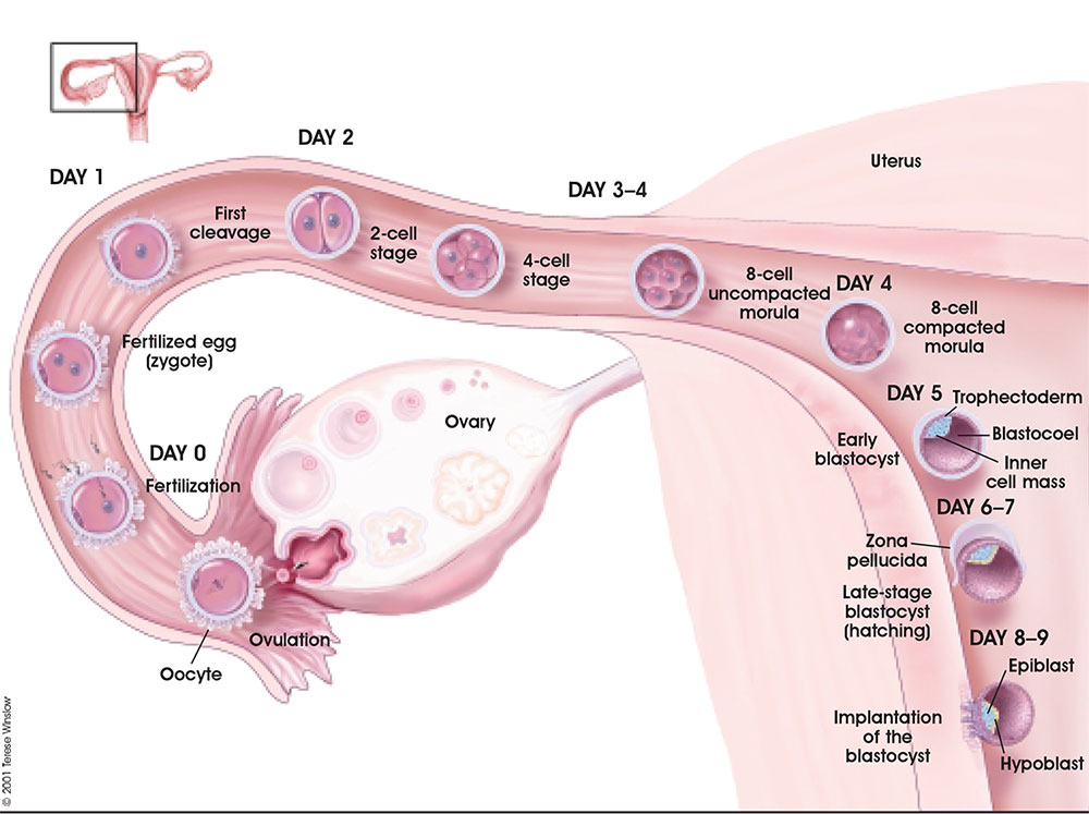
Overview of development of the 1-chambered conceptus (blastocyst)
- Day 0-1 - zygote, the first diploid cell.
- Day 2 - 2 then 4 blasomeres, the first cells formed by mitotic cell division.
- Day 3 - morula.
- Day 4 - compacting morula.
- Day 5 - blastocyst.
- Day 6 - hatching blastocyst from zona pellucida shell, after this event the blastocyst can now attach and implant into the uterine wall.
Ovulation is the initial release of oocyte.
Follicular fluid and fimbriae together aid oocyte movement into infundibulum then the ampulla of the uterine tube.
Sperm deposited in the vagina then enter the uterus, mature (capacitation), then actively migrate along the uterine tube.
Fertilization generally occurs in the ampulla region of the tube.
Following fertilization, repeated rounds of cell division occur without growth forming initially a solid ball of cells (morula), which cavitates to form the 1-chambered conceptus (blastocyst). Liberation of the blastocyst from the zona pellucida the allows attachment (adplantation) to the uterine wall.
Following ovulation the empty follicle within the ovary now forms the corpus luteum.
Note - the day timings shown above for the first week are approximate and may vary by several days for events following fertilization.
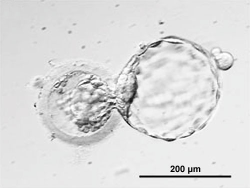
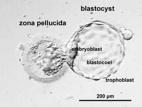
Blastocyst (right) hatching from zona pellucida (left)
Sex Determination
Mark Hill (talk) 16:51, 27 March 2018 (AEDT) These concepts will be covered later in detail with BGDB - Sexual Differentiation.
Mammalian sex determination is regulated by chromosomes.
- Females have two X chromosomes. (XX)
- Males have a single X and a small Y. (XY)
- The X and Y chromosome are morphologically and functionally different from each other.
- Evolutionary studies have shown that the Y was once the homologous pair for X.
- It is only in the last 5 years that we have some idea about how these two types of chromosomes may be regulated and genes of importance located upon them.
X chromosome
|
In females - the main scientific problem was understanding gene dosage, only one copy of X chromosome is needed to be genetically active the other copy is inactivated (More? X Inactivation. About the X Chromosome
|
Y chromosome
Monoygotic Twinning
Monoygotic twins (identical) produced from a single fertilization event (one fertilised egg and a single spermatazoa, form a single zygote), these twins therefore share the same genetic makeup. Occurs in approximately 3-5 per 1000 pregnancies, more commonly with aged mothers. The later the twinning event, the less common are initially separate placental membranes and finally resulting in conjoined twins.
| Week | Week 1 (GA week 3) | Week 2 (GA week 4) | |||||||||||||
| Day | 0 | 1 | 2 | 3 | 4 | 5 | 6 | 7 | 8 | 9 | 10 | 11 | 12 | 13 | 14 |
| Cell Number | 1 | 1 | 2 | 16 | 32 | 128 | bilaminar | ||||||||
| Event | Ovulation | Fertilization | First cell division | Morula | Early blastocyst | Late blastocyst
Hatching |
Implantation starts | X inactivation | |||||||

|

|

|

|
||||||||||||
| Monoygotic
Twin Type |
Diamniotic
Dichorionic |
Diamniotic
Monochorionic |
Monoamniotic
Monochorionic |
Conjoined | |||||||||||
Table based upon recent a recent twinning review.[1] Links: twinning
Glossary Links
- Glossary: A | B | C | D | E | F | G | H | I | J | K | L | M | N | O | P | Q | R | S | T | U | V | W | X | Y | Z | Numbers | Symbols | Term Link
| Practical 3: Oogenesis and Ovulation | Gametogenesis | Fertilization | Early Cell Division | Week 1 | Implantation | Week 2 | Extraembryonic Spaces | Gastrulation | Notochord | Week 3 |
Additional Information
| Additional Information - Content shown under this heading is not part of the material covered in this class. It is provided for those students who would like to know about some concepts or current research in topics related to the current class page. |
Terms
- adplantation - Initial adhesion of blastocyst (released from zona pellucida) to uterine wall. Adplantation is followed by implantation.
- ampulla - longest segment (approximately 2/3 of overall length) of uterine tube (oviduct or Fallopian tube). Medial segment forming the remainder of the tube is called the isthmus.
- antrum- (L. a cave), cavity; a nearly-closed cavity or bulge. In the ovary this refers to the follicular fluid-filled space within the follicle.
- blastocyst - the developmental stage following morula, as this stage matures, the zona pellucia is lost allowing the coceptus to adplant and then implant into the uterine wall.
- capacitation the process by which sperm become capable of fertilizing an egg, requires membrane changes, removal of surface glycoproteins and increased motility.
- cavitates - to form a space within a solid object.
- conceptus the product of conception, that is all the structures derived from the zygote. This includes not only the embryo, but also the placental and membrane components formed from the conceptus.
- fertilization The penetration of the egg by the sperm and the resulting combining of genetic material that develops into an embryo. The union of two haploid gametes to form a diploid cell or zygote.
- fimbriae (Latin, fimbria = a fringe) finger-like projections at the ovarian end of uterine tube. At ovulation they sit over the ovary to aid egg movement into the uterine tube.
- follicular fluid - the fluid found in the antrum of an antral follicle (secondary follicle). Secreted by cells in the wall of the follicle, this fluid is released along with the oocyte at ovulation.
- infundibulum - funnel-shaped initial segment of uterine tube (oviduct or Fallopian tube) opening into peritoneal cavity and connected to the ampulla. The peritoneal opening sitting over the ovary.
- morula (Latin, morula = mulberry) early stage in development (week 1) where the cells have divided to produce a solid mass of cells (12-15 cells) with a "mulberry" appearance. This stage is followed by formation of a cavity in the mass (blastocyst stage).
- oocyte - (egg or ovum) female germ cell.
- ovulation- release of the oocyte from the mature follicle.
- triploidy - in humans, three sets of 23 chromosomes instead of 2 (diploid) combine to form the embryo. This occurs mainly by fertilization of a single egg by two sperm and less frequently by a diploid egg or sperm. Most human triploids abort spontaneously, with very rare survival to term.
- tetraploidy - in humans, four sets of 23 chromosomes instead of 2 (diploid) due to a failure of the first mitotic division after fertilization, these fertilization events do not development.
- uterine tube - (also called oviduct or Fallopian tube) the laterally paired tubes that connect the ovary to the uterus. Is the site for oocyte fertilization and initial development of the conceptus.
- uterine wall - the site of normal blastocyst implantation.
- zona pellucida- glycoprotein shell that surrounds the oocyte through to blastula stage of development
- uterus- site of embryo implantation and development. Uterine wall has 3 layers; endometrium, myometrium, and perimetrium.
- zona pellucida- extracellular layer lying directly around the oocyte underneath follicular cells. Consists of glcosaminoglycans and glycoproteins (ZP1, ZP2, ZP3).
References
BGDA: Lecture 1 | Lecture 2 | Practical 3 | Practical 6 | Practical 12 | Lecture Neural | Practical 14 | Histology Support - Female | Male | Tutorial
Glossary Links
- Glossary: A | B | C | D | E | F | G | H | I | J | K | L | M | N | O | P | Q | R | S | T | U | V | W | X | Y | Z | Numbers | Symbols | Term Link
Cite this page: Hill, M.A. (2024, April 28) Embryology BGDA Practical 3 - Week 1 Summary. Retrieved from https://embryology.med.unsw.edu.au/embryology/index.php/BGDA_Practical_3_-_Week_1_Summary
- © Dr Mark Hill 2024, UNSW Embryology ISBN: 978 0 7334 2609 4 - UNSW CRICOS Provider Code No. 00098G


