ANAT2241 Connective Tissue Types

| ANAT2241 This practical support page content is not part of the virtual science practical class and provides additional information for student self-directed learning purposes. All practical class pages are located on Moodle - ANAT2241 |
General Objective
To recognise various components of connective tissue and understand their functions.
Specific Objectives
- To recognise the histological appearance of mesenchymal and mucoid connective tissue, and revise loose and dense fibrous connective tissue.
- To recognise adipose tissue (white and brown fat) and understand its functions.
- To recognise the histological appearance of the different forms of cartilage: hyaline, elastic and fibrocartilage.
- To understand the role of the perichondrium in the formation of cartilage, and the mechanisms of appositional and interstitial growth.
Learning Activities
Examine the following virtual slides. Identify, draw and label the main features and note their function.
Connective Tissue Types | Histology Drawings
Terms
- CT - connective tissue
- Dense CT - connective tissue mainly occupied by fibres (dermis of skin).
- Loose CT - connective tissue mainly cells, vessels and nerves. Generates a soft and compliant CT (mucous connective tissue, reticular connective tissue and adipose tissue).
- irregular CT - fibres do not show a clear orientation within the tissue and form a densely woven three-dimensional network.
- regular CT - fibres show a clear parallel orientation within the tissue.
Adipose Tissue


White Adipose


unilocular - single lipid drop in cytoplasm. Nucleus flattened on edge of cell.
Brown Adipose

Cartilage
| Developing cartilage | |
|---|---|

|

|
| Mesenchyme forming cartilage | Endochondral ossification |
- Mesenchyme - is an embryonic (undifferentiated) connective tissue
Cartilage Growth
|

isogenic group development |
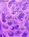
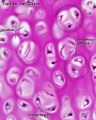
Hyaline
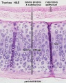
Hyaline cartilage HE
Hyaline cartilage VG
Hyaline cartilage HE
Hyaline cartilage VG


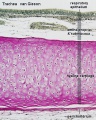
| Articular cartilage | ||
|---|---|---|
|

|

|
Elastic Cartilage
- occurs in tissues where "flexibility" required
- histologically similar to hyaline cartilage with the addition of a elastic fibres
- elastic fibres - dense network of delicately branched fibres, individual fibres are difficult observe
- Examples - epiglottic cartilage, larynx (corniculate and cuneiform cartilage), external ear and auditory tube


Elastic cartilage (elastin stain)
Fibrocartilage
- Transitional between dense connective tissue and hyaline cartilage
- merges into neighbouring tissues (tendons or articular hyaline cartilage)
- difficult identify the perichondrium
- Examples - joints (intra-articular lips, discs and menisci) and intervertebral discs.
Chondrocytes
- form short rows between dense bundles of collagen fibres
- lie singly or in pairs
- collagen type I


Tendon

|
Dense Regular Connective Tissue
Tissue Examples - tendons, ligaments, fasciae and aponeuroses of muscles. |
Electron Microscopy - Collagen
|
Collagen Synthesis
|
Collagen is the major extracellular matrix protein found in connective tissues.
|

- Cartilage Histology: Developing | Hyaline HE | Hyaline VG | Hyaline HE | Hyaline VG | Elastic 1 | Elastic 2 | Fibrous - articular disc | Fibrous - intervertebral disc | Articular 1 | Articular 2
Course Links
- Histology Glossary: A | B | C | D | E | F | G | H | I | J | K | L | M | N | O | P | Q | R | S | T | U | V | W | X | Y | Z | ANAT2241 Support | Histology | Histology Stains | Embryology Glossary
| Common Histology Stains | ||||||||||||||||||||||||||||||||||||||||||||||||||||||||||||||||||||||||||||||||||||||||||||||||||||||||||||||||||||||||||||||||||||||||||||||||
|---|---|---|---|---|---|---|---|---|---|---|---|---|---|---|---|---|---|---|---|---|---|---|---|---|---|---|---|---|---|---|---|---|---|---|---|---|---|---|---|---|---|---|---|---|---|---|---|---|---|---|---|---|---|---|---|---|---|---|---|---|---|---|---|---|---|---|---|---|---|---|---|---|---|---|---|---|---|---|---|---|---|---|---|---|---|---|---|---|---|---|---|---|---|---|---|---|---|---|---|---|---|---|---|---|---|---|---|---|---|---|---|---|---|---|---|---|---|---|---|---|---|---|---|---|---|---|---|---|---|---|---|---|---|---|---|---|---|---|---|---|---|---|---|---|
| ||||||||||||||||||||||||||||||||||||||||||||||||||||||||||||||||||||||||||||||||||||||||||||||||||||||||||||||||||||||||||||||||||||||||||||||||
| ||||||||||||||||||||||||||||||||||||||||||||||||||||||||||||||||||||||||||||||||||||||||||||||||||||||||||||||||||||||||||||||||||||||||||||||||
Practical Support
- Pages can be accessed from any internet connected computer.
ANAT2241 Support Links: The Virtual Microscope | Covering and Lining Epithelia | Glandular Epithelia | CT Components | CT Types | Bone, Bone Formation and Joints | Muscle | Nervous | Blood | Eye | Cardiovascular | Respiratory | Integumentary | Gastrointestinal | Gastrointestinal Organs | Lymphatic and Immune | Endocrine | Urinary | Female Reproductive | Male Reproductive | Histology Stains | Histology Drawings | Practicals Health and Safety 2013 | Moodle - 2019
ANAT2241 This practical support page content is not part of the science practical class and provides only background information for student self-directed learning purposes.
Cite this page: Hill, M.A. (2026, July 22) Embryology ANAT2241 Connective Tissue Types. Retrieved from https://embryology.med.unsw.edu.au/embryology/index.php/ANAT2241_Connective_Tissue_Types
- © Dr Mark Hill 2026, UNSW Embryology ISBN: 978 0 7334 2609 4 - UNSW CRICOS Provider Code No. 00098G