2010 Lecture 9
Endoderm Development
Introduction

This lecture will cover the early development of the endoderm layer of the trilaminar embryo as it contributes to the lining, glands and organs of the gastrointestinal tract (GIT). Note that we will be returning in the laboratory and later to discuss the gastrointestinal tract, associated organs and physical growth changes.
Gastrulation, or gut formation, was historically the easiest observable feature of frog development. In human development, during the 4th week the 3 distinct portions (fore-, mid- and hind-gut) extend the length of the embryo and will contribute different components of the GIT. The large mid-gut is generated by lateral embryonic folding which "pinches off" a pocket of the yolk sac, the 2 compartments continue to communicate through the vitelline duct.
On this current page there is a brief developmental overview and stage 13/14 embryo overview. The oral cavity (mouth) is formed following breakdown of the buccopharyngeal membrane (=oropharyngeal or oral) and contributed to mainly by the pharynx lying within the pharyngeal arches. The opening of the GIT means that it contains amniotic fluid, which is also swallowed later in development. From the oral cavity the next portion of the foregut is initially a single gastrointestinal (oesophagus) and respiratory (trachea) common tube, the pharynx which lies behind the heart. Note that the respiratory tract will also form from a ventral bud arising at this level.
Lecture Objectives

- Understanding of germ layer contributions to the early gastrointestinal tract (GIT)
- Understanding of the folding of the GIT
- Understanding of three main GIT embryonic divisions
- Understanding of associated organ development (liver, pancreas, spleen)
- Brief understanding of mechanical changes (rotations) during GIT development
- Brief understanding of gastrointestinal abnormalities
Gastrointestinal Tract Movies
| Endoderm | Yolk Sac | Tract Growth | Stomach Rotation | Lesser Sac | Greater Omentum |
| Quicktime version | Quicktime version | Quicktime version | Quicktime version | Quicktime version | Quicktime version |
Germ Layer Contributions
- Endoderm - epithelium and associated glands
- Mesoderm (splanchnic) - mesentry, connective tissues, smooth muscle, blood vessels
- Ectoderm (neural crest) - enteric nervous system
Both endoderm and mesoderm will contribute to associated organs.
Folding

Folding of the embryonic disc occurs ventrally around the notochord, which forms a rod-like region running rostro-caudally in the midline.
In relation to the notochord:
- Laterally (either side of the notochord) lies mesoderm.
- Rostrally (above the notochord end) lies the buccopharyngeal membrane, above this again is the mesoderm region forming the heart.
- Caudally (below the notochord end) lies the primitive streak (where gastrulation occurred), below this again is the cloacal membrane.
- Dorsally (above the notochord) lies the neural tube then ectoderm.
- Ventrally (beneath the notochord) lies the mesoderm then endoderm.
The ventral endoderm (shown yellow) has grown to line a space called the yolk sac. Folding of the embryonic disc "pinches off" part of this yolk sac forming the first primative GIT.
| Endoderm | Yolk Sac |
The cartoon above is a section through the trunk of the trilaminar embryo showing the further development of the 3 layers and the space (coelom) that forms in the mesoderm (only the righhand side is shown).
Within the embryonic disc lateral plate mesoderm a space (coelom) forms, it lies within the embryo and so is called the intraembryonic coelom. This single "horseshoe-shaped" space will form the 3 major body cavities: pericardial (around the heart), pleural (around the lungs) and peritoneal (around the GIT and visceral organs).
The mesoderm adjacennt to the endoderm is now called the splanchnic mesoderm which forms the connective tissue and muscular wall of the GIT.
Note intraembryonic coelomic cavity communicates with extraembryonic coelom (space outside the embryo) through portals (holes) initially on lateral margin of embryonic disc.
Canalization
| Beginning at week 5 endoderm in the GIT wall proliferates to the extent of totally blocking (occluding) by week 6, over the next two weeks this tissue degenerates reforming a hollow gut tube. The process is called recanalization (hollow, then solid, then hollow again), abnormalities in this process can lead to duplications or stenosis. By the end of week 8 the GIT endoderm tube is a tube once more. | |
| Tract Growth |
3 GIT divisions

During the 4th week the 3 distinct portions (fore-, mid- and hind-gut) extend the length of the embryo and will contribute different components of the GIT. These 3 divisions are also later defined by the vascular (artery) supply to each of theses divisions.
The large mid-gut is generated by lateral embryonic folding which "pinches off" a pocket of the yolk sac, the 2 compartments continue to communicate through the vitelline duct.
The oral cavity (mouth) is formed following breakdown of the buccopharyngeal membrane (oropharyngeal, oral membrane) and contributed to mainly by the pharynx lying within the pharyngeal arches. The opening of the GIT means that it contains amniotic fluid, which is also swallowed later in development.
Foregut

From the oral cavity the next portion of the foregut is initially a single gastrointestinal (oesophagus) and respiratory (trachea) common tube, the pharynx which lies behind the heart. Note that the respiratory tract will form from a ventral bud arising at this level.
- Oral cavity
- Pharynx (esophagus, trachea)
- Respiratory tract
- Stomach
Midgut
From beneath the stomach the initial portion of the small intestine, the duodenum, and the associated pancreas now lie.
Much of the midgut is herniated at the umbilicus external to the abdomen through development. A key step in development is the rotation of this midgut that must occur to place the GIT in the correct abdominal position with its associated mesentry. The GIT itself differentiates to form significantly different structures along its length: oesophagus, stomach, duodenum, jejunum, iliem (small intestine), colon (large intestine). (More? [git13.htm Intestine Development])
The mesentries of the GIT are generated from the common dorsal mesentry, with the ventral mesentry contributing to the lesser omentum and falciform ligament.
Hindgut
The distral transverse colon, descending colon, sigmoid colon, rectum and cloaca. The cloaca is the common urogenital sinus which will later become partitioned into an anterior urinary and posterior GIT rectal component.
Stage 13
The images below provide an overview of the mid-embryonic period (end week 4) stage 13 embryo gastrointestinal tract.

Stomach

During week 4 where the stomach will form the tube begins to dilate, forming an enlarged lumen in the tube. Dorsal border grows more rapidly than ventral, which establishes the greater curvature of the stomach. A second rotation (of 90 degrees) occurs on the longitudinal axis establishing the adult orientation of the stomach.
Gastrointestinal Tract - Stomach Development
Greater Omentum

The greater omentum hangs like an apron over the small intestine and transverse colon. It begins attacted to the inferior end of the stomach as a fold of the dorsal mesogastrium which later fuses to form the structure we recognise anatomically. The figure shows a lateral view of this process comparing the early second trimester arrangement with the newborn structure.
| Greater Omentum | Lesser Sac |
Duodenum/Pancreas Rotation
After the stomach the initial portion of the GIT tube is the duodenum which initially lies in the midline within the peritoneal cavity, but then (along with the attached pancreas) undergoes rotation to become a retroperitoneal structure.
The diagram shows this rotation with spinal cord at the top, vertebral body then dorsal aorta then pertioneal wall and cavity.
Midgut Herniation

Gastrointestinal Tract Associated Organs
Liver
The transverse septum (septum transversum) arises at an embryonic junctional site. The junctional region externally is where the ectoderm of the amnion meets the endoderm of the yolk sac. The junctional region internally is where the foregut meets the midgut. The mesenchymal structure of the transverse septum provides a support within which both blood vessels and the liver begin to form.
Gastrointestinal Tract - Liver Development
Spleen
Mesoderm within the dorsal mesogastrium form a long strip of cells adjacent to the forming stomach above the developing pancreas.
The spleen is located on the left side of the abdomen and has a role initially in blood and then immune system development. The spleen's haematopoietic function (blood cell formation) is lost with embryo development and lymphoid precursor cells migrate into the developing organ. Vascularization of the spleen arises initially by branches from the dorsal aorta.
Cardiovascular System - Spleen Development
Pancreas
At the foregut/midgut junction the septum transversum generates 2 pancreatic buds (dorsal and ventral endoderm) which will fuse to form the pancreas. The dorsal bud arises first and generates most of the pancreas. The ventral bud arises beside the bile duct and forms only part of the head and uncinate process of the pancreas.
Gastrointestinal Tract - Pancreas Development | Endocrine - Pancreas Development | pancreas notes
Gastrointestinal Tract Abnormalities
Gastrointestinal Tract - Abnormalities
Lumen Abnormalities
There are several types of abnormalities that impact upon the continuity of the gastrointestinal tract lumen.
- Atresia - interuption of the lumen (esophageal atresia, duodenal atresia, extrahepatic biliary atresia, anorectal atresia)
- Stenosis - narrowing of the lumen (duodenal stenosis, pyloric stenosis).
- Duplication - incomplete recanalization resulting in parallel lumens, this is really a specialized form of stenosis.
Meckel's Diverticulum
This GIT abnormality is a very common and results from improper closure and absorption of the omphalomesenteric duct (vitelline duct) in development. This transient developmental duct connects the yolk to the primitive GIT.
Intestinal Malrotation

Presents clinically in symptomatic malrotation as:
- Neonates - bilious vomiting and bloody stools.
- Newborn - bilious vomiting and failure to thrive.
- Infants - recurrent abdominal pain, intestinal obstruction, malabsorption/diarrhea, peritonitis/septic shock, solid food intolerance, common bile duct obstruction, abdominal distention, and failure to thrive.
Ladd's Bands - are a series of bands crossing the duodenum which can cause duodenal obstruction.
Intestinal Aganglionosis
(intestinal aganglionosis, Hirschsprung's disease, aganglionic colon, megacolon, congenital aganglionic megacolon, congenital megacolon) A condition caused by the lack of enteric nervous system (neural ganglia) in the intestinal tract responsible for gastric motility (peristalsis).
MH - will cover again in neural crest lecture.
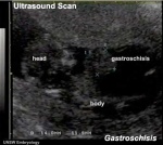
Gastroschisis
| Gastroschisis (omphalocele, paraomphalocele, laparoschisis, abdominoschisis, abdominal hernia) is a congenital abdominal wall defect which results in herniation of fetal abdominal viscera (intestines and/or organs) into the amniotic cavity.
Incidence of gastroschisis has been reported at 1.66/10,000, occuring more frequently in young mothers (less than 20 years old). By definition, it is a body wall defect, not a gastrointestinal tract defect, which in turn impacts upon GIT development. |
|
| Gastroschisis |
Images


UNSW Embryology Links
- Lecture 9 2008
- Gastrointestinal Tract Notes Overview of GIT Formation | Abnormalities | Overview Stage 13/14 Overview | Stage 13/14 | Stage 22 | Selected stage 22 | Histology | Liver | Gall Bladder | Spleen | Pancreas | Stomach | Folding | Intestine | Sensory Development - Taste | Head and Neck Development - Tongue
External Links
- Embryo Images by Drs. Kathleen K. Sulik and Peter R. Bream Jr. notes/images sections on Gut Development
- Anatomy of the Human Body 1918 Henry Gray 2. The Digestive Apparatus | liver
Terms
allantois - An extraembryonic membrane, endoderm in origin extension from the early hindgut, then cloaca into the connecting stalk of placental animals, connected to the superior end of developing bladder. In reptiles and birds, acts as a reservoir for wastes and mediates gas exchange. In mammals is associated/incorporated with connecting stalk/placental cord fetal-maternal interface.
amnion - An extraembryonic membrane]ectoderm and extraembryonic mesoderm in origin and forms the innermost fetal membrane, produces amniotic fluid. This fluid-filled sac initially lies above the trilaminar embryonic disc and with embryoic disc folding this sac is drawn ventrally to enclose (cover) the entire embryo, then fetus. The presence of this membane led to the description of reptiles, bird, and mammals as amniotes.
amniotic fluid - The fluid that fills amniotic cavity totally encloses and cushions the embryo. Amniotic fluid enters both the gastrointestinal and respiratory tract following rupture of the buccopharyngeal membrane. The late fetus swallows amniotic fluid.
buccal - (Latin, bucca = cheek) A term used to relate to the mouth (oral cavity).
buccopharyngeal membrane - (oral membrane) (Latin, bucca = cheek) A membrane which forms the external upper membrane limit (cranial end) of the early gastrointestinal tract (GIT). This membrane develops during gastrulation by ectoderm and endoderm without a middle (intervening) layer of mesoderm. The membrane lies at the floor of the ventral depression (stomadeum) where the oral cavity will open and will breakdown to form the initial "oral opening" of the gastrointestinal tract. The equivilent membrane at the lower end of the gastrointestinal tract is the cloacal membrane.
cloacal membrane - Forms the external lower membrane limit (caudal end) of the early gastrointestinal tract (GIT). This membrane is formed during gastrulation by ectoderm and endoderm without a middle (intervening) layer of mesoderm. The membrane breaks down to form the initial "anal opening" of the gastrointestinal tract.
coelom - Term used to describe a space. There are extraembryonic and intraembryonic coeloms that form during vertebrate development. The single intraembryonic coelom will form the 3 major body cavities: pleural, pericardial and peritoneal.
foregut - The first of the three part/division (foregut - midgut - hindgut) of the early forming gastrointestinal tract. The foregut runs from the buccopharyngeal membrane to the midgut and forms all the tract (esophagus and stomach) from the oral cavity to beneath the stomach. In addition, a ventral bifurcation of the foregut will also form the respiratory tract epithelium.
gastrula - (Greek, gastrula = little stomach) A stage of an animal embryo in which the three germ layers have just formed.
gastrulation - The process of differentiation forming a gastrula. Term means literally means "to form a gut" but is more in development, as this process converts the bilaminar embryo (epiblast/hypoblast) into the trilaminar embryo ([E.htm#endoderm endoderm]/mesoderm/ectoderm) establishing the 3 germ layers that will form all the future tissues of the entire embryo. This process also establishes the the initial body axes.
hindgut - The last of the three part/division foregut - midgut - hindgut) of the early forming gastrointestinal tract. The hindgut forms all the tract from the distral transverse colon to the cloacal membrane and extends into the connecting stalk (placental cord) as the allantois. In addition, a ventral of the hindgut will also form the urinary tract (bladder, urethra) epithelium.
intraembryonic coelom - The "horseshoe-shaped" space (cavity) that forms initially in the third week of development in the lateral plate mesoderm that will eventually form the 3 main body cavities: pericardial, pleural, peritoneal. The intraembryonic coelom communicates transiently with the extraembryonic coelom.
neuralation - The general term used to describe the early formation of the nervous system. It is often used to describe the early events of differentiation of the central ectoderm region to form the neural plate, then neural groove, then neural tube. The nervous system includes the central nervous system (brain and spinal cord) from the neural tube and the peripheral nervous system (peripheral sensory and sympathetic ganglia) from neural crest. In humans, early neuralation begins in week 3 and continues through week 4.
pharynx - uppermost end of gastrointestinal and respiratory tract, in the embryo beginning at the buccopharyngeal membrane and forms a major arched cavity within the phrayngeal arches.
somitogenesis The process of segmentation of the paraxial mesoderm within the trilaminar embryo body to form pairs of somites, or balls of mesoderm. A somite is added either side of the notochord (axial mesoderm) to form a somite pair. The segmentation does not occur in the head region, and begins cranially (head end) and extends caudally (tailward) adding a somite pair at regular time intervals. The process is sequential and therefore used to stage the age of many different species embryos based upon the number visible somite pairs. In humans, the first somite pair appears at day 20 and adds caudally at 1 somite pair/90 minutes until on average 44 pairs eventually form.
splanchnic mesoderm - Gastrointestinal tract (endoderm) associated mesoderm formed by the separation of the lateral plate mesoderm into two separate components by a cavity, the intraembryonic coelom. Splanchnic mesoderm is the embryonic origin of the gastrointestinal tract connective tissue, smooth muscle, blood vessels and contribute to organ development (pancreas, spleen, liver). The intraembryonic coelom will form the three major body cavities including the space surrounding the gut, the peritoneal cavity. The other half of the lateral plate mesoderm (somatic mesoderm) is associated with the ectoderm of the body wall.
stomadeum - (stomadeum) A ventral surface depression on the early embryo head surrounding the buccopharyngeal membrane, which lies at the floor of this depression. This surface depression lies between the maxillary and mandibular components of the first pharyngeal arch.
Glossary Links
- Glossary: A | B | C | D | E | F | G | H | I | J | K | L | M | N | O | P | Q | R | S | T | U | V | W | X | Y | Z | Numbers | Symbols | Term Link
Course Content 2010
Embryology Introduction | Cell Division/Fertilization | Lab 1 | Week 1&2 Development | Week 3 Development | Lab 2 | Mesoderm Development | Ectoderm, Early Neural, Neural Crest | Lab 3 | Early Vascular Development | Placenta | Lab 4 | Endoderm, Early Gastrointestinal | Respiratory Development | Lab 5 | Head Development | Neural Crest Development | Lab 6 | Musculoskeletal Development | Limb Development | Lab 7 | Kidney | Genital | Lab 8 | Sensory | Stem Cells | Stem Cells | Endocrine | Lab 10 | Late Vascular Development | Integumentary | Lab 11 | Birth, Postnatal | Revision | Lab 12 | Lecture Audio | Course Timetable
Cite this page: Hill, M.A. (2026, July 18) Embryology 2010 Lecture 9. Retrieved from https://embryology.med.unsw.edu.au/embryology/index.php/2010_Lecture_9
- © Dr Mark Hill 2026, UNSW Embryology ISBN: 978 0 7334 2609 4 - UNSW CRICOS Provider Code No. 00098G