2010 Lab 6
Introduction

This laboratory will studies head and neural crest development. Note that neural crest will mainly focus on the head region.
Human Head Development

Firstly, examine the external appearance of the human head at each stage of embryonic development starting at Carnegie stage 11 at the beginning of Week 4. Making note of the relative head size, appearance of external head features and position of these feature (relative to the adult position). Note that many of the stages have larger versions available and should be opened alone for best detailed viewing.
About Carnegie Stages: 12 | 13 | 14 | 15 | 16 | 17 | 18 | 19 | 20 | 21 | 22 | 23
Buccopharyngeal Membrane
Identify: position of the stomodeum and the buccopharyngeal membrane.
These images of the Stage 11 embryo show the breakdown of the buccopharyngeal membrane, lying at the floor of the stomodeum.
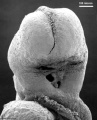
Low power ventral view of the Buccopharyngeal Membrane
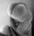
Higher power ventrolateral view of the Buccopharyngeal Membrane
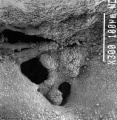
Close up view of the degenerating Buccopharyngeal Membrane


Pharyngeal Arches and Sensory Placodes

Identify: each pharyngeal arch position and relative size, sensory placode position and note the disappearance of the otic placode from the surface. Note that we will be looking at sensory placodes again when we study sensory development.

|

|

|

|

|

|

|
| 11 | 12 | 13 | 14 | 15 | 16 | 17 |
 This is a scanning EM of the embryo superior dorsal view showing the paired otic placodes sinking into the surface at the level of the hindbrain. Features: surface ectoderm, paired otic placodes, pharyngeal arches heart, 25 days, 19 somite pairs |
 This is a scanning EM of the embryo looks inside the pharyngeal arches at the floor of the Carnegie stage 13 pharynx. |
Stage 13 Head

|

|

|

|

| ||
| A1L | A2L | A3L | A4L | A5L | A6L | A7L |

|

|

|

|

|

|

|
| B1L | B2L | B3L | B4L | B5L | B6L | B7L |
Stage 22 Head

|

|

|

|

|

|

|
| A1L | A2L | A3L | A4L | A5L | A6L | A7L |

|

|

|

|

|

|

|
| B1L | B2L | B3L | B4L | B5L | B6L | B7L |
Fetal
Begin by working through the features that have developed and are present in the early 10 week female fetus head region. Head - most lateral | lateral | medial | midline

|

|

|

|
Then look in detail at the head development in a 12 week fetus showing both forms of ossification in the skull.

|

|

|
Pharyngeal Arch - Adult Structures
| Arch | Nerve | Muscles | Skeletal | Artery
|
| 1
(maxillary/mandibular) |
trigeminal (V) | mastication
(temporalis, masseter, medial pterygoid, lateral pterygoid) |
Meckel's cartilage
mandible model malleus, incus |
maxillary
(terminal branches) |
| 2
(hyoid) |
facial (VII) | facial expression
( buccinator, platysma, stapedius, stylohyoid, digastric posterior belly) |
Reichert's cartilage
stapes, styloid process, lesser cornu of hyoid, upper part of body of hyoid bone |
stapedial (embryonic)
corticotympanic (adult)
|
| 3 | glossopharyngeal (IX) | Stylopharyngeus | greater cornu of hyoid, lower part of body of hyoid bone | common carotid, internal carotid (root) |
| 4 and 6 | vagus (X)
superior laryngeal and recurrent laryngeal branches |
intrinsic muscles of larynx, pharynx; levator palati | thyroid, cricoid, arytenoid, corniculate and cuneform cartilages | 4 - aortic arch, right subclavian
6 - ductus arteriosus, pulmonary (roots) |
Structures derived from Pouches
| Pouch | Overall Structure | Specific Structures |
| 1 | tubotympanic recess | tympanic membrane, tympanic cavity, mastoid antrum, auditory tube |
| 2 | intratonsillar cleft | crypts of palatine tonsil, lymphatic nodules of palatine tonsil |
| 3 | inferior parathyroid gland, thymus gland | |
| 4 | superior parathyroid gland, ultimobranchial body | |
| 5 | becomes part of 4th pouch |
Grooves - 1st groove forms part of the external acoustic meatus
Membranes - 1st membrane forms the tympanic membrane
More: The above table is not for you to learn by heart but an indication of the structures formed from each arch.
It is easy to remember that:
- 1st arch - about the mouth (chewing, jaw) and the external and middle ear.
- 2nd arch - about the face, hyoid and external and middle ear.
- 3rd arch - about the neck and endocrine.
- 4th arch- about the neck and endocrine.
Face
Now look again at the animation from the lecture that show how each pharyngeal arch contributes to the final fetal face.
| <Flowplayer width="420" height="500" autoplay="true">Face_001.flv</Flowplayer> |
Development of the Face
The separate embryonic components that contribute to the face have been colour coded.
The stomodeum is the primordial mouth region and a surface central depression lying between the forebrain bulge and the heart bulge. At the floor of the stomodeum indentation is the buccopharyngeal membrane (oral membrane). Note the complex origin of the maxillary region (upper jaw) requiring the fusion of several embryonic elements, abnormalities of this process lead to cleft lip and cleft palate. See also the movie (Quicktime | Flash) showing a similar view of human embryo faces between Carnegie stage 16 to 18.
|
Palate and Tongue

Cleft lip and palate develop between the 4th and 8th week of gestation and is dominated by changes resulting in the formation of the nose and maxilla. Between stage 17 and 18 a key step in primary palate is the transforms the developing palate going from an epithelial seam to the mesenchymal bridge.
Cleft Palate development occurs between the 7th and 12th week of gestation and is divided into the formation of the primary palate (prolabium), premaxilla and cartilaginous septum) and formation of the secondary palate (hard and soft palate).
| Palate 1 | Palate 2 | Tongue |
Links: Ultrasound Movie - Facial Cleft 1 | Ultrasound Movie - Facial Cleft 2
Group Project
Group Projects (20% of your final mark). It is now week 7 and the project is due for peer assessment in the week following the mid-semester break.
Peer Assessment
The Peer assessment process will evaluate only what is on the Project page before Lab 7 (16th September) and this should be as close to the final format that you intend to submit for the course coordinator assessment later this semester. The peer assessment process that all students will participate in as part of their ongoing individual assessment (20% of your final mark).
Briefly, each student will need to assess all other projects (not your own) and add comments to the appropriate Project talk page, using the same assessment criteria that the assessor will use as outlined in the first Laboratory and reproduced below. These comments will need to be signed and should be balanced in their critical assessment.
Your feedback will be used for each group to update their project before the final course coordinator assessment. Your own comments are up to you. Your assessment should be objective, avoid inappropriate terms (not "gushing" not "damning").
If you are having trouble getting started here are some examples:
- Did you learn something about that prenatal diagnostic technique
- Was it overall clearly structured and organised
- Was it missing something
- Was there too much of one particular concept
- Did it lack a "scientific" feel suitable for an undergraduate university level student.
- If possible include an example of what you found good or bad about the project.
Finally and most importantly include a section "What would improve this project....".
When you have completed each assessment you should add a copy of your comments for each to your own student page for my assessment. All peer assessments must be completed before the following weeks Lab (23rd September). I remind you that you are supposed to be working on these projects in your own time and the time I make available within the labs is for project tutorial and co-ordination only.
Group Assessment Criteria
- The key points relating to the topic that your group allocated are clearly described.
- The choice of content, headings and sub-headings, diagrams, tables, graphs show a good understanding of the topic area.
- Content is correctly cited and referenced.
- The wiki has an element of teaching at a peer level using the student's own innovative diagrams, tables or figures and/or using interesting examples or explanations.
- Evidence of significant research relating to basic and applied sciences that goes beyond the formal teaching activities.
- Relates the topic and content of the Wiki entry to learning aims of embryology.
- Clearly reflects on editing/feedback from group peers and articulates how the Wiki could be improved (or not) based on peer comments/feedback. Demonstrates an ability to review own work when criticised in an open edited wiki format. Reflects on what was learned from the process of editing a peer's wiki.
- Evaluates own performance and that of group peers to give a rounded summary of this wiki process in terms of group effort and achievement.
- The content of the wiki should demonstrate to the reader that your group has researched adequately on this topic and covered the key areas necessary to inform your peers in their learning.
- Develops and edits the wiki entries in accordance with the above guidelines.
Glossary Links
- Glossary: A | B | C | D | E | F | G | H | I | J | K | L | M | N | O | P | Q | R | S | T | U | V | W | X | Y | Z | Numbers | Symbols | Term Link
Cite this page: Hill, M.A. (2026, July 25) Embryology 2010 Lab 6. Retrieved from https://embryology.med.unsw.edu.au/embryology/index.php/2010_Lab_6
- © Dr Mark Hill 2026, UNSW Embryology ISBN: 978 0 7334 2609 4 - UNSW CRICOS Provider Code No. 00098G