Palate Development
Introduction
The palate has two key stages of development during embryonic and an early fetal involving the fusion of structures (epithelia to mesenchymal).
Some Recent Findings
|
Textbooks

- The Developing Human: Clinically Oriented Embryology (8th Edition) by Keith L. Moore and T.V.N Persaud - Moore & Persaud Chapter Chapter 10 The Pharyngeal Apparatus pp201 - 240.
- Larsen’s Human Embryology by GC. Schoenwolf, SB. Bleyl, PR. Brauer and PH. Francis-West - Chapter 12 Development of the Head, the Neck, the Eyes, and the Ears pp349 - 418.
Movies

| ||||
| Face Development | Palate 1 | Palate 2 | Tongue | Ultrasound - Cleft Lip |
| Quicktime | Flash | Quicktime | Flash | Quicktime | Flash | Quicktime | Flash | Quicktime | Flash |
- Links: Quicktime Movies | Flash Movies | Ultrasound
Development Overview
Pharyngeal Arch Components
Major features to identify for each: arch, pouch, groove and membrane. Contribute to the formation of head and neck and in the human appear at the 4th week. The first arch contributes the majority of upper and lower jaw structures.
Early Face and Pharynx
- Pharynx - begins at the buccopharyngeal membrane (oral membrane), apposition of ectoderm with endoderm (no mesoderm between)
Pharyngeal Arch Development


- branchial arch (Gk. branchia= gill)
- arch consists of all 3 trilaminar embryo layers
- ectoderm- outside
- mesoderm- core of mesenchyme
- endoderm- inside
Neural Crest
- Mesenchyme invaded by neural crest generating connective tissue components
- cartilage, bone, ligaments
- arises from midbrain and hindbrain region
Face Development

Begins week 4 centered around stomodeum, external depression at oral membrane
5 initial primordia from neural crest mesenchyme
- single frontonasal prominence (FNP) - forms forehead, nose dorsum and apex
- nasal placodes develop later bilateral, pushed medially
- paired maxillary prominences - form upper cheek and upper lip
- paired mandibular prominences - lower cheek, chin and lower lip
Frontonasal Process
The frontonasal process (FNP) forms the majority of the superior part of the early face primordia. It later fuses with the maxillary component of the first pharyngeal arch to form the upper jaw. Failure of this fusion event during the embryonic period leads to cleft lip. Under the surface ectoderm the process mesenchyme consists of two cell populations; neural crest cells, forming the connective tissues; and the mesoderm forming the endothelium of the vascular network.
A chicken developmental model study has identified a specific surface region, the Frontonasal Ectodermal Zone (FEZ), initially induced by bone morphogenetic proteins that appears to regulate the future growth and patterning of the frontonasal process. The specific frontonasal ectodermal zone was located in the frontonasal process ectoderm flanking a boundary between Sonic hedgehog (Shh) and Fibroblast growth factor 8 (Fgf8) expression domains.[3]
Head Growth
- continues postnatally - fontanelle allow head distortion on birth and early growth
- bone plates remain unfused to allow growth, puberty growth of face
Embryonic Palate
Primary palate, fusion in the human embryo between stage 17 and 18, from an epithelial seam to the mesenchymal bridge.

Fetal Palate
Secondary palate, fusion in the human embryo in week 9. This requires the early palatal shelves growth, elevation and fusion during the early embryonic period. The fusion event is to both each other and the primary palate. palatal shelf elevation | secondary palate
Abnormalities

The way in which the upper jaw forms from fusion of the smaller upper prominence of the first pharyngeal arch leads to a common congenital defect in this region called "clefting", which may involve either the upper lip, the palate or both structures.
Ten most frequently reported birth anomalies
- Hypospadias (More? Development Animation - Genital Male External | Genital Abnormalities - Hypospadia)
- Obstructive Defects of the Renal Pelvis (More? Renal System - Abnormalities)
- Ventricular Septal Defect (More? Cardiovascular Abnormalities - Ventricular Septal Defect)
- Congenital Dislocated Hip (More? Musculoskelal Abnormalities - Congenital Dislocation of the Hip (CDH))
- Trisomy 21 or Down syndrome - (More? Trisomy 21)
- Hydrocephalus
- Cleft Palate
- Trisomy 18 or Edward Syndrome - multiple abnormalities of the heart, diaphragm, lungs, kidneys, ureters and palate 86% discontinued (More? (More? Trisomy 18)
- Renal Agenesis/Dysgenesis- reduction in neonatal death and stillbirth since 1993 may be due to the more severe cases being identified in utero and being represented amongst the increased proportion of terminations (approximately 31%).
- Cleft Lip and Palate - occur with another defect in 33.7% of cases.
From the Victorian Perinatal Data Collection Unit in Victoria between 2003-2004.
Embryonic Human Cleft Palate

|
File:Stage18 cleft palate.jpg | File:Stage19 cleft palate.jpg |
| Stage16 | Stage18 | Stage19 |
Cleft Lip

|
|
Cleft Palate

|
(Data: Congenital Malformations Australia 1981-1992 P. Lancaster and E. Pedisich ISSN 1321-8352) Links: Development Animation - Palate 1 | Development Animation - Palate 2 | Orofacial Cleft with or without cleft palate Search Pubmed Now: cleft lip | cleft palate |
References
Reviews
Articles
Search PubMed
Search Pubmed: palate development | cleft palate development |
Additional Images
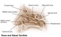
Nasal cavities and palate
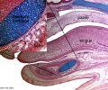
Palate, tongue and Meckel's cartilage
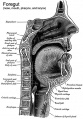
Historic Pharynx cartoon
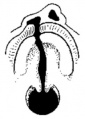
Unilateral cleft palate
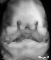
Stage16 cleft palate
- Stage18 cleft palate.jpg
Stage18 cleft_palate
- Stage19 cleft palate.jpg
Stage19 cleft palate


Terms
Glossary Links
- Glossary: A | B | C | D | E | F | G | H | I | J | K | L | M | N | O | P | Q | R | S | T | U | V | W | X | Y | Z | Numbers | Symbols | Term Link
Cite this page: Hill, M.A. (2024, May 7) Embryology Palate Development. Retrieved from https://embryology.med.unsw.edu.au/embryology/index.php/Palate_Development
- © Dr Mark Hill 2024, UNSW Embryology ISBN: 978 0 7334 2609 4 - UNSW CRICOS Provider Code No. 00098G