Neural System Development: Difference between revisions
| Line 49: | Line 49: | ||
== Human Early Neural Development == | == Human Early Neural Development == | ||
The stages below refer to specific Carneigie stages of development. | The stages below refer to specific Carneigie stages of development. Data and text modified from O'Rahilly and Müller 1994.<ref>Neurulation in the normal human embryo. O'Rahilly R, Müller F. Ciba Found Symp. 1994;181:70-82; discussion 82-9. Review. [http://www.ncbi.nlm.nih.gov/pubmed/8005032 PMID: 8005032]</ref> | ||
* '''stage 8 '''(about 18 postovulatory days) neural groove and folds are first seen | * '''stage 8 '''(about 18 postovulatory days) neural groove and folds are first seen | ||
| Line 64: | Line 64: | ||
* is the differentiation of the caudal part of the neural tube from the caudal eminence (or end-bud) without the intermediate phase of a neural plate. | * is the differentiation of the caudal part of the neural tube from the caudal eminence (or end-bud) without the intermediate phase of a neural plate. | ||
== Late Neural Development == | |||
Three-dimensional magnetic resonance imaging and image-processing algorithms have been used to quantitate between 29-41 weeks volumes of: total brain, cerebral gray matter, unmyelinated white matter, myelinated, and cerebrospinal fluid (grey matter- mainly neuronal cell bodies; white matter- mainly neural processes and glia). A study of 78 premature and mature newborns showed that total brain tissue volume increased linearly over this period at a rate of 22 ml/week. Total grey matter also showed a linear increase in relative intracranial volume of approximately 1.4% or 15 ml/week. The rapid increase in total grey matter is mainly due to a fourfold increase in cortical grey matter. Quantification of extracerebral and intraventricular CSF was found to change only minimally. (text modified from Huppi etal., (1998) Quantitative magnetic resonance imaging of brain development in premature and mature newborns.Ann Neurol 43(2):224-235.) [[Late Neural Development]] | |||
== Gliogenesis and Myelination == | |||
Glial cells have many different types and roles in central and peripheral neural development, though they are typically described as "supportive", and have the same early embryonic origins as neurons. (More? [neuron7.htm Gliogenesis and Myelination]) | |||
Early in neural development a special type of developmental glia, '''radial glia''', provide pathway for developing neuron (neuroblasts) migration out from the proliferating ventricular layer and are involved in the subsequent '''lamination''' and '''columnar organization''' of the central nervous system. | |||
'''Types of glia:''' radial glia, astroglia, oligodendroglia, microglia and Schwann cells. | |||
== References == | == References == | ||
Revision as of 13:40, 22 April 2010
Introduction
Neural development is one of the earliest systems to begin and the last to be completed after birth. This development generates the most complex structure within the embryo and the long time period of development means in utero insult during pregnancy may have consequences to development of the nervous system.
The early central nervous system begins as a simple neural plate that folds to form a groove then tube, open initially at each end. Failure of these opening to close contributes a major class of neural abnormalities (neural tube defects).
Sonic Hedgehog expression (white) in both the notocord (pale circular) and neural tube floorplate (bright triangle). (Image- Lance Davidson)
Within the neural tube stem cells generate the 2 major classes of cells that make the majority of the nervous system : neurons and glia. Both these classes of cells differentiate into many different types generated with highly specialized functions and shapes. This section covers the establishment of neural populations, the inductive influences of surrounding tissues and the sequential generation of neurons establishing the layered structure seen in the brain and spinal cord.
- Neural development beginnings quite early, therefore also look at notes covering Week 3- neural tube and Week 4-early nervous system.
- Development of the neural crest and sensory systems (hearing/vision/smell) are only introduced in these notes and are covered in other notes sections.
| System Links: Introduction | Cardiovascular | Coelomic Cavity | Endocrine | Gastrointestinal Tract | Genital | Head | Immune | Integumentary | Musculoskeletal | Neural | Neural Crest | Placenta | Renal | Respiratory | Sensory | Birth |
--Mark Hill 10:09, 22 April 2010 (EST) Page content from original site and currently being updated and reformatted.
Some Recent Findings
Reading
- Human Embryology (3rd ed.) Chapter 5 p107-125
- The Developing Human: Clinically Oriented Embryology (6th ed.) Moore and Persaud Chapter 18 p451-489
- Essentials of Human Embryology Larson Chapter 5 p69-79
- Before We Are Born (5th ed.) Moore and Persaud Chapter 19 p423-458
- Human Embryology, Fitzgerald and Fitzgerald
- History of Science- Brain and Mind, Brain Structure, Camille Golgi, S. Ramon y Cajal
Objectives
- Understand early neural development.
- Understand the formation of spinal cord.
- Understand the formation of the brain; grey and white matter from the neural tube.
- Understand the role of migration of neurons during neural development.
- To know the main derivatives of the brain vesicles and their walls.
- To know how the nervous system is modelled, cell death etc.
- To understand the contribution of the neural crest.
- Understand the developmental basis of certain congenital anomalies of the nervous system, including hydrocephalus, spina bifida, anencephaly and encephalocele.
Animal Neural Development
A number of different animal models of neural development, both normal and abnormal, have been established.
Human Early Neural Development
The stages below refer to specific Carneigie stages of development. Data and text modified from O'Rahilly and Müller 1994.[1]
- stage 8 (about 18 postovulatory days) neural groove and folds are first seen
- stage 9 the three main divisions of the brain, which are not cerebral vesicles, can be distinguished while the neural groove is still completely open.
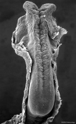
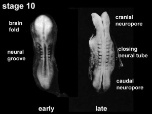
- stage 10 (two days later) neural folds begin to fuse near the junction between brain and spinal cord, when neural crest cells are arising mainly from the neural ectoderm
- stage 11 (about 24 days) the rostral (or cephalic) neuropore closes within a few hours; closure is bidirectional, it takes place from the dorsal and terminal lips and may occur in several areas simultaneously. The two lips, however, behave differently.
- stage 12 (about 26 days) The caudal neuropore takes a day to close
- the level of final closure is approximately at future somitic pair 31
- corresponds to the level of sacral vertebra 2
- stage 13 (4 weeks) the neural tube is normally completely closed
Secondary neurulation begins at stage 12
- is the differentiation of the caudal part of the neural tube from the caudal eminence (or end-bud) without the intermediate phase of a neural plate.
Late Neural Development
Three-dimensional magnetic resonance imaging and image-processing algorithms have been used to quantitate between 29-41 weeks volumes of: total brain, cerebral gray matter, unmyelinated white matter, myelinated, and cerebrospinal fluid (grey matter- mainly neuronal cell bodies; white matter- mainly neural processes and glia). A study of 78 premature and mature newborns showed that total brain tissue volume increased linearly over this period at a rate of 22 ml/week. Total grey matter also showed a linear increase in relative intracranial volume of approximately 1.4% or 15 ml/week. The rapid increase in total grey matter is mainly due to a fourfold increase in cortical grey matter. Quantification of extracerebral and intraventricular CSF was found to change only minimally. (text modified from Huppi etal., (1998) Quantitative magnetic resonance imaging of brain development in premature and mature newborns.Ann Neurol 43(2):224-235.) Late Neural Development
Gliogenesis and Myelination
Glial cells have many different types and roles in central and peripheral neural development, though they are typically described as "supportive", and have the same early embryonic origins as neurons. (More? [neuron7.htm Gliogenesis and Myelination])
Early in neural development a special type of developmental glia, radial glia, provide pathway for developing neuron (neuroblasts) migration out from the proliferating ventricular layer and are involved in the subsequent lamination and columnar organization of the central nervous system.
Types of glia: radial glia, astroglia, oligodendroglia, microglia and Schwann cells.
References
- ↑ Neurulation in the normal human embryo. O'Rahilly R, Müller F. Ciba Found Symp. 1994;181:70-82; discussion 82-9. Review. PMID: 8005032
Reviews
Articles
Search PubMed
Search Pubmed: Neural System Development
| System Links: Introduction | Cardiovascular | Coelomic Cavity | Endocrine | Gastrointestinal Tract | Genital | Head | Immune | Integumentary | Musculoskeletal | Neural | Neural Crest | Placenta | Renal | Respiratory | Sensory | Birth |
Glossary Links
- Glossary: A | B | C | D | E | F | G | H | I | J | K | L | M | N | O | P | Q | R | S | T | U | V | W | X | Y | Z | Numbers | Symbols | Term Link
Cite this page: Hill, M.A. (2024, May 1) Embryology Neural System Development. Retrieved from https://embryology.med.unsw.edu.au/embryology/index.php/Neural_System_Development
- © Dr Mark Hill 2024, UNSW Embryology ISBN: 978 0 7334 2609 4 - UNSW CRICOS Provider Code No. 00098G

