Neural Crest Development: Difference between revisions
mNo edit summary |
mNo edit summary |
||
| Line 80: | Line 80: | ||
* Chapter 16 - Development of the Pharyngeal Apparatus and Face | * Chapter 16 - Development of the Pharyngeal Apparatus and Face | ||
|- | |- | ||
| Additional Resources | | colspan=2|Additional Resources | ||
* Developmental Biology. 6th edition. [http://www.ncbi.nlm.nih.gov/books/NBK10065 The Neural Crest] | * Developmental Biology. 6th edition. [http://www.ncbi.nlm.nih.gov/books/NBK10065 The Neural Crest] | ||
* Nelms BL, Labosky PA. Transcriptional Control of Neural Crest Development. San Rafael (CA): Morgan & Claypool Life Sciences; 2010. Available from: http://www.ncbi.nlm.nih.gov/books/NBK53145/ | * Nelms BL, Labosky PA. Transcriptional Control of Neural Crest Development. San Rafael (CA): Morgan & Claypool Life Sciences; 2010. Available from: http://www.ncbi.nlm.nih.gov/books/NBK53145/ | ||
* Dupin E, Creuzet S, Le Douarin NM. The Contribution of the Neural Crest to the Vertebrate Body. In: Madame Curie Bioscience Database [Internet]. Austin (TX): Landes Bioscience; 2000-. Available from: http://www.ncbi.nlm.nih.gov/books/NBK6098/ | |||
|} | |} | ||
Revision as of 19:14, 6 September 2015
| Embryology - 26 Apr 2024 |
|---|
| Google Translate - select your language from the list shown below (this will open a new external page) |
|
العربية | català | 中文 | 中國傳統的 | français | Deutsche | עִברִית | हिंदी | bahasa Indonesia | italiano | 日本語 | 한국어 | မြန်မာ | Pilipino | Polskie | português | ਪੰਜਾਬੀ ਦੇ | Română | русский | Español | Swahili | Svensk | ไทย | Türkçe | اردو | ייִדיש | Tiếng Việt These external translations are automated and may not be accurate. (More? About Translations) |
Introduction

The neural crest are bilaterally paired strips of cells arising in the ectoderm at the margins of the neural tube. These cells migrate to many different locations and differentiate into many cell types within the embryo. This means that many different systems (neural, skin, teeth, head, face, heart, adrenal glands, gastrointestinal tract) will also have a contribution fron the neural crest cells.
In the body region, neural crest cells also contribute the peripheral nervous system (both neurons and glia) consisting of sensory ganglia (dorsal root ganglia), sympathetic and parasympathetic ganglia and neural plexuses within specific tissues/organs.
In the head region, neural crest cells migrate into the pharyngeal arches (as shown in movie below) forming ectomesenchyme contributing tissues which in the body region are typically derived from mesoderm (cartilage, bone, and connective tissue). General neural development is also covered in Neural Notes.
Historically identified as "neural crest" by Arthur Marshall in 1879.[1] (see history below and original article).
| Neural Crest Links: neural crest | Lecture - Early Neural | Lecture - Neural Crest Development | Lecture Movie | Schwann cell | adrenal | melanocyte | peripheral nervous system | enteric nervous system | cornea | cranial nerve neural crest | head | skull | cardiac neural crest | Nicole Le Douarin | Neural Crest Movies | neural crest abnormalities | Category:Neural Crest | |||
|
Some Recent Findings

|
| More recent papers |
|---|
 This table allows an automated computer search of the external PubMed database using the listed "Search term" text link.
More? References | Discussion Page | Journal Searches | 2019 References | 2020 References Search term: Neural Crest Embryology <pubmed limit=5>Neural Crest Embryology</pubmed> |

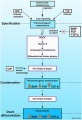
Neural crest formation stages and gene regulatory networks[7]
| System | Cell Type |
|---|---|
| Peripheral Nervous System (PNS) | Neurons - sensory ganglia, sympathetic and parasympathetic ganglia, enteric nervous system, and plexuses
Glia (neuroglial cells) - Schwann cells[8], satellite cells, olfactory ensheathing cells[9] |
| endocrine | Adrenal medulla Calcitonin-secreting cells Carotid body type I cells |
| integumentary | Epidermal pigment cells melanocyte |
| Facial cartilage and bone | Facial and anterior ventral skull cartilage and bones |
| Sensory | inner ear, cornea endothelium and stroma |
| Connective tissue | tooth odontoblast
smooth muscle, and adipose tissue of skin in head and neck Connective tissue of meninges, salivary, lachrymal, thymus, thyroid, and pituitary glands Connective tissue and smooth muscle in arteries of aortic arch origin |
| Links: neural crest | Category:Neural Crest | Neural Crest collapsible table | |
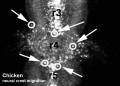
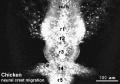
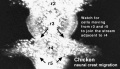
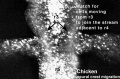
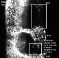
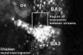
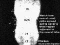
Neural Crest Migration
| <html5media height="380" width="410">File:Chicken-neural crest migration 01.mp4</html5media> | 
Chicken embryo sequence shows the migration of DiI-labeled neural crest cells towards the branchial arches as the embryo. White rings indicate migration of individual cells. Each image represents 10 confocal sections separated by 10 microns. |
Movie Source: Original Neural Crest movies kindly provided by Paul Kulesa.[10]
| Neural crest migration Chicken Head (movies overview) | |||||||||||||||||||||||||||
|---|---|---|---|---|---|---|---|---|---|---|---|---|---|---|---|---|---|---|---|---|---|---|---|---|---|---|---|
|
|
|
|
|
|
| |||||||||||||||||||||
- Neural Crest Movies: Migration 01 | Migration 02 | Migration 03 | Migration 04 | Migration 05 | Migration 06 | Migration 07
Textbooks


|
Hill, M.A. (2020). UNSW Embryology (20th ed.) Retrieved April 26, 2024, from https://embryology.med.unsw.edu.au
| |||||

|
Moore, K.L. & Persuad, T.V.N. (2008). The Developing Human: clinically oriented embryology (8th ed.). Philadelphia: Saunders. (chapter links only work with a UNSW connection). | |||||

|
Schoenwolf, G.C., Bleyl, S.B., Brauer, P.R. and Francis-West, P.H. (2009). Larsen’s Human Embryology (4th ed.). New York; Edinburgh: Churchill Livingstone. The following chapter links only work with a UNSW Library subscription
| |||||
Additional Resources
| ||||||
Objectives

- Understand the structures derived from ectoderm.
- Understand the formation of neural folds.
- Identify the initial location of neural crest cells in the trilaminar embryo.
- Identify pathways of neural crest migration throughout the embryo.
- To know the major tissues to which neural crest cells contribute.
- To know how abnormalities in development that result from abnormal neural crest cell migration.
- Understand how neural crest cells contribute to the pharyngeal arches and the head structures they form.
Neural Crest Derivatives
A key feature of neural crest is the migration into other embryonic tissues to form specific neural and non-neural populations and structures.
Cranial neural crest
- migration - dorsolaterally and into pharyngeal arches
- craniofacial mesenchyme - cartilage, bone, cranial neurons, glia, and connective tissues of the face
- pharyngeal arches and pouches - thymic cells, tooth odontoblasts, middle ear bones (ossicles) and jaw (mandible)
Cardiac neural crest
- migration - located between the cranial and trunk neural crests, overlapping the anterior portion of the vagal neural crest.
- pharyngeal arches - (3,4,6) melanocytes, neurons, cartilage, and connective tissue
- heart outflow tract - aortic arch/pulmonary artery septum, large arteries wall musculoconnective tissue
Trunk neural crest
- migration - two major pathways over somites (dorsolaterally) and between somite and neural tube (ventrolaterally)
- dorsolateral - skin melanocytes
- ventrolaterally - dorsal root ganglia, sympathetic ganglia, adrenal medulla, aortic nerve clusters
Vagal and sacral neural crest
- migration - ventrally into surrounding splanchnic mesenchyme of gastrointestinal tract
- splanchnic mesenchyme - parasympathetic (enteric) ganglia of the gut
Development Overview
The following cranial and trunk data is based upon 185 serially sectioned staged (Carnegie) human embryos.[11]
Cranial Neural Crest
- stage 9 - an indication of mesencephalic neural crest
- stage 10 - trigeminal, facial, and postotic components
- stage 11 - crest-free zones are soon observable in rhombomere 1, 3, and 5
- stage 12 - rhombomeres 6 and 7 neural crest migrate to pharyngeal arch 3 and then rostrad to the truncus arteriosus
- stage 13 - nasal crest and the terminalis-vomeronasal complex are last of the cranial crest to appear
stages 9-14 - otic vesicle primordium descends
Vagal Neural Crest
Recent research suggests that the vagal neural crest cells are a transitional population that has evolved between the head and the trunk, taking separate pathways to the both the heart and to the gut.[12][13]
Trunk Neural Crest
Spinal ganglia increase in number over time and are in phase with the somites, though not their centre. There are 3 migratory pathways: ventrolateral between dermatomyotome and sclerotome, ventromedial between neural tube and sclerotomes, and lateral between surface ectoderm and dermatomyotome.
- stage 13 - about 19 present
- stage 14 - about 33 present
- stage 15-23 - 30–35 ganglia
Neck and Shoulder
A mouse study using individually labelled cells of postotic neural crest followed the development of the shoulder girdle (clavicle and scapula) that connects the upper limb to the axial skeleton.[14]
- Clavicle is a neural crest-mesodermal structure, posterior dermal clavicle mesoderm.
- Cryptic cell boundaries traverse apparently homogeneous skeleton of the neck and shoulders.
- Bones and muscles code of connectivity that mesenchymal stem cells of both neural crest and mesodermal origin obey
- Neural crest anchors the head onto the anterior lining of the shoulder girdle
- Hox-gene-controlled mesoderm links trunk muscles to the posterior neck and shoulder skeleton.
- Skeleton identified as neural crest-derived is affected in human Klippel-Feil syndrome, Sprengel's deformity and Arnold-Chiari I/II malformation.
Skin Melanocytes

|
|
| Mouse melanocyte migration[15] | Movie Mouse Skin - Melanoblast Migration E14.5[16] |
Historic
The paper by Marshall, Morphology of the Vertebrate Olfactory Organ (1879)[1], was historically the first time the term "neural crest" was used. In his own earlier papers he had referred to this as a "neural ridge" in describing development of the chicken embryo neural tube.
See paper text and his referenced comment:
- "I take this opportunity to make a slight alteration in the nomenclature adopted in my former paper. I have there suggested the term neural ridge for the longitudinal ridge of cells which grows out from the reentering angle between the external epiblast and the neural canal, and from which the nerves, whether cranial or spinal, arise. Since this ridge appears before closure of the neural canal is effected, there are manifestly two neural ridges, one on either side ; but I have also applied the same term, neural ridge, to the single outgrowth formed by the fusion of the neural ridges of the two sides after complete closure of the neural canal is effected, and after the external epiblast has become completely separated from the neural canal. I propose in future to speak of this single median outgrowth as the neural crest, limiting the term neural ridge to the former acceptation. Thus, while there are two neural ridges, there is only one neural crest, a distinction that will be at once evident on reference to my former figures."
- Links: Embryology History
References
- ↑ 1.0 1.1 Marshall, A. Morphology of Vertebrate Olfactory Organ Quarterly Journal of Microscopic Science (1879) Vol. 19: 300–340.
- ↑ 2.0 2.1 <pubmed>23028350</pubmed>| PLoS Genet.
- ↑ <pubmed>21552538</pubmed>
- ↑ <pubmed>20478300</pubmed>
- ↑ <pubmed>20399765</pubmed>
- ↑ <pubmed>20360764</pubmed>
- ↑ <pubmed>25903629</pubmed>| Nature
- ↑ Woodhoo A & Sommer L. (2008). Development of the Schwann cell lineage: from the neural crest to the myelinated nerve. Glia , 56, 1481-90. PMID: 18803317 DOI.
- ↑ Barraud P, Seferiadis AA, Tyson LD, Zwart MF, Szabo-Rogers HL, Ruhrberg C, Liu KJ & Baker CV. (2010). Neural crest origin of olfactory ensheathing glia. Proc. Natl. Acad. Sci. U.S.A. , 107, 21040-5. PMID: 21078992 DOI.
- ↑ <pubmed>10683170</pubmed>
- ↑ <pubmed>17848161</pubmed>| PMC2375817 | J Anat.
- ↑ <pubmed>20962585</pubmed>
- ↑ Bryan R. Kuo, Carol A. Erickson Vagal neural crest cell migratory behavior: A transition between the cranial and trunk crest. Volume 240, Issue 9, pages 2084–2100, September 2011 Dev Dynamics
- ↑ <pubmed>16034409</pubmed>| PMC1352163| Nature
- ↑ <pubmed>16277556</pubmed>| PLoS Biol.
- ↑ <pubmed>20067551</pubmed>| PMC2859249
Books
Trainor, P. (ed) Neural crest cells: evolution, development and disease. ISBN: 978-0-12-401730-6 ScienceDirect Nelms BL, Labosky PA. Transcriptional Control of Neural Crest Development. San Rafael (CA): Morgan & Claypool Life Sciences; 2010. PMID 21452438
Reviews
<pubmed></pubmed> <pubmed>25903629</pubmed> <pubmed>21309066</pubmed> <pubmed>21309065</pubmed> <pubmed>21295685</pubmed> <pubmed>21452438</pubmed>
Articles
<pubmed></pubmed> <pubmed></pubmed> <pubmed></pubmed>
Search PubMed
Search Pubmed: Neural Crest Development
Additional Images
Model neural crest mesenchymal condensation
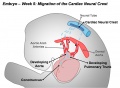
Cardiac Neural Crest Migration
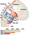
Hindbrain neural crest migration
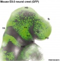
Mouse head E9 neural crest GFP
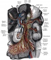
Great plexuses of the sympathetic system


Glossary Links
- Glossary: A | B | C | D | E | F | G | H | I | J | K | L | M | N | O | P | Q | R | S | T | U | V | W | X | Y | Z | Numbers | Symbols | Term Link
Cite this page: Hill, M.A. (2024, April 26) Embryology Neural Crest Development. Retrieved from https://embryology.med.unsw.edu.au/embryology/index.php/Neural_Crest_Development
- © Dr Mark Hill 2024, UNSW Embryology ISBN: 978 0 7334 2609 4 - UNSW CRICOS Provider Code No. 00098G