Neural Crest - Cranial Nerve Development: Difference between revisions
mNo edit summary |
mNo edit summary |
||
| Line 18: | Line 18: | ||
{{Neural Crest Links}} | {{Neural Crest Links}} | ||
== Some Recent Findings == | == Some Recent Findings == | ||
[[File:Zebrafish_neural_crest_model.jpg|thumb|Zebrafish neura crest model | [[File:Zebrafish_neural_crest_model.jpg|thumb|Zebrafish neura crest model{{#pmid:23028350|PMID23028350}}]] | ||
{| | {| | ||
|-bgcolor="F5FAFF" | |-bgcolor="F5FAFF" | ||
| | | | ||
* '''An essential role of variant histone h3.3 for ectomesenchyme potential of the cranial neural crest''' | * '''An essential role of variant histone h3.3 for ectomesenchyme potential of the cranial neural crest'''{{#pmid:23028350|PMID23028350}} "The neural crest (NC) is a vertebrate-specific cell population that exhibits remarkable multipotency. Although derived from the neural plate border (NPB) ectoderm, cranial NC (CNC) cells contribute not only to the peripheral nervous system but also to the ectomesenchymal precursors of the head skeleton. ...Surprisingly, embryo-wide expression of dominant mutant H3.3 had little effect on embryonic development outside CNC, indicating an unexpectedly specific sensitivity of CNC to defects in H3.3 incorporation. Whereas previous studies had implicated H3.3 in large-scale histone replacement events that generate totipotency during germ line development, our work has revealed an additional role of H3.3 in the broad potential of the ectoderm-derived CNC, including the ability to make the mesoderm-like ectomesenchymal precursors of the head skeleton." | ||
* '''Dbx1-expressing cells are necessary for the survival of the mammalian anterior neural and craniofacial structures''' | |||
* '''Analysis of early human neural crest development''' | * '''Dbx1-expressing cells are necessary for the survival of the mammalian anterior neural and craniofacial structures'''{{#pmid:21552538|PMID21552538}} "Development of the vertebrate forebrain and craniofacial structures are intimately linked processes, the coordinated growth of these tissues being required to ensure normal head formation. In this study, we identify five small subsets of progenitors expressing the transcription factor dbx1 in the cephalic region of developing mouse embryos at E8.5. ... Our results demonstrate that dbx1-expressing cells have a unique function during head development, notably by controlling cell survival in a non cell-autonomous manner." | ||
* '''Cranial neural crest migration: new rules for an old road.''' | |||
* '''Derivation of neural crest cells from human pluripotent stem cells.''' | * '''Analysis of early human neural crest development'''{{#pmid:20478300|PMID20478300}} "The outstanding migration and differentiation capacities of neural crest cells (NCCs) have fascinated scientists since Wilhelm His described this cell population in 1868. Today, after intense research using vertebrate model organisms, we have gained considerable knowledge regarding the origin, migration and differentiation of NCCs. However, our understanding of NCC development in human embryos remains largely uncharacterized, despite the role the neural crest plays in several human pathologies. Here, we report for the first time the expression of a battery of molecular markers before, during, or following NCC migration in human embryos from Carnegie Stages (CS) 12 to 18. Our work demonstrates the expression of Sox9, Sox10 and Pax3 transcription factors in premigratory NCCs, while actively migrating NCCs display the additional transcription factors Pax7 and AP-2alpha. Importantly, while HNK-1 labels few migrating NCCs, p75(NTR) labels a large proportion of this population. However, the broad expression of p75(NTR) - and other markers - beyond the neural crest stresses the need for the identification of additional markers to improve our capacity to investigate human NCC development, and to enable the generation of better diagnostic and therapeutic tools." | ||
* '''Cranial neural crest migration: new rules for an old road.'''{{#pmid:20399765|PMID20399765}} "In this review, we discuss recent cellular and molecular discoveries of the CNCC migratory pattern. We focus on events from the time when CNCCs encounter the tissue adjacent to the neural tube and their travel through different microenvironments and into the branchial arches. We describe the patterning of discrete cell migratory streams that emerge from the hindbrain, rhombomere (r) segments r1-r7, and the signals that coordinate directed migration." | |||
* '''Derivation of neural crest cells from human pluripotent stem cells.'''{{#pmid:20360764|PMID20360764}} "Here we provide protocols for the step-wise differentiation of human embryonic stem cells (hESCs) or human induced pluripotent stem cells (hiPSCs) into neuroectodermal and NC cells using either the MS5 coculture system or a novel defined culture method based on pharmacological inhibition of bone morphogenetic protein and transforming growth factor-beta signaling pathways." (More? [[Stem Cells]]) | |||
|} | |} | ||
{| class="wikitable mw-collapsible mw-collapsed" | {| class="wikitable mw-collapsible mw-collapsed" | ||
! More recent papers | ! More recent papers | ||
|- | |- | ||
| [[File:Mark_Hill.jpg|90px|left]] {{Most_Recent_Refs}} | | [[File:Mark_Hill.jpg|90px|left]] {{Most_Recent_Refs}} | ||
| Line 36: | Line 40: | ||
Search term: [http://www.ncbi.nlm.nih.gov/pubmed/?term=Neural+Crest+Embryology ''Neural Crest Embryology''] | Search term: [http://www.ncbi.nlm.nih.gov/pubmed/?term=Neural+Crest+Embryology ''Neural Crest Embryology''] | ||
|} | |} | ||
==Neural Crest Migration== | ==Neural Crest Migration== | ||
| Line 49: | Line 52: | ||
|} | |} | ||
Movie Source: Original Neural Crest movies kindly provided by Paul Kulesa. | Movie Source: Original Neural Crest movies kindly provided by Paul Kulesa.{{#pmid:10683170|PMID10683170}} | ||
{{Chicken neural crest movies}} | {{Chicken neural crest movies}} | ||
| Line 78: | Line 81: | ||
==Objectives== | ==Objectives== | ||
[[File:Mouse-E10.5_ganglia_Sox10.jpg|thumb|Mouse neural crest ( | [[File:Mouse-E10.5_ganglia_Sox10.jpg|thumb|Mouse neural crest ({{ME10.5}} ganglia Sox10)]] | ||
* Understand the structures derived from ectoderm. | * Understand the structures derived from ectoderm. | ||
* Understand the formation of neural folds. | * Understand the formation of neural folds. | ||
| Line 109: | Line 112: | ||
== Development Overview == | == Development Overview == | ||
The following cranial and trunk data is based upon 185 serially sectioned staged (Carnegie) human embryos. | The following cranial and trunk data is based upon 185 serially sectioned staged (Carnegie) human embryos.{{#pmid:17848161|PMID17848161}} | ||
===Cranial Neural Crest=== | ===Cranial Neural Crest=== | ||
| Line 122: | Line 125: | ||
===Vagal Neural Crest=== | ===Vagal Neural Crest=== | ||
Recent research suggests that the vagal neural crest cells are a transitional population that has evolved between the head and the trunk, taking separate pathways to the both the heart and to the gut. | Recent research suggests that the vagal neural crest cells are a transitional population that has evolved between the head and the trunk, taking separate pathways to the both the heart and to the gut.{{#pmid:20962585|PMID20962585}}<ref>Bryan R. Kuo, Carol A. Erickson '''Vagal neural crest cell migratory behavior: A transition between the cranial and trunk crest.''' Volume 240, Issue 9, pages 2084–2100, September 2011 [http://onlinelibrary.wiley.com/doi/10.1002/dvdy.22715/abstract Dev Dynamics]</ref> | ||
===Trunk Neural Crest=== | ===Trunk Neural Crest=== | ||
| Line 132: | Line 135: | ||
===Neck and Shoulder=== | ===Neck and Shoulder=== | ||
A mouse study using individually labelled cells of postotic neural crest followed the development of the shoulder girdle (clavicle and scapula) that connects the upper limb to the axial skeleton. | A mouse study using individually labelled cells of postotic neural crest followed the development of the shoulder girdle (clavicle and scapula) that connects the upper limb to the axial skeleton.{{#pmid:16034409|PMID16034409}} | ||
* Clavicle is a neural crest-mesodermal structure, posterior dermal clavicle mesoderm. | * Clavicle is a neural crest-mesodermal structure, posterior dermal clavicle mesoderm. | ||
| Line 146: | Line 149: | ||
| [[File:Mouse-melanoblast migration icon.jpg|200px|link=Quicktime Movie_-_Mouse_Melanoblast_Migration]] | | [[File:Mouse-melanoblast migration icon.jpg|200px|link=Quicktime Movie_-_Mouse_Melanoblast_Migration]] | ||
|- | |- | ||
| Mouse melanocyte migration | | Mouse melanocyte migration{{#pmid:16277556|PMID16277556}} | ||
| Movie Mouse Skin - Melanoblast Migration | | Movie Mouse Skin - Melanoblast Migration {{ME14.5}}{{#pmid:20067551|PMID20067551}} | ||
|} | |} | ||
| Line 157: | Line 159: | ||
===Reviews=== | ===Reviews=== | ||
{{#pmid:21309066}} | |||
{{#pmid:21309065}} | |||
{{#pmid:21295685}} | |||
{{#pmid:21452438}} | |||
===Articles=== | ===Articles=== | ||
Latest revision as of 13:46, 19 February 2019
| Embryology - 26 May 2024 |
|---|
| Google Translate - select your language from the list shown below (this will open a new external page) |
|
العربية | català | 中文 | 中國傳統的 | français | Deutsche | עִברִית | हिंदी | bahasa Indonesia | italiano | 日本語 | 한국어 | မြန်မာ | Pilipino | Polskie | português | ਪੰਜਾਬੀ ਦੇ | Română | русский | Español | Swahili | Svensk | ไทย | Türkçe | اردو | ייִדיש | Tiếng Việt These external translations are automated and may not be accurate. (More? About Translations) |
Introduction

The neural crest are bilaterally paired strips of cells arising in the ectoderm at the margins of the neural tube. These cells migrate to many different locations and differentiate into many cell types within the embryo. This means that many different systems (neural, skin, teeth, head, face, heart, adrenal glands, gastrointestinal tract) will also have a contribution fron the neural crest cells.
In the body region, neural crest cells also contribute the peripheral nervous system (both neurons and glia) consisting of sensory ganglia (dorsal root ganglia), sympathetic and parasympathetic ganglia and neural plexuses within specific tissues/organs.
In the head region, neural crest cells migrate into the pharyngeal arches (as shown in movie below) forming ectomesenchyme contributing tissues which in the body region are typically derived from mesoderm (cartilage, bone, and connective tissue).General neural development is also covered in Neural Notes.
Draft Page (Notice removed when complete)
| Cranial Nerve Links: Neural | Neural Crest | CN I | CN II | CN III| CN IV | CN V | CN VI | CN VII | CN VIII | CN IX | CN X | CN XI | CN XII | placodes | Category:Cranial Nerve |
| Neural Crest Links: neural crest | Lecture - Early Neural | Lecture - Neural Crest Development | Lecture Movie | Schwann cell | adrenal | melanocyte | peripheral nervous system | enteric nervous system | cornea | cranial nerve neural crest | head | skull | cardiac neural crest | Nicole Le Douarin | Neural Crest Movies | neural crest abnormalities | Category:Neural Crest | |||
|
Some Recent Findings

|
| More recent papers |
|---|
 This table allows an automated computer search of the external PubMed database using the listed "Search term" text link.
More? References | Discussion Page | Journal Searches | 2019 References | 2020 References Search term: Neural Crest Embryology |
Neural Crest Migration
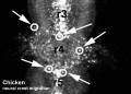
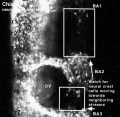
| <mediaplayer width='410' height='340' image="http://php.med.unsw.edu.au/embryology/images/7/7d/Chicken-neural-crest-migration-01.jpg">File:Chicken-neural crest migration 01.mp4</mediaplayer> | 
Chicken embryo sequence shows the migration of DiI-labeled neural crest cells towards the branchial arches as the embryo. White rings indicate migration of individual cells. Each image represents 10 confocal sections separated by 10 microns. |
Movie Source: Original Neural Crest movies kindly provided by Paul Kulesa.[6]
| Neural crest migration Chicken Head (movies overview) | |||||||||||||||||||||||||||
|---|---|---|---|---|---|---|---|---|---|---|---|---|---|---|---|---|---|---|---|---|---|---|---|---|---|---|---|
|
|
|
|
|
|
| |||||||||||||||||||||
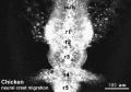
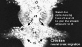
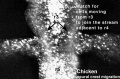
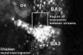
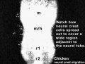
- Neural Crest Movies: Migration 01 | Migration 02 | Migration 03 | Migration 04 | Migration 05 | Migration 06 | Migration 07
Textbooks


Objectives

- Understand the structures derived from ectoderm.
- Understand the formation of neural folds.
- Identify the initial location of neural crest cells in the trilaminar embryo.
- Identify pathways of neural crest migration throughout the embryo.
- To know the major tissues to which neural crest cells contribute.
- To know how abnormalities in development that result from abnormal neural crest cell migration.
- Understand how neural crest cells contribute to the pharyngeal arches and the head structures they form.
Neural Crest Derivatives
A key feature of neural crest is the migration into other embryonic tissues to form specific neural and non-neural populations and structures.
Cranial neural crest
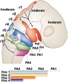
- migration - dorsolaterally and into pharyngeal arches
- craniofacial mesenchyme - cartilage, bone, cranial neurons, glia, and connective tissues of the face
- pharyngeal arches and pouches - thymic cells, tooth odontoblasts, middle ear bones (ossicles) and jaw (mandible)
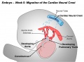
Cardiac neural crest
- migration - located between the cranial and trunk neural crests, overlapping the anterior portion of the vagal neural crest.
- pharyngeal arches - (3,4,6) melanocytes, neurons, cartilage, and connective tissue
- heart outflow tract - aortic arch/pulmonary artery septum, large arteries wall musculoconnective tissue
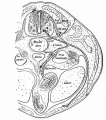
Trunk neural crest
- migration - two major pathways over somites (dorsolaterally) and between somite and neural tube (ventrolaterally)
- dorsolateral - skin melanocytes
- ventrolaterally - dorsal root ganglia, sympathetic ganglia, adrenal medulla, aortic nerve clusters
Vagal and sacral neural crest
- migration - ventrally into surrounding splanchnic mesenchyme of gastrointestinal tract
- splanchnic mesenchyme - parasympathetic (enteric) ganglia of the gut
Development Overview
The following cranial and trunk data is based upon 185 serially sectioned staged (Carnegie) human embryos.[7]
Cranial Neural Crest
- stage 9 - an indication of mesencephalic neural crest
- stage 10 - trigeminal, facial, and postotic components
- stage 11 - crest-free zones are soon observable in rhombomere 1, 3, and 5
- stage 12 - rhombomeres 6 and 7 neural crest migrate to pharyngeal arch 3 and then rostrad to the truncus arteriosus
- stage 13 - nasal crest and the terminalis-vomeronasal complex are last of the cranial crest to appear
stages 9-14 - otic vesicle primordium descends
Vagal Neural Crest
Recent research suggests that the vagal neural crest cells are a transitional population that has evolved between the head and the trunk, taking separate pathways to the both the heart and to the gut.[8][9]
Trunk Neural Crest
Spinal ganglia increase in number over time and are in phase with the somites, though not their centre. There are 3 migratory pathways: ventrolateral between dermatomyotome and sclerotome, ventromedial between neural tube and sclerotomes, and lateral between surface ectoderm and dermatomyotome.
- stage 13 - about 19 present
- stage 14 - about 33 present
- stage 15-23 - 30–35 ganglia
Neck and Shoulder
A mouse study using individually labelled cells of postotic neural crest followed the development of the shoulder girdle (clavicle and scapula) that connects the upper limb to the axial skeleton.[10]
- Clavicle is a neural crest-mesodermal structure, posterior dermal clavicle mesoderm.
- Cryptic cell boundaries traverse apparently homogeneous skeleton of the neck and shoulders.
- Bones and muscles code of connectivity that mesenchymal stem cells of both neural crest and mesodermal origin obey
- Neural crest anchors the head onto the anterior lining of the shoulder girdle
- Hox-gene-controlled mesoderm links trunk muscles to the posterior neck and shoulder skeleton.
- Skeleton identified as neural crest-derived is affected in human Klippel-Feil syndrome, Sprengel's deformity and Arnold-Chiari I/II malformation.
Skin Melanocytes

|
|
| Mouse melanocyte migration[11] | Movie Mouse Skin - Melanoblast Migration E14.5[12] |
References
- ↑ 1.0 1.1 Cox SG, Kim H, Garnett AT, Medeiros DM, An W & Crump JG. (2012). An essential role of variant histone H3.3 for ectomesenchyme potential of the cranial neural crest. PLoS Genet. , 8, e1002938. PMID: 23028350 DOI.
- ↑ Causeret F, Ensini M, Teissier A, Kessaris N, Richardson WD, Lucas de Couville T & Pierani A. (2011). Dbx1-expressing cells are necessary for the survival of the mammalian anterior neural and craniofacial structures. PLoS ONE , 6, e19367. PMID: 21552538 DOI.
- ↑ Betters E, Liu Y, Kjaeldgaard A, Sundström E & García-Castro MI. (2010). Analysis of early human neural crest development. Dev. Biol. , 344, 578-92. PMID: 20478300 DOI.
- ↑ Kulesa PM, Bailey CM, Kasemeier-Kulesa JC & McLennan R. (2010). Cranial neural crest migration: new rules for an old road. Dev. Biol. , 344, 543-54. PMID: 20399765 DOI.
- ↑ Lee G, Chambers SM, Tomishima MJ & Studer L. (2010). Derivation of neural crest cells from human pluripotent stem cells. Nat Protoc , 5, 688-701. PMID: 20360764 DOI.
- ↑ Kulesa PM & Fraser SE. (2000). In ovo time-lapse analysis of chick hindbrain neural crest cell migration shows cell interactions during migration to the branchial arches. Development , 127, 1161-72. PMID: 10683170
- ↑ O'Rahilly R & Müller F. (2007). The development of the neural crest in the human. J. Anat. , 211, 335-51. PMID: 17848161 DOI.
- ↑ Kuo BR & Erickson CA. (2010). Regional differences in neural crest morphogenesis. Cell Adh Migr , 4, 567-85. PMID: 20962585
- ↑ Bryan R. Kuo, Carol A. Erickson Vagal neural crest cell migratory behavior: A transition between the cranial and trunk crest. Volume 240, Issue 9, pages 2084–2100, September 2011 Dev Dynamics
- ↑ Matsuoka T, Ahlberg PE, Kessaris N, Iannarelli P, Dennehy U, Richardson WD, McMahon AP & Koentges G. (2005). Neural crest origins of the neck and shoulder. Nature , 436, 347-55. PMID: 16034409 DOI.
- ↑ Millar SE. (2005). An ideal society? Neighbors of diverse origins interact to create and maintain complex mini-organs in the skin. PLoS Biol. , 3, e372. PMID: 16277556 DOI.
- ↑ Mort RL, Hay L & Jackson IJ. (2010). Ex vivo live imaging of melanoblast migration in embryonic mouse skin. Pigment Cell Melanoma Res , 23, 299-301. PMID: 20067551 DOI.
Reviews
Lee YH & Saint-Jeannet JP. (2011). Sox9 function in craniofacial development and disease. Genesis , 49, 200-8. PMID: 21309066 DOI.
Kish PE, Bohnsack BL, Gallina D, Kasprick DS & Kahana A. (2011). The eye as an organizer of craniofacial development. Genesis , 49, 222-30. PMID: 21309065 DOI.
Jiang M, Stanke J & Lahti JM. (2011). The connections between neural crest development and neuroblastoma. Curr. Top. Dev. Biol. , 94, 77-127. PMID: 21295685 DOI.
PubmedParser error: The PubmedParser extension received invalid XML data. ()
Articles
Search PubMed
Search April 2010 "Neural Crest Development" - All (4354) Review (843) Free Full Text (1621)
Search Pubmed: Neural Crest Development
Additional Images
Cardiac Neural Crest Migration
Hindbrain neural crest migration
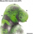
Mouse head E9 neural crest GFP
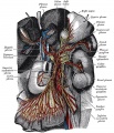
Great plexuses of the sympathetic system


Terms
Glossary Links
- Glossary: A | B | C | D | E | F | G | H | I | J | K | L | M | N | O | P | Q | R | S | T | U | V | W | X | Y | Z | Numbers | Symbols | Term Link
Cite this page: Hill, M.A. (2024, May 26) Embryology Neural Crest - Cranial Nerve Development. Retrieved from https://embryology.med.unsw.edu.au/embryology/index.php/Neural_Crest_-_Cranial_Nerve_Development
- © Dr Mark Hill 2024, UNSW Embryology ISBN: 978 0 7334 2609 4 - UNSW CRICOS Provider Code No. 00098G