Musculoskeletal System - Muscle Development: Difference between revisions
| Line 11: | Line 11: | ||
|-bgcolor="F5FAFF" | |-bgcolor="F5FAFF" | ||
| | | | ||
* '''Jamb and jamc are essential for vertebrate myocyte fusion'''<ref><pubmed>22180726</pubmed></ref> "Cellular fusion is required in the development of several tissues, including skeletal muscle. In vertebrates, this process is poorly understood and lacks an in vivo-validated cell surface heterophilic receptor pair that is necessary for fusion. Identification of essential cell surface interactions between fusing cells is an important step in elucidating the molecular mechanism of cellular fusion. We show here that the zebrafish orthologues of JAM-B and JAM-C receptors are essential for fusion of myocyte precursors to form syncytial muscle fibres. Both jamb and jamc are dynamically co-expressed in developing muscles and encode receptors that physically interact." | * '''Jamb and jamc are essential for vertebrate myocyte fusion'''<ref><pubmed>22180726</pubmed></ref> "Cellular fusion is required in the development of several tissues, including skeletal muscle. In vertebrates, this process is poorly understood and lacks an in vivo-validated cell surface heterophilic receptor pair that is necessary for fusion. Identification of essential cell surface interactions between fusing cells is an important step in elucidating the molecular mechanism of cellular fusion. We show here that the zebrafish orthologues of JAM-B and JAM-C receptors are essential for fusion of myocyte precursors to form syncytial muscle fibres. Both jamb and jamc are dynamically co-expressed in developing muscles and encode receptors that physically interact." JAM-B = [http://omim.org/entry/606870 JAM2] JAM-C = [http://omim.org/entry/606871 JAM3] | ||
* '''Origin of vertebrate limb muscle: the role of progenitor and myoblast populations'''<ref><pubmed>21621065</pubmed></ref> (review) "Muscle development, growth, and regeneration take place throughout vertebrate life. In amniotes, myogenesis takes place in four successive, temporally distinct, although overlapping phases. Understanding how embryonic, fetal, neonatal, and adult muscle are formed from muscle progenitors and committed myoblasts is an area of active research. In this review we examine recent expression, genetic loss-of-function, and genetic lineage studies that have been conducted in the mouse, with a particular focus on limb myogenesis." | * '''Origin of vertebrate limb muscle: the role of progenitor and myoblast populations'''<ref><pubmed>21621065</pubmed></ref> (review) "Muscle development, growth, and regeneration take place throughout vertebrate life. In amniotes, myogenesis takes place in four successive, temporally distinct, although overlapping phases. Understanding how embryonic, fetal, neonatal, and adult muscle are formed from muscle progenitors and committed myoblasts is an area of active research. In this review we examine recent expression, genetic loss-of-function, and genetic lineage studies that have been conducted in the mouse, with a particular focus on limb myogenesis." | ||
* '''The histone methyltransferase Set7/9 promotes myoblast differentiation and myofibril assembly''' <ref><pubmed>21859860</pubmed></ref> Together, our experiments define a biological function for Set7 in muscle differentiation and provide a molecular mechanism by which Set7 modulates myogenic transcription factors during muscle differentiation. | * '''The histone methyltransferase Set7/9 promotes myoblast differentiation and myofibril assembly''' <ref><pubmed>21859860</pubmed></ref> Together, our experiments define a biological function for Set7 in muscle differentiation and provide a molecular mechanism by which Set7 modulates myogenic transcription factors during muscle differentiation. | ||
Revision as of 06:02, 5 January 2012

Introduction
There are 3 different types of muscle: skeletal, cardiac and smooth. This page describes skeletal muscle development, descriptions of cardiac muscle and smooth muscle development can be found in other notes. Skeletal muscle forms by fusion of mononucleated myoblasts to form mutinucleated myotubes.
Differentiation/determination of mesoderm into muscle cells is thought to involve a family of basic Helix-Loop-Helix transcription factors, the first of which discovered was MyoD1. MyoD1 needs to form a dimer to be active and is maintained in an inactive state by binding of an inhibitor, Id.
Some Recent Findings
|
Recent References | References
Myogenesis

Three different types of muscle form in the body.
- Skeletal muscle - cells originate from the paraxial mesoderm, forming somites, then dermamyotome and finally the myotome. Myoblasts undergo frequent divisions and coalesce with the formation of a multinucleated, syncytial muscle fibre or myotube. The nuclei of the myotube are still located centrally in the muscle fibre. In the course of the synthesis of the myofilaments/myofibrils, the nuclei are gradually displaced to the periphery of the cell.
- Cardiac muscle - cells originate from the prechordal splanchnic mesoderm.
- Smooth muscle - cells originate from undifferentiated mesenchymal cells. These cells differentiate first into mitotically active cells, myoblasts, which contain a few myofilaments. Myoblasts give rise to the cells which will differentiate into mature smooth muscle cells.


Muscle Groups
Epaxial Muscle
Anatomical term describing skeletal muscles which lie dorsal (posterior) to the vertebral column developing from the somite myotome. In humans, this is only a small muscle group formed by the transversospinalis, longissimus, and iliocostalis muscles. Also at the ribcage level the levatores costarum muscles involved with rib elevation during respiration. The body muscles lying ventral (anterior) to the vertebral column are the hypaxial muscles.
Hypaxial Muscle
(hypomere) Anatomical term describing skeletal muscles which lie ventral (anterior) to the vertebral column developing from the somite myotome. These muscles contribute both body (trunk) and limb skeletal muscle.
- In the trunk, these form the three anterior body muscle layers.
- In the limb, these form the extensor and flexor muscle groups.
Head Muscle
- jaw associated muscles mainly from cranial mesoderm.
- jaw, connective tissues and tendons from neural crest cells.
For head muscle and connective tissue see.[6]
Skeletal Muscle Stages


Myoblast - individual progenitor cells
Myotube - multinucleated, but undifferentiated contractile apparatus (sarcomere)
Myofibre (myofiber, muscle cell) - multinucleated and differentiated sarcomeres
- primary myofibres - first-formed myofibres, act as a structural framework upon which myoblasts proliferate, fuse in linear sequence
- secondary myofibers - second later population of myofibres that form surrounding the primary fibres.
Muscle Fibre Types

Muscle fiber types
- type IIB, IIA, IIX, and I fibres - based only on the myosin ATPase activity.
- Type I fibres appear red, due to the presence of myoglobin.
- Type II fibres appear white, due to the absence of myoglobin and their glycolytic nature.
- A group of individual myofibres within a muscle will be innervated by a single motor neuron (motor unit).
- The electrical properties of the motor neuron will regulate the contractile properties of all associated myofibres.
| Fibre Type | Type I fibres | Type II a fibres | Type II x fibres | Type II b fibres |
|---|---|---|---|---|
| Contraction time | Slow | Moderately Fast | Fast | Very fast |
| Size of motor neuron | Small | Medium | Large | Very large |
| Resistance to fatigue | High | Fairly high | Intermediate | Low |
| Activity Used for | Aerobic | Long-term anaerobic | Short-term anaerobic | Short-term anaerobic |
| Maximum duration of use | Hours | <30 minutes | <5 minutes | <1 minute |
| Power produced | Low | Medium | High | Very high |
| Mitochondrial density | High | High | Medium | Low |
| Capillary density | High | Intermediate | Low | Low |
| Oxidative capacity | High | High | Intermediate | Low |
| Glycolytic capacity | Low | High | High | High |
| Major storage fuel | Triglycerides | Creatine phosphate, glycogen | Creatine phosphate, glycogen | Creatine phosphate, glycogen |
| Myosin heavy chain, human genes |
MYH7 | MYH2 | MYH1 | MYH4 |
Muscle Contraction

Individual myoblasts in the developing muscle bed initial fuse together to form multi-nucleated myotubes. These myotubes then express the contractile proteins, that are organized into sarcomeres in series along the length of the myotube.
This animation shows the molecular interactions that occur within the skeletal muscle sarcomere between actin and myosin during skeletal muscle contraction.
|
Legend
|

|
Myotome
In both development and the adult, the group of skeletal muscles supplied by a specific segmental spinal nerve is referred to as a myotome. The muscle arises from a specific somite and the spinal nerve arises from a specific level of the spinal cord (identified by veretebral column).
In humans this corresponds to the following spinal nerves (from top to bottom) and muscular functions:
- C3,4 and 5 supply the diaphragm for breathing.
- C5 supply shoulder muscles and muscles to bend our elbow.
- C6 for bending the wrist back.
- C7 for straightening the elbow.
- C8 bends the fingers.
- T1 spreads the fingers.
- T1 –T12 supplies the chest wall and abdominal muscles.
- L2 bends the hip.
- L3 straightens the knee.
- L4 pulls the foot up.
- L5 wiggles the toes.
- S1 pulls the foot down.
- S3,4 and 5 supply the bladder, bowel, sex organs, anal and other pelvic muscles.
Histology Images
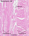
Human HE x4 longitudinal and transverse
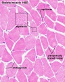
Human HE x40 transverse
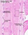
Human HE x40 longitudinal
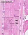
Human HE x40 longitudinal
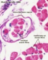


- Muscle Histology: Muscle Development | Human HE x4 longitudinal and transverse | Human HE x40 transverse | Human HE x40 longitudinal | Human HE x40 longitudinal | Human HE x4 longitudinal and transverse | Muscle Spindle HE x40 | Human HE x40 | Human HE x40 | Human HE x40 | Human HE x100 | Human HE x100 | Fetal human muscle | Myotendinous junction label | Myotendinous junction HE x40 | Whipf 1 | Whipf 2 | Whipf 3 | Tongue HE x10 transverse | Tongue x100 | Muscle spindle HE x20 | Muscle spindle HE x40
Puberty
- Musculoskeletal mass doubles by the end of puberty
- regulated growth by - sex steroid hormones, growth hormone, insulin-like growth factors
- accumulation of (peak) bone mass during puberty relates to future osteoporosis in old age
Abnormalities
There can be abnormalities associated directly with muscle differentiation and function as well as those mediated indirectly by abnormalities of innervation or skeletal development and other associated systems.
Duchenne Muscular Dystrophy
The most common occuring in Boys and in Duchenne Muscular Dystrophy (DMD). This cause of the disease was discovered in 1988 as a mutation in dystrophin, a protein that lies under the muscle fiber membrane and maintains the cell's integrity. As skeletal muscles have little prenatal load or use it is not until postnatally that muscle wasting occurs, usually in the anti-gravity muscles first. This is a progressive disease usually detected between 3-5 years old.
- X-linked dystrophy
- large gene encoding cytoskeletal protein - Dystrophin
- progressive wasting of muscle, die late teens
Becker Muscular Dystrophy
A milder adult (30-40 years old) onset form of the disease Becker's Muscular Dystrophy (BMD) that involves mutations in the same dystrophin gene.
Autosomal Recessive Muscular Dystrophy
Dystroglycan, a protein that associates with both dystrophin and membrane molecules, is a candidate gene for the site of the mutation in autosomal recessive muscular dystrophies. A knockout mouse has been generated that has early developmental abnormalities.
Myotonic Dystrophy
An inherited disorder in which the muscles contract but have decreasing power to relax. With this condition, the muscles also become weak and waste away. The myotonic dystrophy gene, found on chromosome 19, codes for a protein kinase that is found in skeletal muscle, where it likely plays a regulatory role. The disease is "amplified" through generations probably by a similar GC expansion associated with Huntington disease.
Facioscapulohumeral muscular dystrophy (FSHD)
- characterized by the progressive weakness and atrophy of a specific subset of skeletal muscles.
- mostly affects the muscles of the face, scapula, and upper arms.
- involvement of specific muscles that it is often used clinically to distinguish FSHD from other forms of muscular dystrophy.
References
- ↑ <pubmed>22180726</pubmed>
- ↑ <pubmed>21621065</pubmed>
- ↑ <pubmed>21859860</pubmed>
- ↑ <pubmed>20888930</pubmed>
- ↑ <pubmed>20946953</pubmed>
- ↑ <pubmed>19198652</pubmed>
- ↑ 7.0 7.1 <pubmed>18945372</pubmed>| PMC2596796 | BMC Syst Biol.
Reviews
<pubmed>21621065</pubmed> <pubmed>21183656</pubmed> <pubmed>20553711</pubmed> <pubmed>19762225</pubmed> <pubmed>16118057</pubmed>
Articles
<pubmed>21859860</pubmed> <pubmed>20195544</pubmed> <pubmed>20037161</pubmed> <pubmed>19198652</pubmed>
Search PubMed
June 2010 " Skeletal Muscle Development" All (19316) Review (2515) Free Full Text (5587) Manage Filters Search Pubmed: Skeletal Muscle Development
Additional Images

Endochondral bone
Terms
External Links
Glossary Links
- Glossary: A | B | C | D | E | F | G | H | I | J | K | L | M | N | O | P | Q | R | S | T | U | V | W | X | Y | Z | Numbers | Symbols | Term Link
Cite this page: Hill, M.A. (2024, April 26) Embryology Musculoskeletal System - Muscle Development. Retrieved from https://embryology.med.unsw.edu.au/embryology/index.php/Musculoskeletal_System_-_Muscle_Development
- © Dr Mark Hill 2024, UNSW Embryology ISBN: 978 0 7334 2609 4 - UNSW CRICOS Provider Code No. 00098G