Musculoskeletal System - Limb Development: Difference between revisions
| Line 23: | Line 23: | ||
== Some Recent Findings == | == Some Recent Findings == | ||
:"First, we assessed the sequence of events following limb amputation in chick embryos and compared the features of limb development and regeneration in amphibians and chicks. Based on our findings, we attempted to re-induce the AER. When wnt-2b/fgf-10-expressing cells were inserted concurrently with wounding, successful re-induction of the AER occurred." | |||
== Textbooks == | == Textbooks == | ||
Revision as of 02:23, 26 April 2010

Introduction

The mesoderm forms nearly all the connective tissues of the musculoskeletal system. Each tissue (cartilage, bone, and muscle) goes through many different mechanisms of differentiation.
The musculoskeletal system consists of skeletal muscle, bone, and cartilage and is mainly mesoderm in origin with some neural crest contribution.
The intraembryonic mesoderm can be broken into paraxial, intermediate and lateral mesoderm relative to its midline position. During the 3rd week the paraxial mesoderm forms into "balls" of mesoderm paired either side of the neural groove, called somites.
Somites appear bilaterally as pairs at the same time and form earliest at the cranial (rostral,brain) end of the neural groove and add sequentially at the caudal end. This addition occurs so regularly that embryos are staged according to the number of somites that are present. Different regions of the somite differentiate into dermomyotome (dermal and muscle component) and sclerotome (forms vertebral column). An example of a specialized musculoskeletal structure can be seen in the development of the limbs.
Skeletal muscle forms by fusion of mononucleated myoblasts to form mutinucleated myotubes. Bone is formed through a lengthy process involving ossification of a cartilage formed from mesenchyme. Two main forms of ossification occur in different bones, intramembranous (eg skull) and endochondrial (eg limb long bones) ossification. Ossification continues postnatally, through puberty until mid 20s. Early ossification occurs at the ends of long bones.
Musculoskeletal and limb abnormalities are one of the largest groups of congenital abnormalities.
| System Links: Introduction | Cardiovascular | Coelomic Cavity | Endocrine | Gastrointestinal Tract | Genital | Head | Immune | Integumentary | Musculoskeletal | Neural | Neural Crest | Placenta | Renal | Respiratory | Sensory | Birth |
--Mark Hill 09:25, 14 April 2010 (EST) Page Template only - content from original UNSW Embryology site currently being edited and updated.
Some Recent Findings
- "First, we assessed the sequence of events following limb amputation in chick embryos and compared the features of limb development and regeneration in amphibians and chicks. Based on our findings, we attempted to re-induce the AER. When wnt-2b/fgf-10-expressing cells were inserted concurrently with wounding, successful re-induction of the AER occurred."
Textbooks
- The Developing Human: Clinically Oriented Embryology (8th Edition) by Keith L. Moore and T.V.N Persaud - Moore & Persaud Chapter 15 the skeletal system
- Larsen’s Human Embryology by GC. Schoenwolf, SB. Bleyl, PR. Brauer and PH. Francis-West - Chapter 11 Limb Dev (bone not well covered in this textbook)
- Before we Are Born (5th ed.) Moore and Persaud Chapter 16,17: p379-397, 399-405
- Essentials of Human Embryology Larson Chapter 11 p207-228
Objectives
- Identify the components of a somite and the adult derivatives of each component.
- Give examples of sites of (a) endochondral and (b) intramembranous ossification and to compare these two processes.
- Identify the general times (a) of formation of primary and (b) of formation of secondary ossification centres, and (c) of fusion of such centres with each other.
- Briefly summarise the development of the limbs.
- Describe the developmental abnormalities responsible for the following malformations: selected growth plate disorders; congenital dislocation of the hip; scoliosis; arthrogryposis; and limb reduction deformities.
Computer Activities
Development Overview
Below is a very brief overview using simple figures of 3 aspects of early musculoskeletal development. More detailed overviews are shown on other notes pages Mesoderm and Somite, Vertebral Column, Limb in combination with serial sections and Carnegie images.
Mesoderm Development

|
Cells migrate through the primitive streak to form mesodermal layer. Extraembryonic mesoderm lies adjacent to the trilaminar embryo totally enclosing the amnion, yolk sac and forming the connecting stalk. |

|
Paraxial mesoderm accumulates under the neural plate with thinner mesoderm laterally. This forms 2 thickened streaks running the length of the embryonic disc along the rostrocaudal axis. In humans, during the 3rd week, this mesoderm begins to segment. The neural plate folds to form a neural groove and folds. |

|
Segmentation of the paraxial mesoderm into somites continues caudally at 1 somite/90minutes and a cavity (intraembryonic coelom) forms in the lateral plate mesoderm separating somatic and splanchnic mesoderm.
Note intraembryonic coelomic cavity communicates with extraembryonic coelom through portals (holes) initially on lateral margin of embryonic disc. |

|
Somites continue to form. The neural groove fuses dorsally to form a tube at the level of the 4th somite and "zips up cranially and caudally and the neural crest migrates into the mesoderm. |
Somite Development

|
Mesoderm beside the notochord (axial mesoderm, blue) thickens, forming the paraxial mesoderm as a pair of strips along the rostro-caudal axis. |

|
Paraxial mesoderm towards the rostral end, begins to segment forming the first somite. Somites are then sequentially added caudally. The somitocoel, is a cavity forming in early somites, which is lost as the somite matures. |

|
Cells in the somite differentiate medially to form the sclerotome (forms vertebral column) and dorsolaterally to form the dermomyotome. |

|
The dermomyotome then forms the dermotome (forms dermis) and myotome (forms muscle).
Neural crest cells migrate beside and through somite. |

|
The myotome differentiates to form 2 components dorsally the epimere and ventrally the hypomere, which in turn form epaxial and hypaxial muscles respectively. The bulk of the trunk and limb muscle coming from the Hypaxial mesoderm. Different structures will be contributed depending upon the somite level. |
Limb Axis Formation
Four Concepts - much of the work has been carried out using the chicken and more recently the mouse model of development.
- Limb Initiation
- Proximodistal Axis
- Dorsoventral Axis
- Anteroposterior Axis
Limb Initiation
- Fibroblast growth factor (FGF) coated beads can induce additional limb
- FGF10 , FGF8 (lateral plate intermediate mesoderm) prior to bud formation
- FGF8 (limb ectoderm) FGFR2
- FGF can respecify Hox gene expression (Hox9- limb position)
- Hox could then activate FGF expression
Note that during the embryonic period there is a rostrocaudal (anterior posterior) timing difference between the upper and lower limb development
- this means that developmental changes in the upper limb can precede similar changes in the lower limb (2-5 day difference in timing)
Limb Identity
Forelimb and hindlimb (mouse) identity appears to be regulated by T-box (Tbx) genes, which are a family of transcription factors.
- hindlimb Tbx4 is expressed.
- forelimb Tbx5 is expressed.
- Tbx2 and Tbx3 are expressed in both limbs.
Related Research - PMID: 12490567 | Development 2003 Figures | Scanning electron micrographs of E9 Limb bud wild-type and Tbx5del/del A model for early stages of limb bud growth | PMID: 12736217 | Development 2003 Figures
Body Axes
- Anteroposterior - (Rostrocaudal, Craniocaudal, Cephalocaudal) from the head end to opposite end of body or tail.
- Dorsoventral - from the spinal column (back) to belly (front).
- Proximodistal - from the tip of an appendage (distal) to where it joins the body (proximal).
Proximodistal Axis
- Apical Ectodermal Ridge (AER) formed by Wnt7a
- then AER secretes FGF2, 4, 8
- stimulates proliferation and outgrowth
apical ectodermal ridge | AER and vascular channel
Dorsoventral Axis
- Somites - provides dorsal signal to mesenchyme which dorsalizes ectoderm
- Ectoderm - then in turn signals back (Wnt7a) to mesenchyme to pattern limb
Wnt7a
- name was derived from 'wingless' and 'int’
- Wnt gene first defined as a protooncogene, int1
- Humans have at least 4 Wnt genes
- Wnt7a gene is at 3p25 encoding a 349aa secreted glycoprotein
- patterning switch with different roles in different tissues
- mechanism of Wnt and receptor distribution still being determined (free diffusion, restricted diffusion and active transport)
One WNT receptor is Frizzled (FZD)
- Frizzled gene family encodes a 7 transmembrane receptor
Fibroblast growth factors (FGF)
- Family of at least 17 secreted proteins
- bind membrane tyrosine kinase receptors
- Patterning switch with many different roles in different tissues
- FGF8 = androgen-induced growth factor, AIGF
FGF receptors
- comprise a family of at least 4 related but individually distinct tyrosine kinase receptors (FGFR1- 4) similar protein structure
- 3 immunoglobulin-like domains in extracellular region
- single membrane spanning segment
- cytoplasmic tyrosine kinase domain
Anteroposterior Axis
- Zone of polarizing activity (ZPA)
- a mesenchymal posterior region of limb
- secretes sonic hedgehog (SHH)
- apical ectodermal ridge (AER), which has a role in patterning the structures that form within the limb
- majority of cell division (mitosis) occurs just deep to AER in a region known as the progress zone
- A second region at the base of the limbbud beside the body, the zone of polarizing activity (ZPA) has a similar patterning role to the AER, but in determining another axis of the limb
Wing as Limb Model
- chicken wing easy to manipulate
- removal, addition and rotation of limb regions
- grafting additional AER, ZPA
- implanting growth factor secreting structures
UNSW Embryology - Axes Formation - Limb | Signal Factors - Wnt
References
Reviews
- How to make a zone of polarizing activity: insights into limb development via the abnormality preaxial polydactyly. Hill RE. Dev Growth Differ. 2007 Aug;49(6):439-48. Review. PMID: 17661738
Articles
- The apical ectodermal ridge (AER) can be re-induced by wounding, wnt-2b, and fgf-10 in the chicken limb bud. Satoh A, Makanae A, Wada N. Dev Biol. 2010 Mar 27. PMID: 20347761
- Modification of the zone of polarizing activity signal by trypsin. Stefanov EK, Ferrage JM, Parchim NF, Lee CE, Reginelli AD, Taché M, Anderson RA. Dev Growth Differ. 2009 Feb;51(2):123-33. PMID: 19207183
Search PubMed
Search April 2010
- Limb Development - All (776) Review (108) Free Full Text (196)
- apical ectodermal ridge - All (95) Review (2) Free Full Text (31)
- zone polarizing activity - All (15) Review (2) Free Full Text (7)
Search Pubmed: Limb Development | apical ectodermal ridge | zone polarizing activity]
Additional Images

Adult appendicular skeleton

Bone structure
Developing vertebra

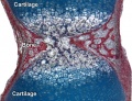
Endochondral bone


Terms
Glossary Links
- Glossary: A | B | C | D | E | F | G | H | I | J | K | L | M | N | O | P | Q | R | S | T | U | V | W | X | Y | Z | Numbers | Symbols | Term Link
Cite this page: Hill, M.A. (2024, May 2) Embryology Musculoskeletal System - Limb Development. Retrieved from https://embryology.med.unsw.edu.au/embryology/index.php/Musculoskeletal_System_-_Limb_Development
- © Dr Mark Hill 2024, UNSW Embryology ISBN: 978 0 7334 2609 4 - UNSW CRICOS Provider Code No. 00098G