Mouse E13 microCT Movie: Difference between revisions
mNo edit summary |
mNo edit summary |
||
| (One intermediate revision by the same user not shown) | |||
| Line 22: | Line 22: | ||
:'''Links:''' [[Media:Mouse_embryo_E13_microCT_06.mp4|MP4 version | :'''Links:''' [[Media:Mouse_embryo_E13_microCT_06.mp4|MP4 version]] | [[Movies]] | [[Computed Tomography]] | {{mouse}} | ||
<br> | |||
{{Mouse links}} | |||
|} | |} | ||
| Line 28: | Line 30: | ||
===Reference=== | ===Reference=== | ||
{{#pmid:19545439}} | |||
====Copyright==== | ====Copyright==== | ||
| Line 37: | Line 39: | ||
PTA-stained embryo | PTA-stained embryo | ||
{{Footer}} | |||
[[Category:Mouse]] [[Category:Computed Tomography]] [[Category:Mouse E13]] | [[Category:Mouse]] [[Category:Computed Tomography]] [[Category:Mouse E13]] | ||
[[Category:Movies]] | [[Category:Movies]] | ||
Latest revision as of 16:44, 24 April 2018
| <html5media height="640" width="400">File:Mouse_embryo_E13_microCT_06.mp4</html5media> |
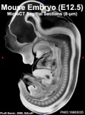
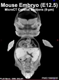
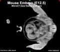
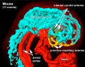
Mouse embryo, Theiler stage 21. Theiler Stage 21 Anterior footplate indented, marked pinna
| ||||||||||||||||||||||||||||||||||||||||||||||||||||||||||||||||||||||||||||||||||||||||||||||||||||||||||||||||||||||||||||||
Reference
Metscher BD. (2009). MicroCT for comparative morphology: simple staining methods allow high-contrast 3D imaging of diverse non-mineralized animal tissues. BMC Physiol. , 9, 11. PMID: 19545439 DOI.
Copyright
© 2009 Metscher; licensee BioMed Central Ltd. This is an Open Access article distributed under the terms of the Creative Commons Attribution License (http://creativecommons.org/licenses/by/2.0), which permits unrestricted use, distribution, and reproduction in any medium, provided the original work is properly cited.
PTA-stained embryo
Cite this page: Hill, M.A. (2024, April 26) Embryology Mouse E13 microCT Movie. Retrieved from https://embryology.med.unsw.edu.au/embryology/index.php/Mouse_E13_microCT_Movie
- © Dr Mark Hill 2024, UNSW Embryology ISBN: 978 0 7334 2609 4 - UNSW CRICOS Provider Code No. 00098G