Medaka Development: Difference between revisions
| Line 19: | Line 19: | ||
Posthatch features observed included: fins, scales and secondary sexual characteristics. | Posthatch features observed included: fins, scales and secondary sexual characteristics. | ||
===Stages=== | |||
Stage 0. Unfertilized eggs | |||
Stage 1 (3 min). Activated egg stage | |||
Stage 2. Blastodisc stage | |||
2.4. Stage 3 (1 h 5 min). 2 cell stage | |||
2.5. Stage 4 (1 h 45 min). 4 cell stage | |||
2.6. Stage 5 (2 h 20 min). 8 cell stage | |||
2.7. Stage 6 (2 h 55 min). 16 cell stage | |||
2.8. Stage 7 (3 h 30 min). 32 cell stage | |||
2.9. Stage 8 (4 h 5 min). Early morula stage | |||
2.10. Stage 9 (5 h 15 min). Late morula stage | |||
2.11. Stage 10 (6 h 30 min). Early blastula stage | |||
2.12. Stage 11 (8 h 15 min). Late blastula stage | |||
2.13. Stage 12 (10 h 20 min). Pre-early gastrula stage | |||
2.14. Stage 13 (13 h). Early gastrula stage | |||
2.15. Stage 14 (15 h). Pre-mid-gastrula stage | |||
2.16. Stage 15 (17 h 30 min). Mid-gastrula stage | |||
2.17. Stage 16 (21 h). Late gastrula stage | |||
2.18. Stage 17 (1 day 1 h). Early neurula stage (head formation) | |||
2.19. Stage 18 (1 day 2 h). Late neurula stage (optic bud formation) | |||
2.20. Stage 19 (1 day 3 h 30 min). 2 somite stage | |||
2.21. Stage 20 (1 day 7 h 30 min). 4 somite stage | |||
2.22. Stage 21 (1 day 10 h). 6 somite stage (brain regionalization and otic vesicle formation) | |||
2.23. Stage 22 (1 day 14 h). 9 somite stage (appearance of heart anlage) | |||
2.24. Stage 23 (1 day 17 h). 12 somite stage (formation of tubular heart) | |||
2.25. Stage 24 (1 day 20 h). 16 somite stage (start of heart beating) | |||
2.26. Stage 25 (2 days 2 h). 18–19 somite stage (onset of blood circulation) | |||
2.27. Stage 26 (2 days 6 h). 22 somite stage (development of guanophores and vacuolization of the notochord) | |||
2.28. Stage 27 (2 days 10 h). 24 somite stage (appearance of pectoral fin bud) | |||
2.29. Stage 28 (2 days 16 h). 30 somite stage (onset of retinal pigmentation) | |||
2.30. Stage 29 (3 days 2 h). 34 somite stage (internal ear formation) | |||
2.31. Stage 30 (3 days 10 h). 35 somite stage (blood vessel development) | |||
2.32. Stage 31 (3 days 23 h). Gill blood vessel formation stage | |||
2.33. Stage 32 (4 days 5 h). Somite completion stage (formation of pronephros and air bladder) | |||
2.34. Stage 33 (4 days 10 h). Stage at which notochord vacuolization is completed | |||
2.35. Stage 34 (5 days 1 h). Pectoral fin blood circulation stage | |||
2.36. Stage 35 (5 days 12 h). Stage at which visceral blood vessels form | |||
2.37. Stage 36 (6 days). Heart development stage | |||
2.38. Stage 37 (7 days). Pericardial cavity formation stage | |||
2.39. Stage 38 (8 days). Spleen development stage (differentiation of caudal fin begins) | |||
2.40. Stage 39 (9 days). Hatching stage | |||
2.41. Stage 40. 1st fry stage | |||
2.42. Stage 41 | |||
2.43. Stage 42 | |||
2.44. Stage 43 | |||
2.45. Stage 44 | |||
2.46. Stage 45 | |||
==References== | ==References== | ||
Revision as of 15:52, 14 July 2010
Introduction
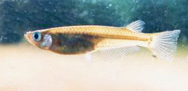
Medaka Oryzias latipes or Japanese rice fish is a member of the killifish family first described in 1846 and has been widely used as a aquarium fish. A modified aquarium version with a genetically modified fluorescent (GFP) version also now available in some countries.
A recent study by Iwamatsu T., 2004[1] has characterised the stages of normal fish development.
Medaka fish were also the first for the first vertebrate animal to mate in space (The International Microgravity Laboratory IML-2/STS-65 mission in 1994) as a developmental model for space experiments. The fish has also been used in studies of pigmentation development.
Links: original Medaka page
Taxon
cellular organisms; Eukaryota; Fungi/Metazoa group; Metazoa; Eumetazoa; Bilateria; Coelomata; Deuterostomia; Chordata; Craniata; Vertebrata; Gnathostomata; Teleostomi; Euteleostomi; Actinopterygii; Actinopteri; Neopterygii; Teleostei; Elopocephala; Clupeocephala; Euteleostei; Neognathi; Neoteleostei; Eurypterygii; Ctenosquamata; Acanthomorpha; Euacanthomorpha; Holacanthopterygii; Acanthopterygii; Euacanthopterygii; Percomorpha; Smegmamorpha; Atherinomorpha; Beloniformes; Adrianichthyoidei; Adrianichthyidae; Oryziinae; Oryzias
Development Overview
Development has been characerised by light microscope observation into 39 prehatch stages and 6 posthatch stages.
Prehatch features observed included: number and size of blastomeres, form of the blastoderm, extent of epiboly, central nervous system, number and form of somites, optic and otic, notochord, heart, blood circulation, the size and movement of the body, tail, membranous fin (fin fold), viscera (liver gallbladder, gut tube), spleen and swim (air) bladder.
Posthatch features observed included: fins, scales and secondary sexual characteristics.
Stages
Stage 0. Unfertilized eggs
Stage 1 (3 min). Activated egg stage
Stage 2. Blastodisc stage 2.4. Stage 3 (1 h 5 min). 2 cell stage 2.5. Stage 4 (1 h 45 min). 4 cell stage 2.6. Stage 5 (2 h 20 min). 8 cell stage 2.7. Stage 6 (2 h 55 min). 16 cell stage 2.8. Stage 7 (3 h 30 min). 32 cell stage 2.9. Stage 8 (4 h 5 min). Early morula stage 2.10. Stage 9 (5 h 15 min). Late morula stage 2.11. Stage 10 (6 h 30 min). Early blastula stage 2.12. Stage 11 (8 h 15 min). Late blastula stage 2.13. Stage 12 (10 h 20 min). Pre-early gastrula stage 2.14. Stage 13 (13 h). Early gastrula stage 2.15. Stage 14 (15 h). Pre-mid-gastrula stage 2.16. Stage 15 (17 h 30 min). Mid-gastrula stage 2.17. Stage 16 (21 h). Late gastrula stage 2.18. Stage 17 (1 day 1 h). Early neurula stage (head formation) 2.19. Stage 18 (1 day 2 h). Late neurula stage (optic bud formation) 2.20. Stage 19 (1 day 3 h 30 min). 2 somite stage 2.21. Stage 20 (1 day 7 h 30 min). 4 somite stage 2.22. Stage 21 (1 day 10 h). 6 somite stage (brain regionalization and otic vesicle formation) 2.23. Stage 22 (1 day 14 h). 9 somite stage (appearance of heart anlage) 2.24. Stage 23 (1 day 17 h). 12 somite stage (formation of tubular heart) 2.25. Stage 24 (1 day 20 h). 16 somite stage (start of heart beating) 2.26. Stage 25 (2 days 2 h). 18–19 somite stage (onset of blood circulation) 2.27. Stage 26 (2 days 6 h). 22 somite stage (development of guanophores and vacuolization of the notochord) 2.28. Stage 27 (2 days 10 h). 24 somite stage (appearance of pectoral fin bud) 2.29. Stage 28 (2 days 16 h). 30 somite stage (onset of retinal pigmentation) 2.30. Stage 29 (3 days 2 h). 34 somite stage (internal ear formation) 2.31. Stage 30 (3 days 10 h). 35 somite stage (blood vessel development) 2.32. Stage 31 (3 days 23 h). Gill blood vessel formation stage 2.33. Stage 32 (4 days 5 h). Somite completion stage (formation of pronephros and air bladder) 2.34. Stage 33 (4 days 10 h). Stage at which notochord vacuolization is completed 2.35. Stage 34 (5 days 1 h). Pectoral fin blood circulation stage 2.36. Stage 35 (5 days 12 h). Stage at which visceral blood vessels form 2.37. Stage 36 (6 days). Heart development stage 2.38. Stage 37 (7 days). Pericardial cavity formation stage 2.39. Stage 38 (8 days). Spleen development stage (differentiation of caudal fin begins) 2.40. Stage 39 (9 days). Hatching stage 2.41. Stage 40. 1st fry stage 2.42. Stage 41 2.43. Stage 42 2.44. Stage 43 2.45. Stage 44 2.46. Stage 45
References
- ↑ <pubmed>15210170</pubmed>
Search Pubmed
Search Pubmed: Medaka Development
| Animal Development: axolotl | bat | cat | chicken | cow | dog | dolphin | echidna | fly | frog | goat | grasshopper | guinea pig | hamster | horse | kangaroo | koala | lizard | medaka | mouse | opossum | pig | platypus | rabbit | rat | salamander | sea squirt | sea urchin | sheep | worm | zebrafish | life cycles | development timetable | development models | K12 |
Glossary Links
- Glossary: A | B | C | D | E | F | G | H | I | J | K | L | M | N | O | P | Q | R | S | T | U | V | W | X | Y | Z | Numbers | Symbols | Term Link
Cite this page: Hill, M.A. (2024, May 19) Embryology Medaka Development. Retrieved from https://embryology.med.unsw.edu.au/embryology/index.php/Medaka_Development
- © Dr Mark Hill 2024, UNSW Embryology ISBN: 978 0 7334 2609 4 - UNSW CRICOS Provider Code No. 00098G
