Magnetic Resonance Imaging
Introduction


Recently there have been several groups preparing developmental embryo atlases of several species, including human[2], based upon imaging of different age embryos. There have been many studies of adult anatomical structures and also some of the placenta.
Magnetic Resonance Imaging (MRI) began in 1977 and uses magnetism, radio waves, and a computer to produce images either as individual slices or reconstructed to give three dimensional (3D) views of specific anatomical regions or structures.
MRI can be used in fetuses at 18 weeks gestational age or later and has been used mainly in brain and spinal diagnosis, and has also been used to investigate other abnormalities of pregnancy.
Diffusion Tensor Imaging (DTI) is a newly developed form of magnetic resonance imaging (MRI). Magnetic field variations of the MRI magnet are applied in at least six different directions generating a three dimensional shape of the diffusion pattern. This technique has be used mainly in neural imaging of white matter, due to the orientation of axon bundles and the associated directional water flow. (More? Neural Development Imaging) Computed Tomography is an alternative method of diagnostic imaging using X-rays.
- Links: Movies | Neural System - Postnatal | Computed Tomography | Category:Magnetic Resonance Imaging
About MRI
A strong magnetic field (up to 1.5 to 4 Tesla) is generated in the machine through which the body is passed (the centre of the "donut ring" seen in the image). The earth's natural magnetic field is about 0.5 Gauss compared to 15,000 Gauss (1.5 Tesla) in the MRI.
Tesla (symbol T) The SI derived unit of magnetic flux density (or magnetic inductivity) was defined in 1960 and named after Nikola Tesla.
Some Recent Findings
|
Human Birth
The images below are from a recent study employing new MRI equipment and methodology that allows more free access to the scanned patient compared to the original methodologies. This should allow study of complex physiological processes, such as childbirth, in relative real-time.

2010.12.07 - Press release | Birth Image | AG OMRT - Radiologie "the birth of a child in an “open” MRI (magnetic resonance imaging) scanner that allows a mother-to-be to fit fully into the machine."

This birth is a vertex (head or cephalic) presentation in an occipito-anterior position. Labor stage 2 is shown with the head already within the pelvis birth canal, and lying between the maternal pubic symphysis (anterior) and the sacrum (posterior).
- Links: Birth | AG OMRT - Radiologie
Species Imaging with MRI
Human
- Whole Embryo[2] "To obtain data on early human development, we used magnetic resonance (MR) imaging and episcopic fluorescence capture (EFIC) to acquire digital images of human embryos spanning the time of dynamic tissue remodeling and organogenesis (Carnegie stages 13 to 23)."
- Brain[1] "Quantitative whole brain 3D validation of tissue labeling performed on a set of 14 fetal MR scans (20.57-22.86 weeks gestational age) demonstrates that this atlas-based EM segmentation approach achieves consistently high DSC performance for the main tissue types in the fetal brain."
- Birth 2010.12.07 - Press release | Birth Image | AG OMRT - Radiologie "the birth of a child in an “open” MRI (magnetic resonance imaging) scanner that allows a mother-to-be to fit fully into the machine."
Baboon
- Brain[4] "We devised a protocol to scan pregnant baboons serially at 3 T for up to 3 h per session. Seven baboons were scanned 1-6 times, beginning as early as 56 days post-conceptional age, and as late as 185 days (term approximately 185 days). Successful scanning of the fetal baboon required careful animal preparation and anesthesia, in addition to optimization of the scanning protocol. We successfully acquired maps of relaxation times (T(1) and T(2)) and high-resolution anatomical images of the brains of fetal baboons at multiple time points during the course of gestation."
Mouse
- Mouse Mutants[5]"Using individual 3D embryo MRI histology, we identified new pituitary phenotypes in Hesx1 mutant mice. Subsequently we use advanced computational techniques to produce a whole-body embryo atlas from 6 CD-1 embryos, creating an average image with a greatly enhanced anatomical detail, particularly in CNS structures."
- A 4D atlas and morphologic database[6] "This work makes magnetic resonance microscopy of the mouse embryo and neonate broadly available with carefully annotated normative data and an extensive environment for collaborations."
Chicken
- Eye development PMID19540232 "We subsequently used the images obtained from the MRI data in order to make precise measurements of chick embryo eye surface area, volume and axial length from E4 to E10."
Xenopus
- Early embryo[7]"Here, we report on the use of microscopic magnetic resonance imaging (mMRI) to noninvasively observe mitotic cell division of early blastomeres in the optically opaque Xenopus laevis embryo."
Embryo Imaging
| MRI 01 | MRI 02 | MRI 03 |
- Links: Movies | Flash Movies | Quicktime Movies
Structure Imaging
Placenta

|
| Magnetic resonance angiography (MRA) of human placenta viewed from the fetal side.[8] |
- Links: Placenta Development
Adult Inner Ear
The 3D reconstructed technique was used to acquire coronal and axial images of the adult inner ear. The coronal section reconstruction was chosen since it increases visibility of the turns of the cochlea.[9]

|

|
Neural
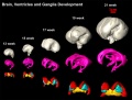
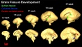
The following images are from human fixed fetal brains scanned with diffusion tensor magnetic resonance imaging.[1]
Diffusion tensor imaging (DTI) A newly developed form of magnetic resonance imaging (MRI). Magnetic field variations of the MRI magnet are applied in at least six different directions generating a three dimensional shape of the diffusion pattern. This technique can be used in neural imaging of white matter due to the orientation of axon bundles and the associated water flow. (More? Magnetic Resonance Imaging)
- Neural DTI Links: Scaled Fissures 13-21 weeks | Fissures 13-21 weeks | Brain Sylvian Fissure | Scaled Brain and Ventricles 13-21 weeks | Scaled Brain, Ventricles and Ganglia 13-21 weeks | Limbic Tract 13-19 weeks | Brain and Ventricles 13-21 weeks | Sylvian Fissure Movie | Neural System Development | Magnetic Resonance Imaging
Brain Tract


Brain Ventricles and Ganglia
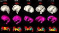
Brain ventricles and ganglia development
Brain ventricles and ganglia development (scaled)
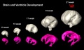
Brain and ventricle development (scaled)


Brain Fissures
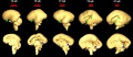
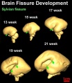
Sylvian Fissure


Prenatal Diagnosis
MRI can be used in fetuses at 18 weeks gestational age or later and has been used mainly in brain and spinal diagnosis, and has also been used to investigate other abnormalities of pregnancy.
- Absence of harmful effects of magnetic resonance exposure at 1.5 T in utero during the third trimester of pregnancy: a follow-up study.[10]"Thirty-five children between 1 and 3 years of age, and nine children between 8 and 9 years of age, that were exposed to MR during the third trimester of pregnancy, were checked for possible adverse effects in a follow-up study. Data on pregnancy and birth, the results of a neurological examination at 3 months, their medical documentary with emphasis on eye and ear functioning, and a questionnaire answered by their mothers were collected and evaluated. In five children abnormal test results were observed, that had no relation to the MR exposure. No harmful effects of prenatal MR exposure in the third trimester of pregnancy were detected in this study."
- Prenatal diagnosis of neurofibromatosis type 1: sonographic and MRI findings.[11] "Prenatal ultrasound and magnetic resonance imaging (MRI) demonstrated a large oropharyngeal tumor, and cardiac and cranial abnormalities consistent with neurofibromatosis type 1 (NF1) in a third-trimester fetus, which were confirmed on postmortem examination. Sonographic features of NF1 are generally nonspecific; MR examination provided significant additional information, facilitating prenatal diagnosis."
- In utero magnetic resonance imaging for brain and spinal abnormalities in fetuses.[12] "In the past eight years magnetic resonance imaging has been used to detect fetal abnormalities in utero at many centres around the world."
Links: Prenatal Diagnosis
References
- ↑ 1.0 1.1 1.2 <pubmed>20108226</pubmed>
- ↑ 2.0 2.1 2.2 <pubmed>20503356</pubmed>
- ↑ <pubmed>19339620</pubmed>
- ↑ <pubmed>18155925</pubmed>
- ↑ <pubmed>20656039</pubmed>
- ↑ <pubmed>18713865</pubmed>| PMC2527911 | PNAS
- ↑ <pubmed>16958098</pubmed>
- ↑ <pubmed>20226038</pubmed>| BMC Physiol.
- ↑ <pubmed>19575114</pubmed>| Braz J Otorhinolaryngol.
- ↑ <pubmed>15234454</pubmed>
- ↑ <pubmed>16981221</pubmed>
- ↑ <pubmed>16150769</pubmed>| BMJ
Reviews
<pubmed>19208672</pubmed> <pubmed>19755601</pubmed> <pubmed>19732621</pubmed> <pubmed>18591320</pubmed> <pubmed>17353684</pubmed>
Articles
<pubmed>20656039</pubmed> <pubmed>19076956</pubmed> <pubmed>18709400</pubmed> <pubmed>15615595</pubmed>
Search PubMed
Search Pubmed: Embryo Magnetic Resonance Imaging | Magnetic Resonance Imaging |
External Links
External Links Notice - The dynamic nature of the internet may mean that some of these listed links may no longer function. If the link no longer works search the web with the link text or name. Links to any external commercial sites are provided for information purposes only and should never be considered an endorsement. UNSW Embryology is provided as an educational resource with no clinical information or commercial affiliation.
- Brookhaven National Laboratory New MicroMRI Facility Expands Lab's Brain-Imaging Capabilities
- 3-D MRI Digital Atlas Database of an Adult C57BL/6J Mouse Brain
- Related internal links: Mouse Development | Neural System Development
- Beth Israel Deaconess Medical Center Atlas of Fetal MRI - head and neck | brain | face | spine | chest | abdomen | genitourinary | extremities
Glossary Links
- Glossary: A | B | C | D | E | F | G | H | I | J | K | L | M | N | O | P | Q | R | S | T | U | V | W | X | Y | Z | Numbers | Symbols | Term Link
Cite this page: Hill, M.A. (2024, May 21) Embryology Magnetic Resonance Imaging. Retrieved from https://embryology.med.unsw.edu.au/embryology/index.php/Magnetic_Resonance_Imaging
- © Dr Mark Hill 2024, UNSW Embryology ISBN: 978 0 7334 2609 4 - UNSW CRICOS Provider Code No. 00098G