Historic Embryology Vignette
| Embryology - 4 May 2024 |
|---|
| Google Translate - select your language from the list shown below (this will open a new external page) |
|
العربية | català | 中文 | 中國傳統的 | français | Deutsche | עִברִית | हिंदी | bahasa Indonesia | italiano | 日本語 | 한국어 | မြန်မာ | Pilipino | Polskie | português | ਪੰਜਾਬੀ ਦੇ | Română | русский | Español | Swahili | Svensk | ไทย | Türkçe | اردو | ייִדיש | Tiếng Việt These external translations are automated and may not be accurate. (More? About Translations) |
Introduction

This page shows the brief historic vignettes that appear on various notes pages introduction and other sections. These are intended to give some historic background to Embryology. These can also appear as a collapsible table.
The links shown below are to full versions of historic embryology textbooks and papers.
| Historic Disclaimer - information about historic embryology pages |
|---|
|
Cerebellum
| Historic Embryology | ||||
| ||||


Ductus Deferens
| Historic Embryology |
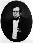
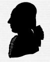  Caspar Friedrich Wolff (1734-1794) was a German embryologist and anatomist best known today for identifying the Wolffian duct (mesonephric duct; ductus deferens, epididymis), Wolffian body (mesonephros) and Wolffian cyst (mesonephric origin uterine broad ligament cyst) that bear his name. Thought also to be a founder of the germ layer theory. His doctorate dissertation Theoria generationis (1774) discarded the developmental theory of preformation. Later in his career, his teaching in Berlin was opposed by the professors of the Medical-Surgical College, who had guild privileges to teach medicine. |
Gastrulation
| Historic Embryology |
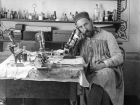
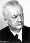 Hans Spemann (1869 - 1941) identified this region in amphibia, also called the "Spemann's organiser". The same region in birds it is known as "Hensen's node" named for Victor Hensen (1835 – 1924) and is also known generally as the primitive node or knot. In humans, it is proposed that similar mechanisms regulate gastrulation to those found in other vertebrates. Currently, the molecular and physical mechanisms that regulate patterning and migration during this key event are being investigated in several different animal models. |
Male Genital
| Historic Embryology |
Caspar Friedrich Wolff (1734-1794) was a German embryologist and anatomist best known today for identifying the Wolffian duct (mesonephric duct; ductus deferens, epididymis), Wolffian body (mesonephros) and Wolffian cyst (mesonephric origin uterine broad ligament cyst) that bear his name. Thought also to be a founder of the germ layer theory. His doctorate dissertation Theoria generationis (1774) discarded the developmental theory of preformation. Later in his career, his teaching in Berlin was opposed by the professors of the Medical-Surgical College, who had guild privileges to teach medicine. |
Neural Crest
| Historic Embryology |
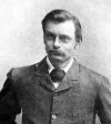 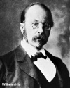 Arthur Milnes Marshall (1852–1893) at Cambridge in 1879 historically first described this embryonic region. In his study of dogfish and chicken brain development, and identified it as "neural crest".[1] See neural crest history and the original 1879 article. Wilhelm His (1831-1904) in 1868 also described in the chick embryo the early neural structure that would form neural crest. |
Ovary
| Historic Embryology |
  |
Pituitary
| Historic Embryology |
|
Uterus
| Historic Embryology |
 |
X Inactivation
| Historic Embryology |
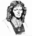
 Mary Lyon (1925-2014) was a UK geneticist who proposed in 1961 the theory of X inactivation, where one of the two X chromosomes in the cells of female mammals is randomly inactivated during early development. In deference to her, this process is also referred to as "Lyonisation". She also worked on other X-linked genetic diseases, such as Duchenne muscular dystrophy and haemophilia. |
References
- ↑ Marshall AM. The morphology of the vertebrate olfactory organ. (1879) Quarterly Journal of Microscopic Science. 19: 300–340.
Glossary Links
- Glossary: A | B | C | D | E | F | G | H | I | J | K | L | M | N | O | P | Q | R | S | T | U | V | W | X | Y | Z | Numbers | Symbols | Term Link
Cite this page: Hill, M.A. (2024, May 4) Embryology Historic Embryology Vignette. Retrieved from https://embryology.med.unsw.edu.au/embryology/index.php/Historic_Embryology_Vignette
- © Dr Mark Hill 2024, UNSW Embryology ISBN: 978 0 7334 2609 4 - UNSW CRICOS Provider Code No. 00098G