Hearing - Inner Ear Development: Difference between revisions
| Line 7: | Line 7: | ||
==Otic Placode== | ==Otic Placode== | ||
The sensory placode associated with hearing and balance. | |||
* Stage 13/14 embryo (shown below) the otic placode has sunk from the surface ectoderm to form a hollow epithelial ball, the otocyst, which now lies beneath the surface surrounded by mesenchyme (mesoderm). | * Stage 13/14 embryo (shown below) the otic placode has sunk from the surface ectoderm to form a hollow epithelial ball, the otocyst, which now lies beneath the surface surrounded by mesenchyme (mesoderm). | ||
Revision as of 12:21, 4 June 2010
Introduction
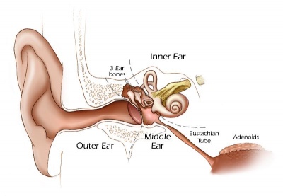
The inner ear is derived from a pair of surface sensory placodes (otic placodes) in the head region. These placodes fold inwards forming a depression, then pinch off entirely from the surface forming a fluid-filled sac or vesicle (otic vesicle, otocyst). The vesicle sinks into the head mesenchyme some of which closely surrounds the otocyst forming the otic capsule. The otocyst finally lies close to the early developing hindbrain (rhombencephalon) and the developing vestibulo-cochlear-facial ganglion complex.
Otic Placode
The sensory placode associated with hearing and balance.
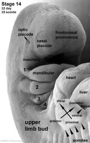
- Stage 13/14 embryo (shown below) the otic placode has sunk from the surface ectoderm to form a hollow epithelial ball, the otocyst, which now lies beneath the surface surrounded by mesenchyme (mesoderm).
- The epithelia of this ball varies in thickness and has begun to distort, it will eventually form the inner ear membranous labyrinth.
Sensory Placodes
- week 4 a series of thickened surface ectodermal patches form in pairs rostro-caudally in the head region.
- Recent research suggests that all sensory placodes may arise from common panplacodal primordium origin around the neural plate, and then differentiate to eventually have different developmental fates. (More? Schlosser G.)
- sensory placodes will later contribute key components of each of our special senses (hearing, vision, smell and taste).
- Otic Placode - one of the first to form and contributes inner ear structures.
- Optic (Lens) Placode - lies on the surface, adjacent to the outpocketing of the nervous system (which will for the retina) and will form the lens.
- Nasal Placode - 2 components (medial and lateral) and will form the nose olefactory epithelium.
- Other species have a number of additional placodes which form other sensory structures (fish, lateral line receptor).
- Note that their initial postion on the developing head is significantly different to their final position in the future sensory system.
Glossary Links
- Glossary: A | B | C | D | E | F | G | H | I | J | K | L | M | N | O | P | Q | R | S | T | U | V | W | X | Y | Z | Numbers | Symbols | Term Link
Cite this page: Hill, M.A. (2024, May 1) Embryology Hearing - Inner Ear Development. Retrieved from https://embryology.med.unsw.edu.au/embryology/index.php/Hearing_-_Inner_Ear_Development
- © Dr Mark Hill 2024, UNSW Embryology ISBN: 978 0 7334 2609 4 - UNSW CRICOS Provider Code No. 00098G

