File:Stage 13 image 001.jpg: Difference between revisions
No edit summary |
mNo edit summary |
||
| (7 intermediate revisions by one other user not shown) | |||
| Line 1: | Line 1: | ||
= | =A2 Image Features= | ||
[[File:Stage 13 A2 plane.gif|right]] | |||
:'''Links:''' A2 | [[:File:Stage 13 image 051.jpg|Labelled]] | [[:File:Stage 13 image 001.jpg|Previous]] | [[:File:Stage 13 image 003.jpg|Next]] | |||
=== | ===Head and Neck=== | ||
[[Head Development]] | |||
[[Category:Head]] | |||
===Neural System=== | |||
[[Neural System Development]] | |||
[[Category:Neural]] | |||
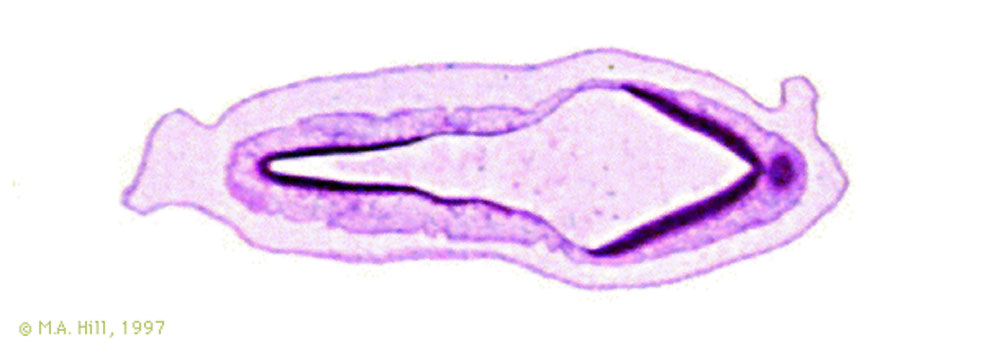
'''A1-A7:''' [[M#midbrain|Midbrain]] and [[H#hindbrain|hindbrain]]. The isthmus can be observed on [[:File:Stage 13 image 052.jpg|52]] - [[:File:Stage 13 image 053.jpg|53]] as a constriction between the mid- and hind-brain. The hindbrain is already subdivided into two parts, the slim myelencephalon and the wider metencephalon. The boundary between these two parts is roughly at the level of the otic vesicles [[:File:Stage 13 image 052.jpg|52]] - [[:File:Stage 13 image 053.jpg|53]]. Identify the optic cup and stalk, which are derived from the forebrain [[:File:Stage 13 image 058.jpg|58]] - [[:File:Stage 13 image 062.jpg|62]]. Caudally one can identify the spinal cord along the entire length of the embryo. | |||
===Sensory System=== | |||
[[Sensory System Development]] | [[Sensory_-_Hearing_and_Balance_Development|Hearing and Balance]] | |||
[[Category:Sensory]] [[Category:Hearing]] | |||
'''A2:''' [[O#otocyst|Otocyst]] (right). Apex of [[O#otocyst|otocyst]] (origin of L endolymphatic sac) | |||
---- | ---- | ||
Latest revision as of 10:02, 13 March 2014
A2 Image Features
Head and Neck
Neural System
A1-A7: Midbrain and hindbrain. The isthmus can be observed on 52 - 53 as a constriction between the mid- and hind-brain. The hindbrain is already subdivided into two parts, the slim myelencephalon and the wider metencephalon. The boundary between these two parts is roughly at the level of the otic vesicles 52 - 53. Identify the optic cup and stalk, which are derived from the forebrain 58 - 62. Caudally one can identify the spinal cord along the entire length of the embryo.
Sensory System
Sensory System Development | Hearing and Balance
A2: Otocyst (right). Apex of otocyst (origin of L endolymphatic sac)
About Stage 13 Embryo Sections - This image is from a serial section of a 6mm CRL pig embryo with some features of the Stage 14 embryo. This embryo is approximately equal to the day 42 human embryo. Use these serial images to identify internal features and relationships that exist within the embryo at this stage. Then compare these images with the later features of the Carnegie stage 22 human embryo.
| Stage 13 Serial unlabeled images | Embryo Stage 13 Serial labeled images |
| System Links: Introduction | Cardiovascular | Coelomic Cavity | Endocrine | Gastrointestinal Tract | Genital | Head | Immune | Integumentary | Musculoskeletal | Neural | Neural Crest | Placenta | Renal | Respiratory | Sensory | Birth |
Cite this page: Hill, M.A. (2024, May 8) Embryology Stage 13 image 001.jpg. Retrieved from https://embryology.med.unsw.edu.au/embryology/index.php/File:Stage_13_image_001.jpg
- © Dr Mark Hill 2024, UNSW Embryology ISBN: 978 0 7334 2609 4 - UNSW CRICOS Provider Code No. 00098G
File history
Click on a date/time to view the file as it appeared at that time.
| Date/Time | Thumbnail | Dimensions | User | Comment | |
|---|---|---|---|---|---|
| current | 17:46, 10 August 2010 | 1,000 × 357 (44 KB) | S8600021 (talk | contribs) | {{Template:Stage13sections}} {{Template:Systems}} {{Template:Footer}} Category:Carnegie Stage 13 |
You cannot overwrite this file.
File usage
The following 5 pages use this file: