Endocrine - Parathyroid Development: Difference between revisions
mNo edit summary |
mNo edit summary |
||
| (3 intermediate revisions by the same user not shown) | |||
| Line 24: | Line 24: | ||
|-bgcolor="F5FAFF" | |-bgcolor="F5FAFF" | ||
| | | | ||
* '''{{Hox}} genes in the pharyngeal region: how Hoxa3 controls early embryonic development of the pharyngeal organs'''{{#pmid:30604847|PMID30604847}} "The pharyngeal organs, namely the {{thyroid}}, {{thymus}}, {{parathyroid}}s, and ultimobranchial bodies, derive from the pharyngeal {{endoderm}} during embryonic development. The pharyngeal region is a segmented structure comprised of a series of reiterated structures: the pharyngeal arches on the exterior surface, the pharyngeal pouches on the interior, and a mesenchymal core. It is well known that {{Hox}} genes control spatial identity along the anterior-posterior axis of the developing vertebrate embryo, and nowhere is this is more evident than in the pharyngeal region. Each of the distinct segmented regions has a unique pattern of Hox expression, which conveys crucial positional information to the cells and tissues within it. In the context of pharyngeal organ development, molecular data suggest that HOXA3 is responsible for specifying organ identity within the third pharyngeal pouch, and in its absence, {{thymus}} and {{parathyroid}} organogenesis fails to proceed normally" | |||
* '''Tissue-specific roles for sonic hedgehog signaling in establishing thymus and parathyroid organ fate'''{{#pmid:27633995|PMID27633995}} "The thymus and parathyroids develop from third pharyngeal pouch (3rd pp) endoderm. Our previous studies show that Shh null mice have smaller, aparathyroid primordia in which thymus fate specification extends into the pharynx. SHH signaling is active in both dorsal pouch endoderm and neighboring neural crest (NC) mesenchyme. It is unclear which target tissue of SHH signaling is required for the patterning defects in Shh mutants. Here, we used a genetic approach to ectopically activate or delete the SHH signal transducer Smo in either pp endoderm or NC mesenchyme. Although no manipulation recapitulated the Shh null phenotype, manipulation of SHH signaling in either the endoderm or NC mesenchyme had direct and indirect effects on both cell types during fate specification and organogenesis. SHH pathway activation throughout pouch endoderm activated ectopic Tbx1 expression and partially suppressed the thymus-specific transcription factor Foxn1, identifying Tbx1 as a key target of SHH signaling in the 3rd pp. However, ectopic SHH signaling was insufficient to expand the GCM2-positive parathyroid domain, indicating that multiple inputs, some of which might be independent of SHH signaling, are required for parathyroid fate specification. These data support a model in which SHH signaling plays both positive and negative roles in patterning and organogenesis of the thymus and parathyroids." | * '''Tissue-specific roles for sonic hedgehog signaling in establishing thymus and parathyroid organ fate'''{{#pmid:27633995|PMID27633995}} "The thymus and parathyroids develop from third pharyngeal pouch (3rd pp) endoderm. Our previous studies show that Shh null mice have smaller, aparathyroid primordia in which thymus fate specification extends into the pharynx. SHH signaling is active in both dorsal pouch endoderm and neighboring neural crest (NC) mesenchyme. It is unclear which target tissue of SHH signaling is required for the patterning defects in Shh mutants. Here, we used a genetic approach to ectopically activate or delete the SHH signal transducer Smo in either pp endoderm or NC mesenchyme. Although no manipulation recapitulated the Shh null phenotype, manipulation of SHH signaling in either the endoderm or NC mesenchyme had direct and indirect effects on both cell types during fate specification and organogenesis. SHH pathway activation throughout pouch endoderm activated ectopic Tbx1 expression and partially suppressed the thymus-specific transcription factor Foxn1, identifying Tbx1 as a key target of SHH signaling in the 3rd pp. However, ectopic SHH signaling was insufficient to expand the GCM2-positive parathyroid domain, indicating that multiple inputs, some of which might be independent of SHH signaling, are required for parathyroid fate specification. These data support a model in which SHH signaling plays both positive and negative roles in patterning and organogenesis of the thymus and parathyroids." | ||
| Line 34: | Line 36: | ||
| [[File:Mark_Hill.jpg|90px|left]] {{Most_Recent_Refs}} | | [[File:Mark_Hill.jpg|90px|left]] {{Most_Recent_Refs}} | ||
Search term: [http://www.ncbi.nlm.nih.gov/pubmed/?term=Parathyroid+Embryology ''Parathyroid Embryology''] | [http://www.ncbi.nlm.nih.gov/pubmed/?term=pharyngeal+pouch+Embryology ''pharyngeal pouch Embryology''] | [http://www.ncbi.nlm.nih.gov/pubmed/?term=Parathyroid+hormone ''Parathyroid hormone''] | Search term: [http://www.ncbi.nlm.nih.gov/pubmed/?term=Parathyroid+Embryology ''Parathyroid Embryology''] | [http://www.ncbi.nlm.nih.gov/pubmed/?term=pharyngeal+pouch+Embryology ''pharyngeal pouch Embryology''] | [http://www.ncbi.nlm.nih.gov/pubmed/?term=Parathyroid+hormone ''Parathyroid hormone''] | [http://www.ncbi.nlm.nih.gov/pubmed/?term=Parathyroidectomy+in+Pregnancy ''Parathyroidectomy in Pregnancy''] | [http://www.ncbi.nlm.nih.gov/pubmed/?term=Hyperparathyroidism ''Hyperparathyroidism''] | ||
|} | |} | ||
| Line 80: | Line 81: | ||
:'''Links:''' [https://www.omim.org/entry/603716 OMIM GCM2] | :'''Links:''' [https://www.omim.org/entry/603716 OMIM - GCM2] | ||
===Semaphorin3d=== | |||
Semaphorin3d (Sema3d) is a secreted glycoprotein expressed in the developing parathyroid gland in mice. Deletion of Sema3d leads to parathyroid hyperplasia, causing PHPT. Therefore Sema3d may act as as a negative regulator of parathyroid growth.{{#pmid:30979723|PMID30979723}} | |||
Human SEMA3D {{Chr7}}q21.11 | |||
:'''Links:''' [https://www.omim.org/entry/609907 OMIM - SEMA3D] | |||
==Abnormalities== | ==Abnormalities== | ||
| Line 105: | Line 113: | ||
===Reviews=== | ===Reviews=== | ||
{{#pmid:30390809}} | {{#pmid:30390809}} | ||
| Line 113: | Line 122: | ||
{{#pmid:10912527}} | {{#pmid:10912527}} | ||
===Articles=== | ===Articles=== | ||
{{#pmid:32133431}} | |||
{{#pmid:30979723}} | |||
{{#pmid:21203493}} | {{#pmid:21203493}} | ||
{{#pmid:17382312}} | {{#pmid:17382312}} | ||
{{#pmid:8378818}} | |||
===Search PubMed=== | ===Search PubMed=== | ||
Latest revision as of 01:21, 10 March 2020
| Embryology - 27 Apr 2024 |
|---|
| Google Translate - select your language from the list shown below (this will open a new external page) |
|
العربية | català | 中文 | 中國傳統的 | français | Deutsche | עִברִית | हिंदी | bahasa Indonesia | italiano | 日本語 | 한국어 | မြန်မာ | Pilipino | Polskie | português | ਪੰਜਾਬੀ ਦੇ | Română | русский | Español | Swahili | Svensk | ไทย | Türkçe | اردو | ייִדיש | Tiếng Việt These external translations are automated and may not be accurate. (More? About Translations) |
Introduction

The parathyroid gland appears in the adult as a pair of inferior and a pair of superior "bumps" on the beside the (dorsal) thyroid (hence the name, "para"). The embryonic origin of this gland is from the endoderm of the third and fourth pharyngeal pouches, and could also have ectoderm and neural crest contributions. This developmental process also generates multiple small parathyroid clusters in addition to the main parathyroid glands.[1]
At 6 weeks a diverticulum elongates from the pouch, initially hollow and then solidifynig with cell proliferation.
Interestingly, the inferior parathyroid originates from the third pharyngeal pouch and the superior arises from the fourth pharyngeal pouch, the adult anatomical position is the opposite of the pharyngeal rostro-caudal order. This occurs due to the third pharyngeal pouch also giving rise to the thymus, the superior pair descend along with the thymus.
Parathyroid hormone (PTH) regulates calcium and phosphate levels in conjunction with parafollicular cells of the thyroid gland (calcitonin) and Vitamin D (dietary or synthesized in the skin).
The fetal parathyroids appear functional as they respond to calcium levels. The fetal calcium levels also higher than maternal levels.
Historically, see also the 1938 paper on the fate of the ultimobranchial body within the human thyroid gland.[2]
Some Recent Findings

|
| More recent papers |
|---|
 This table allows an automated computer search of the external PubMed database using the listed "Search term" text link.
More? References | Discussion Page | Journal Searches | 2019 References | 2020 References Search term: Parathyroid Embryology | pharyngeal pouch Embryology | Parathyroid hormone | Parathyroidectomy in Pregnancy | Hyperparathyroidism |
| Older papers |
|---|
| These papers originally appeared in the Some Recent Findings table, but as that list grew in length have now been shuffled down to this collapsible table.
See also the Discussion Page for other references listed by year and References on this current page.
|
Development Overview


- Endoderm - third and fourth pharyngeal pouches, could also have ectoderm and neural crest
- 3rd Pharyngeal Pouch - inferior parathyroid, initially descends with thymus
- 4th Pharyngeal Pouch - superior parathyroid
- Week 6 - diverticulum elongate, hollow then solid, dorsal cell proliferation
- Fetal parathyroids - respond to calcium levels, fetal calcium levels higher than maternal
Parathyroid Hormone
(PTH, parathormone or parathyrin) A polypeptide (84 amino acids) hormone which increases the concentration of calcium ions in the blood. Its actions oppose the hormone calcitonin from the parafollicular cells (C cells) of the thyroid gland, which decrease calcium. Acts through the parathyroid hormone receptor in bone, kidney and gastrointestinal tract.
- stimulate osteoclasts - degrade bone matrix, releasing calcium
- increase calcium gastrointestinal tract absorption
(PTHrP) Originally identified in the clinical syndrome humoral hypercalcemia of malignancy. It's developmental role is that of a regulatory protein expressed during the formation of many organs.
- mammary gland development - epithelial-mesenchymal interactions[7]
- chondrocyte differentiation[8]
Molecular
Sonic Hedgehog
Signalling by SHH has been shown to restrict the expression of Gcm2 and controls the position of the developing parathyroids.[9]
Glial Cells Missing
The transcription factor, Glial Cells Missing, Drosophila, Homolog Of, 2 (GCM2) is a key to parathyroid molecular development.[10][11]
- Links: OMIM - GCM2
Semaphorin3d
Semaphorin3d (Sema3d) is a secreted glycoprotein expressed in the developing parathyroid gland in mice. Deletion of Sema3d leads to parathyroid hyperplasia, causing PHPT. Therefore Sema3d may act as as a negative regulator of parathyroid growth.[12]
Human SEMA3D 7q21.11
- Links: OMIM - SEMA3D
Abnormalities
Hyperparathyroidism
Postnatal
- Postnatal adult ageing increased parathyroid hormone plasma levels are associated with cognitive decline and dementia.
- Parathyroid carcinoma (cancer) is a rare malignancy, occurring with an incidence of 0.5 to 4% of all cases of primary hyperparathyroidism.
Adult Histology
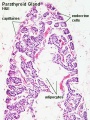
Parathyroid labeled (low power)
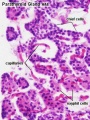
Parathyroid labeled (high power)

Parathyroid (low power)
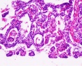
Parathyroid (high power)


References
- ↑ 1.0 1.1 1.2 1.3 Liu Z, Farley A, Chen L, Kirby BJ, Kovacs CS, Blackburn CC & Manley NR. (2010). Thymus-associated parathyroid hormone has two cellular origins with distinct endocrine and immunological functions. PLoS Genet. , 6, e1001251. PMID: 21203493 DOI.
- ↑ Kingsbury BF. On the fate of the ultimobranchial body within the human thyroid gland. (1935) Anat. Rec. 61(2): 155–173.
- ↑ Gordon J. (2018). Hox genes in the pharyngeal region: how Hoxa3 controls early embryonic development of the pharyngeal organs. Int. J. Dev. Biol. , 62, 775-783. PMID: 30604847 DOI.
- ↑ Bain VE, Gordon J, O'Neil JD, Ramos I, Richie ER & Manley NR. (2016). Tissue-specific roles for sonic hedgehog signaling in establishing thymus and parathyroid organ fate. Development , 143, 4027-4037. PMID: 27633995 DOI.
- ↑ Figueiredo M, Silva JC, Santos AS, Proa V, Alcobia I, Zilhão R, Cidadão A & Neves H. (2016). Notch and Hedgehog in the thymus/parathyroid common primordium: Crosstalk in organ formation. Dev. Biol. , 418, 268-82. PMID: 27544844 DOI.
- ↑ Chojnowski JL, Masuda K, Trau HA, Thomas K, Capecchi M & Manley NR. (2014). Multiple roles for HOXA3 in regulating thymus and parathyroid differentiation and morphogenesis in mouse. Development , 141, 3697-708. PMID: 25249461 DOI.
- ↑ Dunbar ME & Wysolmerski JJ. (1999). Parathyroid hormone-related protein: a developmental regulatory molecule necessary for mammary gland development. J Mammary Gland Biol Neoplasia , 4, 21-34. PMID: 10219904
- ↑ Alman BA & Wunder JS. (2008). Parathyroid hormone-related protein regulates glioma-associated oncogene transcriptional activation: lessons learned from bone development and cartilage neoplasia. Ann. N. Y. Acad. Sci. , 1144, 36-41. PMID: 19076361 DOI.
- ↑ Grevellec A, Graham A & Tucker AS. (2011). Shh signalling restricts the expression of Gcm2 and controls the position of the developing parathyroids. Dev. Biol. , 353, 194-205. PMID: 21349263 DOI.
- ↑ Liu Z, Yu S & Manley NR. (2007). Gcm2 is required for the differentiation and survival of parathyroid precursor cells in the parathyroid/thymus primordia. Dev. Biol. , 305, 333-46. PMID: 17382312 DOI.
- ↑ Peissig K, Condie BG & Manley NR. (2018). Embryology of the Parathyroid Glands. Endocrinol. Metab. Clin. North Am. , 47, 733-742. PMID: 30390809 DOI.
- ↑ Singh A, Mia MM, Cibi DM, Arya AK, Bhadada SK & Singh MK. (2019). Deficiency in the secreted protein Semaphorin3d causes abnormal parathyroid development in mice. J. Biol. Chem. , 294, 8336-8347. PMID: 30979723 DOI.
Reviews
Peissig K, Condie BG & Manley NR. (2018). Embryology of the Parathyroid Glands. Endocrinol. Metab. Clin. North Am. , 47, 733-742. PMID: 30390809 DOI.
Zajac JD & Danks JA. (2008). The development of the parathyroid gland: from fish to human. Curr. Opin. Nephrol. Hypertens. , 17, 353-6. PMID: 18660669 DOI.
Parvari R, Diaz GA & Hershkovitz E. (2007). Parathyroid development and the role of tubulin chaperone E. Horm. Res. , 67, 12-21. PMID: 17008776 DOI.
Jüppner H. (2000). Role of parathyroid hormone-related peptide and Indian hedgehog in skeletal development. Pediatr. Nephrol. , 14, 606-11. PMID: 10912527
Articles
Sharma SG, Levine SN, Yatavelli RK, Shaha MA & Nathan CAO. (2020). Parathyroidectomy in First Trimester of Pregnancy. J Endocr Soc , 4, bvaa015. PMID: 32133431 DOI.
Singh A, Mia MM, Cibi DM, Arya AK, Bhadada SK & Singh MK. (2019). Deficiency in the secreted protein Semaphorin3d causes abnormal parathyroid development in mice. J. Biol. Chem. , 294, 8336-8347. PMID: 30979723 DOI.
Liu Z, Farley A, Chen L, Kirby BJ, Kovacs CS, Blackburn CC & Manley NR. (2010). Thymus-associated parathyroid hormone has two cellular origins with distinct endocrine and immunological functions. PLoS Genet. , 6, e1001251. PMID: 21203493 DOI.
Liu Z, Yu S & Manley NR. (2007). Gcm2 is required for the differentiation and survival of parathyroid precursor cells in the parathyroid/thymus primordia. Dev. Biol. , 305, 333-46. PMID: 17382312 DOI.
Mansberger AR & Wei JP. (1993). Surgical embryology and anatomy of the thyroid and parathyroid glands. Surg. Clin. North Am. , 73, 727-46. PMID: 8378818 DOI.
Search PubMed
Search April 2010
- Parathyroid Development - All (3523) Review (768) Free Full Text (741)
Search Pubmed: parathyroid development
Additional Images
Terms
- parathyroid hormone - (PTH, parathormone or parathyrin) A polypeptide (84 amino acids) hormone secreted by the parathyroid gland, which increases the concentration of calcium ions in the blood. Its actions oppose the hormone calcitonin from the thyroid gland parafollicular cells (C cells), which decrease calcium. Acts through the parathyroid hormone receptor located mainly in bone, kidney and gastrointestinal tract. Hormone dual role is to: stimulate osteoclasts in bone to degrade bone matrix releasing calcium; increase gastrointestinal tract absorption of calcium.
| Endocrine Terms (expand to view) |
|---|
|
| Other Terms Lists |
|---|
| Terms Lists: ART | Birth | Bone | Cardiovascular | Cell Division | Endocrine | Gastrointestinal | Genital | Genetic | Head | Hearing | Heart | Immune | Integumentary | Neonatal | Neural | Oocyte | Palate | Placenta | Radiation | Renal | Respiratory | Spermatozoa | Statistics | Tooth | Ultrasound | Vision | Historic | Drugs | Glossary |
Glossary Links
- Glossary: A | B | C | D | E | F | G | H | I | J | K | L | M | N | O | P | Q | R | S | T | U | V | W | X | Y | Z | Numbers | Symbols | Term Link
Cite this page: Hill, M.A. (2024, April 27) Embryology Endocrine - Parathyroid Development. Retrieved from https://embryology.med.unsw.edu.au/embryology/index.php/Endocrine_-_Parathyroid_Development
- © Dr Mark Hill 2024, UNSW Embryology ISBN: 978 0 7334 2609 4 - UNSW CRICOS Provider Code No. 00098G