Category:Neural
From Embryology
This Embryology category shows pages and media related to Neural System Development. This includes related topics and undergraduate classes as well as pages and sub-categories describing specific components formed from the original ectoderm neural tube.
Subcategories
This category has the following 12 subcategories, out of 12 total.
Pages in category 'Neural'
The following 200 pages are in this category, out of 934 total.
(previous page) (next page)P
- Paper - The formation of the cranial subarachnoid spaces
- Paper - The fundamental plan of the vertebrate brain
- Paper - The growth and histogenesis of the cerebro-spinal nerves in mammals
- Paper - The growth of the central nervous system in the human fetus as expressed by graphic analysis and empirical formulae (1921)
- Paper - The histogenesis of the cerebellum (1895)
- Paper - The human brain at stages 18-20 including the choroid plexuses and the amygdaloid and septal nuclei (1990)
- Paper - The hypoglossal nerve in human embryos (1939)
- Paper - The influence of nerve fibers upon taste buds during embryonic development
- Paper - The lamina terminalis and its relation to the fornix system (1911)
- Paper - The mammalian cerebellum - its lobes and fissures 1 (1904)
- Paper - The mammalian cerebellum - its lobes and fissures 2 (1904)
- Paper - The Mammalian Cerebellum part 1 (1895)
- Paper - The morphology and morphogenesis of the choroid plexuses with especial reference to the development of the lateral telencephalic plexus in Chrysemys marginata (1916)
- Paper - The morphology of the diencephalic floor
- Paper - The morphology of the forebrain vesicle in vertebrates
- Paper - The nerve supply of the mammalian ductus arteriosus (1941)
- Paper - The nerve supply to the pituitary body (1913)
- Paper - The oculomotor nucleus in the human fetus (1944)
- Paper - The organ of jacobson in the horse, ox, camel and pig (1925)
- Paper - The origin and development of the carotid body (1924)
- Paper - The origin of the motor neuroblasts of the anterior cornu of the neural tube (1922)
- Paper - The Origin of the Otic and Optic Primordia in Man
- Paper - The origin of the sensory components of the cranial ganglia (1910)
- Paper - The Peripheral Nervous System in the Human Embryo at the End of the First Month (10 mm)
- Paper - The phylogenetic origin of the nervous system (1910)
- Paper - The prechordal plate in a human embryo with small neuropore
- Paper - The prenatal medullation of the sheep's nervous system (1947)
- Paper - The role of the vagi in the development of the sympathetic nervous system (1909)
- Paper - The roof and lateral recesses of the fourth ventricle considered morphologically and embryologically
- Paper - The roots of the facial nerve in human embryos and fetuses
- Paper - The segmental value of the cranial nerves (1882)
- Paper - The segmentation of the primitive vertebrate brain (1890)
- Paper - The sheep neopallium - study of its development and interpretation of its folds (1936)
- Paper - The significance of the prechordal plate - an interpretative study
- Paper - The spinal accessory nerve in human embryos (1938)
- Paper - The status of metamerism in the central nervous system of chick embryos
- Paper - The structure of the spinal cord of the ostrich
- Paper - The structure of the third, fourth, fifth, sixth, ninth, eleventh and twelfth cranial nerves (1916)
- Paper - The subdivisions of the neural folds in man
- Paper - The trochlear nerve in human fetuses (1943)
- Paper - Transitory cavities in the corpus striatum of the human embryo (1915)
- Paper - Ventricular system and choroid plexuses of the human brain during the embryonic period proper
- Paper - Volumetric determinations of the parts of the brain in a human fetus 156 mm long (1915)
- Paper - Wilhelm His - His relation to the institution of learning
- Template:PediNeuroLogic Exam
- Template:Peripheral Nerve Histology
- Template:Peripheral nervous system
- Template:Peripheral Nervous System
- Template:Pia mater
- Template:Pineal
- Template:PNS
- Template:Pons
- Template:Posterior pituitary
- Template:Prosencephalon
- Template:Prosomere
- Template:Purkinje cell
- Template:Purkinje cells
- Template:Purves2001APAcitation
R
- Template:Radial glia
- Template:Ref-Adelman1925
- Template:Ref-Adelmann1922
- Template:Ref-Amin1914
- Template:Ref-Anthony1936
- Template:Ref-Arey1922b
- Template:Ref-Ayers1919
- Template:Ref-Bailey1916
- Template:Ref-Bardeen1903
- Template:Ref-Bardeen1906
- Template:Ref-Bartelmez1923
- Template:Ref-BartelmezDekaban1962
- Template:Ref-BaxterBoyd1938
- Template:Ref-Blake1900
- Template:Ref-BolaflioArtom1924
- Template:Ref-Bossy1981
- Template:Ref-Boyd1941
- Template:Ref-Bradley1903a
- Template:Ref-Bradley1903b
- Template:Ref-Bradley1904a
- Template:Ref-Bradley1904b
- Template:Ref-Bradley1905
- Template:Ref-Bradley1906
- Template:Ref-Bremer1921
- Template:Ref-Brodal1946
- Template:Ref-Cameron1911b
- Template:Ref-CameronMilligan1910
- Template:Ref-Carey1919
- Template:Ref-Coghill1914
- Template:Ref-Coghill1916
- Template:Ref-Coghill1929
- Template:Ref-CohenDavies1938
- Template:Ref-Coronios1933
- Template:Ref-Covell1927b
- Template:Ref-Cunningham1891
- Template:Ref-Dart1924
- Template:Ref-DartShellshear1922
- Template:Ref-DeVries1927
- Template:Ref-Dexter1903
- Template:Ref-Dickson1940
- Template:Ref-Dixon1896
- Template:Ref-Dockeray1915
- Template:Ref-Ellis1919
- Template:Ref-Ellis1920
- Template:Ref-Essick1907
- Template:Ref-Essick1909a
- Template:Ref-Essick1912
- Template:Ref-Essick1915
- Template:Ref-Evrard1993
- Template:Ref-Evrard1996
- Template:Ref-Frazer1921
- Template:Ref-Frazer1928
- Template:Ref-Gage1905
- Template:Ref-Glaser1914
- Template:Ref-Glaser1917
- Template:Ref-Goldby1939
- Template:Ref-GrenellScanimon1943
- Template:Ref-GrosserLewisMcMurrich1912
- Template:Ref-Hardesty1904a
- Template:Ref-Hardesty1904b
- Template:Ref-Harman1922
- Template:Ref-Harrison1906
- Template:Ref-HartleyBurnett1943
- Template:Ref-Hayes1922
- Template:Ref-Heard1936
- Template:Ref-Herrick1895
- Template:Ref-Herrick1948
- Template:Ref-Heuser1913
- Template:Ref-Hewer1935
- Template:Ref-Hilton1912
- Template:Ref-Hines1921
- Template:Ref-Hines1922
- Template:Ref-Hines1923
- Template:Ref-Hochstädter1919
- Template:Ref-HuberCrosby1933
- Template:Ref-HuberGuild1913a
- Template:Ref-HuberGuild1913b
- Template:Ref-Humphrey1940
- Template:Ref-Humphrey1944
- Template:Ref-Hunter1934b
- Template:Ref-Jenkins1919
- Template:Ref-Jenkins1921
- Template:Ref-Johnston1907
- Template:Ref-Johnston1909
- Template:Ref-Johnston1909a
- Template:Ref-Johnston1910
- Template:Ref-Johnston1923
- Template:Ref-KappersHuberCrosby1936a
- Template:Ref-KappersHuberCrosby1936b
- Template:Ref-Keene1931
- Template:Ref-Keene1933
- Template:Ref-Keene1938
- Template:Ref-Keibel1912sensory
- Template:Ref-Keith1932a
- Template:Ref-Keith1932b
- Template:Ref-Kingsbury1920
- Template:Ref-Kingsbury1922
- Template:Ref-Koch1916
- Template:Ref-KolbFernandezNelson2012
- Template:Ref-Kuntz1909a
- Template:Ref-Kuntz1909b
- Template:Ref-Kuntz1910a
- Template:Ref-Kuntz1910b
- Template:Ref-Kuntz1922
- Template:Ref-Kupffer1891a
- Template:Ref-Kupffer1891b
- Template:Ref-Landacre1910
- Template:Ref-Larsell1918
- Template:Ref-Larsell1919
- Template:Ref-leGrosClark1940
- Template:Ref-Lewis1910b
- Template:Ref-Lockard1948
- Template:Ref-Long1901
- Template:Ref-MacArthurDoisy1919
- Template:Ref-Mall1903
- Template:Ref-Mall1904a
- Template:Ref-Mall1905
- Template:Ref-Mall1909
- Template:Ref-Malone1912
- Template:Ref-Marin-Padilla1983
- Template:Ref-Masy1955
- Template:Ref-McClure1890
- Template:Ref-McGaw1924
- Template:Ref-Mellus1911
- Template:Ref-Mellus1912
- Template:Ref-Miller1913
- Template:Ref-MillerDetwiler1936
- Template:Ref-Minot1893
- Template:Ref-MullerO'Rahilly1987
- Template:Ref-MullerO'Rahilly1990
- Template:Ref-Nañagas1925
- Template:Ref-O'Rahilly1984
- Template:Ref-O'RahillyMuller1986b
- Template:Ref-O'RahillyMuller1990b
- Template:Ref-O'RahillyMüller2006
- Template:Ref-O'RahillyMüller2008 PMID18356030
- Template:Ref-O'RahillyMüller2013 PMID23183269
- Template:Ref-Palmgren1921
- Template:Ref-Parker1910
- Template:Ref-Patten 1946b
- Template:Ref-Patten1946
- Template:Ref-Patten1946b
Media in category 'Neural'
The following 200 files are in this category, out of 1,070 total.
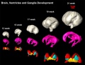
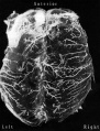
(previous page) (next page)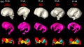 Brain ventricles and ganglia development 01.jpg 994 × 564; 45 KB
 Brain ventricles and ganglia development 02.jpg 1,000 × 760; 49 KB
 Brain ventricles and ganglia development 03.jpg 989 × 583; 36 KB
 Brain week 17 histology.jpg 1,050 × 697; 61 KB
 Brain-tract-development-03.jpg 1,000 × 424; 31 KB
 Brain-tract-development-04.jpg 1,000 × 424; 30 KB
 Brain-tract-development-05.jpg 1,000 × 424; 29 KB
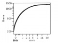
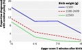
 Brainweight.jpg 312 × 434; 16 KB
 Bremer1906 fig06.jpg 1,300 × 927; 126 KB
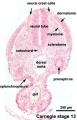 Carnegie stage 13 caudal trunk.jpg 400 × 625; 53 KB
 CDC postnatal milestones.jpg 800 × 616; 90 KB
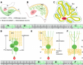
 Cerebellar Bergmann astrocyte cartoon.jpg 979 × 640; 286 KB
 Cerebellar granule cell cartoon.jpg 742 × 644; 232 KB
 Cerebellar oligodendrocyte cartoon.jpg 1,200 × 682; 328 KB
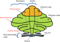 Cerebellum anatomical subdivisions.png 914 × 637; 67 KB
 Cerebral blood supply development 01.jpg 1,200 × 460; 67 KB
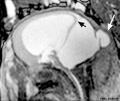
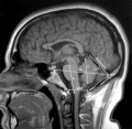
 Chiari II malformation MRI01.jpg 723 × 800; 88 KB
 Chiari II malformation MRI02.jpg 723 × 800; 101 KB
 Chicken neural Plxdc2 expression.jpg 1,000 × 850; 121 KB
 Chicken neural tube thickening.jpg 529 × 600; 69 KB
 Chicken- neural Wnt expression.jpg 600 × 424; 55 KB
 Chicken- rhombomere boundary FGF3 expression.jpg 1,000 × 385; 63 KB
 CNS later development.jpg 961 × 462; 59 KB
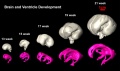
 CNS primary vesicles.jpg 987 × 562; 49 KB
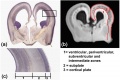
 CNS secondary vesicles.jpg 987 × 562; 81 KB
 Cochlea glial lineage cartoon.jpg 1,000 × 651; 52 KB
 Comparative brain anatomy frog-dog.jpg 1,000 × 835; 112 KB
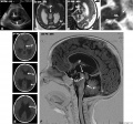
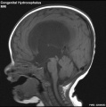 Congenital hydrocephalus MRI01.jpg 595 × 600; 43 KB
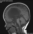 Congenital hydrocephalus MRI02.jpg 595 × 600; 42 KB
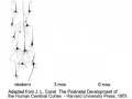
 Corner1929 fig10-11.jpg 1,200 × 1,438; 730 KB
 Csf cartoon1.jpg 600 × 307; 35 KB
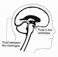 Csf cartoon3.jpg 600 × 588; 37 KB
 Dandy Walker malformation MRI 01.jpg 600 × 506; 37 KB
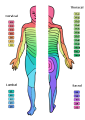 Dermatomes.png 424 × 600; 82 KB
 Dev anat 01.flv ; 288 KB
 Dev anat 01.jpg 500 × 375; 25 KB
- Dev anat 02.flv ; 100 KB
 Dev anat 02.jpg 320 × 240; 9 KB
- Dev anat 03.flv ; 325 KB
 Dev anat 03.jpg 320 × 240; 10 KB
- Dev anat 04.flv ; 532 KB
 Dev anat 04.jpg 320 × 240; 13 KB
- Dev anat 05.flv ; 168 KB
 Dev anat 05.jpg 320 × 240; 16 KB
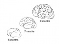
 Developing human cerebellum 01.jpg 1,009 × 1,200; 494 KB
 Developing human cerebellum 02.jpg 492 × 392; 86 KB
 Double meningomyelocele.jpg 504 × 800; 49 KB
 Elizabeth Caroline Crosby.jpg 340 × 600; 43 KB
 FASD-Guide-AUS2016-cover.jpg 587 × 836; 53 KB
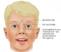 FASface.jpg 320 × 272; 8 KB
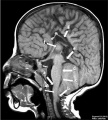
 Fetal brain MRI01.jpg 1,280 × 465; 45 KB
 Fetal brain MRI02.jpg 958 × 708; 22 KB
 Fetal brain MRI03.jpg 958 × 708; 34 KB
 Fetal brain MRI04.jpg 958 × 708; 41 KB
 Fetal head section 01.jpg 1,200 × 821; 186 KB
 Fetal head section.jpg 1,200 × 821; 167 KB
 Fetal pineal gland 01.jpg 700 × 603; 67 KB
 Fly neural development 01.png 515 × 600; 239 KB
 FMR1 gene silencing.jpg 1,280 × 1,124; 103 KB
 Folate Biosynthesis.jpg 787 × 899; 127 KB
 Folate one-carbon metabolism.jpg 600 × 602; 69 KB
 Folate.jpg 338 × 150; 7 KB
 Folatefruit.jpg 263 × 172; 15 KB
 Foster120.jpg 798 × 729; 98 KB
 Foster121.jpg 658 × 539; 59 KB
 Foster122.jpg 608 × 419; 45 KB
 Foster123.jpg 830 × 745; 132 KB
 Foster124.jpg 815 × 799; 149 KB
 Foster126.jpg 614 × 471; 53 KB
 Foster128.jpg 998 × 859; 223 KB
 G. Carl Huber.jpg 530 × 800; 51 KB
 Gestational age and special educational prevalence.jpg 600 × 443; 19 KB
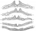
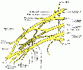
 Gillilan1959-fig01.jpg 1,000 × 1,313; 155 KB
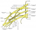
 Gillilan1959-fig02.jpg 905 × 1,000; 159 KB
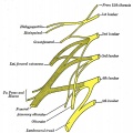
 Gillilan1959-fig03.jpg 1,042 × 1,366; 317 KB
 Granule Cell and Purkinje Cell Migration.png 850 × 673; 399 KB
 Graph- Apgar score and cerebral palsy.jpg 600 × 366; 36 KB
 Gray0015.jpg 800 × 682; 111 KB
 Gray0649.jpg 698 × 700; 72 KB
 Gray0651.jpg 698 × 700; 94 KB
 Gray0652.jpg 698 × 700; 103 KB
 Gray0653.jpg 698 × 700; 108 KB
 Gray0654.jpg 402 × 500; 39 KB
 Gray0655.jpg 500 × 419; 39 KB
 Gray0658.jpg 361 × 450; 29 KB
 Gray0678.jpg 600 × 598; 35 KB
 Gray0697.jpg 500 × 540; 49 KB
 Gray0698.jpg 500 × 518; 47 KB
 Gray0702.jpg 800 × 425; 77 KB
 Gray0704.jpg 800 × 555; 83 KB
 Gray0705.jpg 600 × 449; 45 KB
 Gray0706.jpg 800 × 870; 66 KB
 Gray0708.jpg 650 × 417; 46 KB
 Gray0715.jpg 800 × 591; 127 KB
 Gray0720.jpg 800 × 702; 182 KB
 Gray0732.jpg 600 × 406; 31 KB
 Gray0754.jpg 600 × 779; 135 KB
 Gray0769.jpg 600 × 454; 101 KB
 Gray0770.jpg 700 × 275; 68 KB
 Gray0778.jpg 600 × 630; 101 KB
 Gray0781.jpg 817 × 700; 156 KB
 Gray0784.jpg 851 × 600; 137 KB
 Gray0786.jpg 556 × 400; 52 KB
 Gray0788.jpg 815 × 750; 91 KB
 Gray0789.jpg 800 × 250; 45 KB
 Gray0790.jpg 683 × 800; 160 KB
 Gray0804.jpg 550 × 700; 75 KB
 Gray0806.jpg 600 × 771; 173 KB
 Gray0807.gif 587 × 500; 44 KB
 Gray0807.jpg 704 × 600; 97 KB
 Gray0822.jpg 599 × 600; 69 KB
 Gray0847.jpg 559 × 900; 155 KB
 Gray0848.jpg 800 × 935; 289 KB
 Gray0849.jpg 800 × 885; 258 KB
 Gray1236.jpg 800 × 281; 29 KB
 Gray804.gif 471 × 600; 30 KB
 Hearing sound localization circuits brainstem.jpg 800 × 378; 45 KB
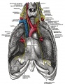
 Heart innervation 01.jpg 1,280 × 599; 92 KB
 Historic-Cerebral-cortex.jpg 483 × 634; 65 KB
 Hochstadter 1919.jpg 845 × 1,200; 0 bytes
 Hochstadter plate 01.jpg 1,408 × 2,000; 255 KB
 Hochstadter plate 02.jpg 1,408 × 2,000; 305 KB
 Hochstadter plate 03.jpg 2,000 × 1,408; 258 KB
 Hochstadter plate 04.jpg 1,408 × 2,000; 273 KB
 Hochstadter plate 05.jpg 1,408 × 2,000; 235 KB
 Hochstadter plate 06.jpg 1,408 × 2,000; 280 KB
 Hochstadter plate 07.jpg 1,626 × 2,000; 261 KB
 Hochstadter plate 08.jpg 1,670 × 2,000; 367 KB
 Hochstadter plate 09.jpg 1,692 × 2,000; 307 KB
 Hochstadter plate 10.jpg 1,626 × 2,000; 551 KB
 Hochstadter plate 11.jpg 1,603 × 2,000; 570 KB
 Hochstadter plate 12.jpg 1,541 × 2,000; 543 KB
 Hochstadter plate 13.jpg 1,461 × 2,000; 531 KB
 Hochstadter plate 14.jpg 1,517 × 2,000; 574 KB
 Hochstadter plate 15.jpg 1,462 × 2,000; 450 KB
 Hochstadter plate 16.jpg 1,507 × 2,000; 640 KB
 Human 15 weeks - terminal nerve and vomeronasal organ nerves.jpg 940 × 403; 306 KB
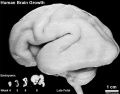 Human brain growth 01.jpg 1,022 × 800; 119 KB
 Human brain white matter tracts.png 1,200 × 490; 289 KB
 Human Carnegie stage 13 GJA1 expression.jpg 706 × 470; 96 KB
 Human Carnegie stage 13 SOX11 MAZ GJA1 expression.jpg 777 × 1,000; 192 KB
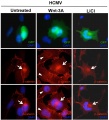 Human cytomegalovirus beta-catenin juxtanuclear region.jpg 908 × 1,000; 132 KB
 Human fetal neural aneuploidy.jpg 1,000 × 1,400; 134 KB
 Human Fetus CRL240mm brain.jpg 1,280 × 1,030; 129 KB
 Human hippocampus cartoon.jpg 1,024 × 881; 62 KB
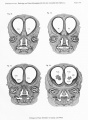
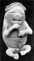
 Human holoprosencephaly cyclopia dissection.jpg 600 × 340; 37 KB
 Human neural crest cell migration-in vitro.jpg 1,280 × 959; 163 KB
 Human Stage13 sagittal upper half01.jpg 1,518 × 2,048; 269 KB
 Human Stage13 sagittal upper half02.jpg 1,518 × 2,048; 289 KB
 Human Stage14 neural01.jpg 1,375 × 2,048; 283 KB
 Human Stage14 neural02.jpg 1,375 × 2,048; 506 KB
 Human Stage14-16 CN5-01.jpg 1,028 × 681; 44 KB
 Human Stage16 neural01.jpg 1,352 × 2,048; 247 KB
 Human Stage16 neural02.jpg 1,352 × 2,048; 286 KB
 Human Stage16 neural03.jpg 1,352 × 2,048; 245 KB
 Human Stage21 neural01.jpg 2,048 × 1,533; 211 KB
 Human Stage21 neural02.jpg 2,048 × 1,533; 230 KB
 Human thyroid system and neural development.jpg 1,032 × 728; 132 KB
 Human trisomy chromosome 7 and 19.jpg 1,000 × 804; 154 KB
 Human week 10 fetus 12.jpg 1,200 × 900; 349 KB
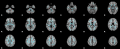
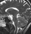
 Human- adult brain MRI.jpg 1,200 × 1,170; 160 KB
 Human- fetal week 10 cerebellum A.jpg 347 × 284; 24 KB
 Human- fetal week 10 cerebellum B.jpg 347 × 284; 21 KB
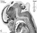
 Human- fetal week 10 cerebellum C.jpg 347 × 284; 25 KB
 Human- fetal week 10 cerebellum D.jpg 347 × 284; 23 KB
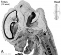
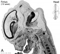 Human- fetal week 10 head A.jpg 600 × 544; 113 KB
 Human- fetal week 10 head A1.jpg 1,200 × 1,088; 159 KB
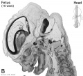 Human- fetal week 10 head B.jpg 600 × 544; 66 KB
 Human- fetal week 10 head C.jpg 600 × 544; 118 KB
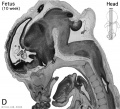 Human- fetal week 10 head D.jpg 600 × 544; 111 KB
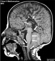
 Human- neural Chiari malformation.jpg 1,200 × 1,344; 212 KB
 Human- ventricular system cartoon 02.jpg 1,179 × 1,254; 115 KB
 Human- ventricular system cartoon.jpg 600 × 638; 47 KB
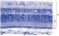 Human-retina-01.jpg 1,000 × 615; 197 KB
 Humphrey1940 fig01.jpg 900 × 383; 34 KB
 Humphrey1940 fig02.jpg 1,000 × 480; 61 KB
 Hunter1934 fig01-02.jpg 1,594 × 1,308; 188 KB
 Hunter1934 fig01.jpg 600 × 1,062; 64 KB
 Hunter1934 fig02.jpg 565 × 1,000; 60 KB
 Huntingtin structure.jpg 595 × 266; 39 KB
 Hydrocephalus aqueduct of Sylvius 01.jpg 779 × 726; 111 KB
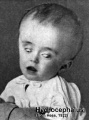 Hydrocephalus.jpg 320 × 432; 29 KB
 Hypothalamus gene interaction model.jpg 1,000 × 695; 60 KB
 Hypothalamus model 01.jpg 1,089 × 796; 119 KB
 Infant lymphocytic choriomeningitis virus CT.jpg 665 × 800; 69 KB
 Interneuron-radial glial interactions.jpg 510 × 720; 33 KB
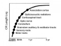
 Jenkins-chart01.jpg 1,217 × 1,400; 173 KB
 Jenkins-table01.jpg 483 × 304; 29 KB
 Jenkins-table02.jpg 308 × 352; 26 KB
 Jenkins-table03.jpg 992 × 1,048; 293 KB
 Jenkins001.jpg 754 × 602; 98 KB
 Jenkins002.jpg 1,230 × 1,102; 321 KB
 Jenkins003-005.jpg 1,555 × 1,555; 550 KB
 Jenkins003.jpg 973 × 653; 204 KB
 Jenkins004.jpg 841 × 707; 129 KB
 Jenkins005.jpg 868 × 766; 154 KB
 Jenkins006.jpg 884 × 637; 142 KB
 Jenkins007-008.jpg 1,836 × 891; 438 KB
 Jenkins007.jpg 836 × 887; 150 KB


