Category:Neural
From Embryology
This Embryology category shows pages and media related to Neural System Development. This includes related topics and undergraduate classes as well as pages and sub-categories describing specific components formed from the original ectoderm neural tube.
Subcategories
This category has the following 12 subcategories, out of 12 total.
Pages in category 'Neural'
The following 200 pages are in this category, out of 934 total.
(previous page) (next page)P
- Paper - On the development and nature of the neuroglia
- Paper - On the development of the blood-vessels of the brain in the human embryo (1905)
- Paper - On the development of the hind-brain of the pig 1
- Paper - On the development of the hind-brain of the pig 2
- Paper - On the embryology of the corpus ponto-bulbare and its relation to the development of the pons
- Paper - On the embryology of the corpus ponto-bulbare and its relation to the development of the pons (1909)
- Paper - On the Mechanism of morphological differentiation in the nervous system 1
- Paper - On the mechanism of morphological differentiation in the nervous system 2. The relation between compression and the development of a series of vesicles (1917)
- Paper - On the nature and mode of origin of the foramen of magendie (1937)
- Paper - On the occurrence of sheath cells and the nature of the axone sheaths in the central nervous system
- Paper - On the pineal region in human embryos
- Paper - On the relation of the head chorda to the pharyngeal epithelium in the pig embryo
- Paper - On the transitory or artificial fissures of the human cerebrum
- Paper - Overgrowth of the neural tube in young human embryos
- Paper - Prenatal growth of the human spinal cord
- Paper - Primary neuromeres and head segmentation (1922)
- Paper - Primitive neurons in the embryonic human central nervous system
- Paper - Recurrent branches of the abducens nerve in human embryos
- Paper - Report on an Anencephalic Embryo
- Paper - Sensory nerves in the skin of human fetuses of 8 to 14 weeks of menstrual age correlated with functional capability (1941)
- Paper - Sequential innervation of the intestinal loop in the human embryo
- Paper - Significant features in the early prenatal development of the human brain
- Paper - Significant superficial anastomoses in the arterial blood supply to the human brain (1959)
- Paper - Some observations on myelination in the human central nervous system (1931)
- Paper - Structural organization of the human cerebral cortex prior to the appearance of the cortical plate (1983)
- Paper - Structural plan of the human brain
- Paper - Studies in the growth and differentiation of the telencephalon in man - the fissura hippocampi
- Paper - Studies on the nervus terminalis - Mammals (1918)
- Paper - Teratological studies (1919)
- Paper - The anterior end of the neural tube and the anterior end of the body (1924)
- Paper - The circle of Willis - An examination of 700 specimens (1905)
- Paper - The connexions of the posterior commissure in the human foetus and young infant
- Paper - The corpus ponto-bulbare - a hitherto undeseribed nuclear mass in the human hind brain (1907)
- Paper - The cortex of the brain in the human embryo during the fourth month with special reference to the so-called Papilla of Retzius
- Paper - The Course of the Phrenic Nerve in the Embryo
- Paper - The developing third nerve nucleus in human embryos
- Paper - The development and myelination of the posterior longitudinal bundle in the human (1933)
- Paper - The development and reduction of the tail and of the caudal end of the spinal cord (1920)
- Paper - The development and significance of the cell columns in the ventral horn of the cervical and upper thoracic spinal cord of the rabbit (1941)
- Paper - The development of a medial motor nucleus and an accessory abducens nucleus in the pig (1934)
- Paper - The development of cerebro-spinal fluid pathway in human embryos (1977)
- Paper - The development of nerve endings in the human foetus
- Paper - The development of the auditory nerve in vertebrates (1910)
- Paper - The development of the cerebral cortex
- Paper - The development of the cerebral ventricles in the pig (1913)
- Paper - The Development of the Cranial and Spinal Nerves in the Occipital Region of the Human Embryo
- Paper - The development of the gyri and sulci on the surface of the island of Reil of the human brain (1891)
- Paper - The development of the human brain stage 12
- Paper - The development of the hypoglossal ganglia of pig embryos
- Paper - The development of the meninges
- Paper - The development of the nervous tissues of the human embryo (1877)
- Paper - The development of the nervus terminali in man (1941)
- Paper - The development of the neural folds and cranial ganglia of the rat
- Paper - The development of the nuclei pontis and the nucleus arcuatus in man (1912)
- Paper - The development of the olfactory and the accessory olfactory formations in human embryos and fetuses
- Paper - The development of the olfactory nerve in man (1941)
- Paper - The development of the subcutaneous vascular plexus in the head of the human embryo
- Paper - The development of the sympathetic nervous system in mammals (1910)
- Paper - The development of the sympathetic system in birds (1910)
- Paper - The development of the venous sinuses of the dura mater in the human embryo
- Paper - The developmental alterations in the vascular system of the brain of the human embryo (1921)
- Paper - The developmental origin of the notochord
- Paper - The early development of the meninges of the spinal cord in human embryos (1951)
- Paper - The embryological development of the commissura posterior in the human spinal cord
- Paper - The embryonic cerebral hemisphere in man (1921)
- Paper - The evolution of the cerebral cortex (1910)
- Paper - The extent of the floor-plate of His and its significance (1920)
- Paper - The formation of the cranial subarachnoid spaces
- Paper - The fundamental plan of the vertebrate brain
- Paper - The growth and histogenesis of the cerebro-spinal nerves in mammals
- Paper - The growth of the central nervous system in the human fetus as expressed by graphic analysis and empirical formulae (1921)
- Paper - The histogenesis of the cerebellum (1895)
- Paper - The human brain at stages 18-20 including the choroid plexuses and the amygdaloid and septal nuclei (1990)
- Paper - The hypoglossal nerve in human embryos (1939)
- Paper - The influence of nerve fibers upon taste buds during embryonic development
- Paper - The lamina terminalis and its relation to the fornix system (1911)
- Paper - The mammalian cerebellum - its lobes and fissures 1 (1904)
- Paper - The mammalian cerebellum - its lobes and fissures 2 (1904)
- Paper - The Mammalian Cerebellum part 1 (1895)
- Paper - The morphology and morphogenesis of the choroid plexuses with especial reference to the development of the lateral telencephalic plexus in Chrysemys marginata (1916)
- Paper - The morphology of the diencephalic floor
- Paper - The morphology of the forebrain vesicle in vertebrates
- Paper - The nerve supply of the mammalian ductus arteriosus (1941)
- Paper - The nerve supply to the pituitary body (1913)
- Paper - The oculomotor nucleus in the human fetus (1944)
- Paper - The organ of jacobson in the horse, ox, camel and pig (1925)
- Paper - The origin and development of the carotid body (1924)
- Paper - The origin of the motor neuroblasts of the anterior cornu of the neural tube (1922)
- Paper - The Origin of the Otic and Optic Primordia in Man
- Paper - The origin of the sensory components of the cranial ganglia (1910)
- Paper - The Peripheral Nervous System in the Human Embryo at the End of the First Month (10 mm)
- Paper - The phylogenetic origin of the nervous system (1910)
- Paper - The prechordal plate in a human embryo with small neuropore
- Paper - The prenatal medullation of the sheep's nervous system (1947)
- Paper - The role of the vagi in the development of the sympathetic nervous system (1909)
- Paper - The roof and lateral recesses of the fourth ventricle considered morphologically and embryologically
- Paper - The roots of the facial nerve in human embryos and fetuses
- Paper - The segmental value of the cranial nerves (1882)
- Paper - The segmentation of the primitive vertebrate brain (1890)
- Paper - The sheep neopallium - study of its development and interpretation of its folds (1936)
- Paper - The significance of the prechordal plate - an interpretative study
- Paper - The spinal accessory nerve in human embryos (1938)
- Paper - The status of metamerism in the central nervous system of chick embryos
- Paper - The structure of the spinal cord of the ostrich
- Paper - The structure of the third, fourth, fifth, sixth, ninth, eleventh and twelfth cranial nerves (1916)
- Paper - The subdivisions of the neural folds in man
- Paper - The trochlear nerve in human fetuses (1943)
- Paper - Transitory cavities in the corpus striatum of the human embryo (1915)
- Paper - Ventricular system and choroid plexuses of the human brain during the embryonic period proper
- Paper - Volumetric determinations of the parts of the brain in a human fetus 156 mm long (1915)
- Paper - Wilhelm His - His relation to the institution of learning
- Template:PediNeuroLogic Exam
- Template:Peripheral Nerve Histology
- Template:Peripheral nervous system
- Template:Peripheral Nervous System
- Template:Pia mater
- Template:Pineal
- Template:PNS
- Template:Pons
- Template:Posterior pituitary
- Template:Prosencephalon
- Template:Prosomere
- Template:Purkinje cell
- Template:Purkinje cells
- Template:Purves2001APAcitation
R
- Template:Radial glia
- Template:Ref-Adelman1925
- Template:Ref-Adelmann1922
- Template:Ref-Amin1914
- Template:Ref-Anthony1936
- Template:Ref-Arey1922b
- Template:Ref-Ayers1919
- Template:Ref-Bailey1916
- Template:Ref-Bardeen1903
- Template:Ref-Bardeen1906
- Template:Ref-Bartelmez1923
- Template:Ref-BartelmezDekaban1962
- Template:Ref-BaxterBoyd1938
- Template:Ref-Blake1900
- Template:Ref-BolaflioArtom1924
- Template:Ref-Bossy1981
- Template:Ref-Boyd1941
- Template:Ref-Bradley1903a
- Template:Ref-Bradley1903b
- Template:Ref-Bradley1904a
- Template:Ref-Bradley1904b
- Template:Ref-Bradley1905
- Template:Ref-Bradley1906
- Template:Ref-Bremer1921
- Template:Ref-Brodal1946
- Template:Ref-Cameron1911b
- Template:Ref-CameronMilligan1910
- Template:Ref-Carey1919
- Template:Ref-Coghill1914
- Template:Ref-Coghill1916
- Template:Ref-Coghill1929
- Template:Ref-CohenDavies1938
- Template:Ref-Coronios1933
- Template:Ref-Covell1927b
- Template:Ref-Cunningham1891
- Template:Ref-Dart1924
- Template:Ref-DartShellshear1922
- Template:Ref-DeVries1927
- Template:Ref-Dexter1903
- Template:Ref-Dickson1940
- Template:Ref-Dixon1896
- Template:Ref-Dockeray1915
- Template:Ref-Ellis1919
- Template:Ref-Ellis1920
- Template:Ref-Essick1907
- Template:Ref-Essick1909a
- Template:Ref-Essick1912
- Template:Ref-Essick1915
- Template:Ref-Evrard1993
- Template:Ref-Evrard1996
- Template:Ref-Frazer1921
- Template:Ref-Frazer1928
- Template:Ref-Gage1905
- Template:Ref-Glaser1914
- Template:Ref-Glaser1917
- Template:Ref-Goldby1939
- Template:Ref-GrenellScanimon1943
- Template:Ref-GrosserLewisMcMurrich1912
- Template:Ref-Hardesty1904a
- Template:Ref-Hardesty1904b
- Template:Ref-Harman1922
- Template:Ref-Harrison1906
- Template:Ref-HartleyBurnett1943
- Template:Ref-Hayes1922
- Template:Ref-Heard1936
- Template:Ref-Herrick1895
- Template:Ref-Herrick1948
- Template:Ref-Heuser1913
- Template:Ref-Hewer1935
- Template:Ref-Hilton1912
- Template:Ref-Hines1921
- Template:Ref-Hines1922
- Template:Ref-Hines1923
- Template:Ref-Hochstädter1919
- Template:Ref-HuberCrosby1933
Media in category 'Neural'
The following 200 files are in this category, out of 1,070 total.
(previous page) (next page) 03mo 01.jpg 320 × 240; 9 KB
 03mo 02.jpg 320 × 240; 8 KB
 03mo 03.jpg 320 × 240; 10 KB
 03mo 04.jpg 320 × 240; 11 KB
 03mo 05.jpg 320 × 240; 11 KB
 10wkcerebellumB.jpg 347 × 284; 21 KB
 1899 Cajal 01.jpg 307 × 1,200; 121 KB
 1899 Cajal 02.jpg 1,200 × 646; 177 KB
 1899 Cajal 03.jpg 952 × 1,000; 204 KB
 1899 Cajal 04.jpg 909 × 1,000; 253 KB
 1899 Cajal 05.jpg 683 × 1,000; 164 KB
 1899 Cajal 06.jpg 812 × 1,000; 163 KB
 1899 Cajal 07.jpg 837 × 1,000; 175 KB
 1899 Cajal 08.jpg 329 × 1,000; 101 KB
 2017BGDALecture-Neural.mp4 ; 52.32 MB
 Abnormal81-92-neuron.png 481 × 344; 9 KB
 Adult brain 01.mov ; 1.59 MB
- Adult brain 02.mov ; 434 KB
 Adult brain animation 01.gif 280 × 224; 396 KB
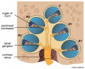 Adult cochlea cartoon 01.jpg 986 × 800; 123 KB
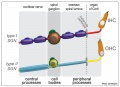 Adult cochlea nerve glia cartoon.jpg 1,000 × 725; 85 KB
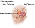 Adult diencephalon.jpg 470 × 376; 20 KB
 Adult human brain movie icon.jpg 717 × 575; 28 KB
 Adult human brain MRI01.jpg 700 × 607; 81 KB
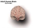
 Adult human brain.jpg 984 × 735; 104 KB
 Adult mouse brain - prosomeric model.jpg 964 × 414; 91 KB
- AEB Histology Prac 171012-3 Ganglion.mp3 ; 1.05 MB
- AEB Histology Prac 171012-5 Nerve.mp3 ; 1.07 MB
 Amin1914 fig01.jpg 1,000 × 652; 120 KB
 Amin1914 fig02.jpg 1,000 × 681; 152 KB
 Amin1914 fig03.jpg 1,000 × 689; 134 KB
 Amin1914 fig04.jpg 1,000 × 727; 139 KB
 Amin1914 fig05.jpg 1,000 × 646; 91 KB
 Anencephaly ultrasound.jpg 900 × 658; 108 KB
 Astrocytes and neonatal hypoxia ischemia.jpg 484 × 705; 309 KB
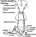 Auditory neural pathway.jpg 450 × 457; 46 KB
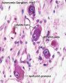 Autonomic ganglion histology 01.jpg 641 × 800; 56 KB
 Baboon- fetal brain.jpg 1,000 × 733; 127 KB
 Bailey358.jpg 854 × 560; 66 KB
 Bailey359.jpg 708 × 572; 46 KB
 Bailey360.jpg 543 × 404; 23 KB
 Bailey361.jpg 815 × 662; 86 KB
 Bailey362.jpg 801 × 354; 49 KB
 Bailey363.jpg 876 × 373; 46 KB
 Bailey364.jpg 809 × 465; 53 KB
 Bailey365.jpg 847 × 595; 94 KB
 Bailey366.jpg 558 × 633; 97 KB
 Bailey367.jpg 1,034 × 440; 101 KB
 Bailey368.jpg 1,074 × 523; 134 KB
 Bailey369.jpg 529 × 446; 33 KB
 Bailey370.jpg 975 × 1,084; 242 KB
 Bailey371.jpg 687 × 997; 97 KB
 Bailey372.jpg 803 × 690; 79 KB
 Bailey373.jpg 894 × 426; 100 KB
 Bailey374.jpg 880 × 490; 75 KB
 Bailey375.jpg 929 × 785; 130 KB
 Bailey376.jpg 922 × 862; 70 KB
 Bailey377.jpg 1,032 × 904; 90 KB
 Bailey378.jpg 1,199 × 700; 69 KB
 Bailey379-382.jpg 671 × 988; 199 KB
 Bailey383.jpg 859 × 455; 118 KB
 Bailey384.jpg 1,532 × 770; 245 KB
 Bailey385.jpg 787 × 683; 165 KB
 Bailey386.jpg 491 × 410; 53 KB
 Bailey387.jpg 507 × 442; 50 KB
 Bailey388.jpg 514 × 438; 68 KB
 Bailey389.jpg 829 × 561; 65 KB
 Bailey390.jpg 583 × 667; 50 KB
 Bailey391.jpg 633 × 485; 56 KB
 Bailey392.jpg 949 × 632; 113 KB
 Bailey393.jpg 680 × 527; 89 KB
 Bailey394.jpg 614 × 563; 70 KB
 Bailey395.jpg 787 × 741; 94 KB
 Bailey396.jpg 829 × 845; 123 KB
 Bailey397.jpg 779 × 995; 162 KB
 Bailey398.jpg 872 × 581; 78 KB
 Bailey399.jpg 832 × 704; 65 KB
 Bailey400.jpg 724 × 749; 66 KB
 Bailey401.jpg 788 × 718; 61 KB
 Bailey402.jpg 640 × 483; 116 KB
 Bailey403.jpg 495 × 617; 118 KB
 Bailey404.jpg 627 × 695; 84 KB
 Bailey405.jpg 745 × 820; 141 KB
 Bailey406.jpg 868 × 763; 116 KB
 Bailey407.jpg 793 × 695; 117 KB
 Bailey408.jpg 862 × 732; 124 KB
 Bailey409.jpg 448 × 408; 32 KB
 Bailey410.jpg 1,412 × 802; 169 KB
 Bailey411.jpg 790 × 809; 104 KB
 Bailey412.jpg 723 × 251; 21 KB
 Bailey413.jpg 674 × 412; 47 KB
 Bailey414.jpg 900 × 717; 123 KB
 Bailey415.jpg 935 × 723; 147 KB
 Bailey416.jpg 815 × 606; 142 KB
 Bailey417.jpg 940 × 494; 116 KB
 Bailey418.jpg 232 × 583; 30 KB
 Bailey419.jpg 805 × 388; 43 KB
 Bailey420.jpg 504 × 348; 34 KB
 Bailey421.jpg 625 × 538; 81 KB
 Bailey422.jpg 495 × 568; 95 KB
 Bailey423.jpg 670 × 581; 56 KB
 Bailey424.jpg 804 × 534; 61 KB
 Bailey425.jpg 957 × 490; 84 KB
 Bailey426.jpg 948 × 758; 80 KB
 Bailey427.jpg 907 × 527; 72 KB
 Bailey428.jpg 756 × 557; 67 KB
 Bailey429.jpg 844 × 484; 81 KB
 Bailey430.jpg 1,072 × 791; 121 KB
 Bailey431.jpg 832 × 535; 75 KB
 Bailey432.jpg 794 × 635; 92 KB
 Bailey433.jpg 723 × 536; 77 KB
 Bailey434.jpg 426 × 436; 44 KB
 Bailey435.jpg 788 × 456; 48 KB
 Bailey436.jpg 552 × 443; 40 KB
 Bailey437.jpg 526 × 397; 50 KB
 Bailey438.jpg 778 × 530; 70 KB
 Bailey439.jpg 932 × 1,016; 125 KB
 Bailey440.jpg 820 × 434; 54 KB
 Bailey441.jpg 786 × 520; 70 KB
 Bailey442.jpg 525 × 504; 38 KB
 Bailey443.jpg 666 × 539; 49 KB
 Bailey444.jpg 777 × 590; 127 KB
 Bailey445.jpg 900 × 655; 89 KB
 Bailey446.jpg 709 × 437; 40 KB
 Bailey447.jpg 795 × 423; 45 KB
 Bailey448.jpg 722 × 416; 55 KB
 Bailey449.jpg 777 × 374; 45 KB
 Bailey450.jpg 680 × 419; 45 KB
 Bailey451-452.jpg 753 × 862; 151 KB
 Bailey453.jpg 461 × 740; 69 KB
 Bailey454.jpg 732 × 527; 119 KB
 Bailey455.jpg 744 × 547; 124 KB
 Bailey456.jpg 591 × 168; 20 KB
 Bailey457.jpg 659 × 335; 40 KB
 Bailey458-459.jpg 741 × 386; 42 KB
 Bailey460.jpg 718 × 423; 55 KB
 Bailey461.jpg 751 × 394; 54 KB
 Bailey462.jpg 463 × 411; 45 KB
 Bailey463.jpg 679 × 345; 52 KB
 Bailey464.jpg 869 × 592; 67 KB
 Bailey465.jpg 806 × 931; 142 KB
 Bailey466.jpg 859 × 683; 139 KB
 Bailey467.jpg 946 × 515; 159 KB
 Bailey468.jpg 774 × 519; 60 KB
 Bailey469.jpg 484 × 401; 50 KB
 Bailey470.jpg 537 × 644; 88 KB
 Bailey475.jpg 1,302 × 852; 213 KB
 Bailey476.jpg 688 × 648; 110 KB
 Bailey477.jpg 593 × 509; 57 KB
 Bailey478.jpg 809 × 620; 81 KB
 Bailey479.jpg 742 × 363; 56 KB
 Bailey480.jpg 799 × 553; 61 KB
 Bailey481.jpg 842 × 752; 109 KB
 Bailey482.jpg 709 × 419; 57 KB
 Baileytable08.jpg 968 × 570; 76 KB
 Baileytable09.jpg 606 × 150; 18 KB
 Bardeen1906-fig02.jpg 1,598 × 1,183; 228 KB
 Bardeen1906-fig03.jpg 1,598 × 1,166; 231 KB
 Bardeen1906-plate01.jpg 1,565 × 2,322; 238 KB
 Bardeen1906-plate02.jpg 1,719 × 2,302; 512 KB
 Bardeen1906-plate06.jpg 1,568 × 2,299; 379 KB
 Bardeen1906-plate31.jpg 1,571 × 2,330; 257 KB
 Bardeen1906-plate32.jpg 1,588 × 2,341; 292 KB
 Bardeen1906-plate41.jpg 1,555 × 2,323; 261 KB
 Bardeen1906-plate42.jpg 1,570 × 2,331; 240 KB
 Bardeen1906-plate51.jpg 1,570 × 2,330; 392 KB
 Bardeen1906-plate52.jpg 1,596 × 2,350; 404 KB
 Bartelmez1922-fig01.jpg 900 × 770; 131 KB
 Bartelmez1922-fig02.jpg 1,203 × 1,700; 317 KB
 Bartelmez1922-fig03.jpg 885 × 1,000; 169 KB
 Bartelmez1922-fig04.jpg 1,300 × 750; 108 KB
 Bartelmez1922-fig05.jpg 800 × 750; 164 KB
 Bartelmez1922-fig06.jpg 749 × 1,000; 179 KB
 Bartelmez1922-fig07.jpg 1,200 × 620; 116 KB
 Bartelmez1922-fig08.jpg 1,000 × 1,255; 251 KB
 Bartelmez1922-fig09.jpg 1,200 × 1,558; 287 KB
 Bartelmez1922-fig10.jpg 1,000 × 1,136; 209 KB
 Bartelmez1923 fig01.jpg 1,312 × 1,919; 245 KB
 Bartelmez1923 fig02.jpg 1,295 × 2,189; 218 KB
 Bartelmez1923 fig03.jpg 1,105 × 1,350; 202 KB
 Bartelmez1923 fig04.jpg 1,416 × 1,155; 170 KB
 Bartelmez1923 fig05.jpg 1,102 × 815; 195 KB
 Bartelmez1923 fig06.jpg 1,280 × 809; 136 KB
 Bat - neural development 01.jpg 733 × 498; 33 KB
 BAW icon 2012.jpg 200 × 280; 17 KB
 Blood-brain barrier cartoon.jpg 765 × 1,000; 75 KB
 Blood-brain barrier EM01.jpg 1,656 × 810; 250 KB
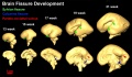
 Brain fissure development 01.jpg 1,157 × 502; 47 KB
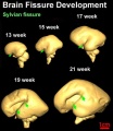
 Brain fissure development 02.jpg 1,000 × 583; 52 KB
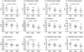
 Brain fissure development 03.jpg 600 × 691; 33 KB
 Brain growth and birth size.jpg 800 × 492; 70 KB
 Brain histology 01.jpg 480 × 600; 125 KB
 Brain histology 02.jpg 480 × 600; 51 KB
 Brain stem subdivisions 01.jpg 1,530 × 520; 168 KB
 Brain tract development 01.jpg 720 × 1,023; 76 KB
 Brain tract development 02.jpg 1,000 × 424; 29 KB
 Brain tract development 06.jpg 1,000 × 605; 35 KB


