Category:Limb: Difference between revisions
From Embryology
mNo edit summary |
mNo edit summary |
||
| Line 1: | Line 1: | ||
This page lists {{Embryology}} content related to limb development and limb abnormalities. The limb is also an excellent model for how tissue pattern is established. | This page lists {{Embryology}} content related to limb development and limb abnormalities. The limb is also an excellent model for how tissue pattern is established. | ||
<br> | |||
{{Limb Links}} | |||
<br> | <br> | ||
{{Musculoskeletal Links}} | {{Musculoskeletal Links}} | ||
Latest revision as of 12:33, 16 March 2020
This page lists Embryology content related to limb development and limb abnormalities. The limb is also an excellent model for how tissue pattern is established.
| Musculoskeletal Links: Introduction | mesoderm | somitogenesis | limb | cartilage | bone | bone timeline | bone marrow | shoulder | pelvis | axial skeleton | skull | joint | skeletal muscle | muscle timeline | tendon | diaphragm | Lecture - Musculoskeletal | Lecture Movie | musculoskeletal abnormalities | limb abnormalities | developmental hip dysplasia | cartilage histology | bone histology | Skeletal Muscle Histology | Category:Musculoskeletal | ||
|
Pages in category 'Limb'
The following 148 pages are in this category, out of 148 total.
B
- Template:Bardeen1906 figures
- BGDA Practical 7 - Week 6
- Book - An Atlas of Topographical Anatomy 19
- Book - An Atlas of Topographical Anatomy 20
- Book - An Atlas of Topographical Anatomy 21
- Book - An Atlas of Topographical Anatomy 22
- Book - An Atlas of Topographical Anatomy 23
- Book - An Atlas of Topographical Anatomy 24
- Book - An Atlas of Topographical Anatomy 26
- Book - An Atlas of Topographical Anatomy 27
- Book - An Atlas of Topographical Anatomy 28
- Book - Human Embryology and Morphology 20
- Book - Manual of Human Embryology 11D
- Book - Manual of Human Embryology 12
C
D
L
- Lecture - Limb Development
- Template:Limb
- Template:Limb abnormalities
- Template:Limb axis
- Template:Limb Links
- Template:Limb Rotation table
- Template:Limb skeleton mesenchyme-cartilage-bone table
- Template:Lower limb ossification collapse table
- Template:Lower limb ossification collapsible table
- Template:Lower limb ossification table
- Template:Lower limb ossification timeline
M
P
- Paper - A contribution to the embryology of the fore-limb
- Paper - A study of the development of the mammalian pelvis
- Paper - An unusual form of brachyphalangy and syndactyly with double proximal phalanx in the middle fingers (1932)
- Paper - Anatomy of a seven months foetus exhibiting bilateral absence of the ulna accompanied by monodactyly
- Paper - Chondrification in the hands and feet of staged human embryos
- Paper - Development and variation of the nerves and the musculature of the inferior extremity and of the neighboring regions of the trunk in man
- Paper - Development of the innervation pattern in the upper limb of staged human embryos (1990)
- Paper - Extra digits and digital reductions
- Paper - Familial abnormalities of the middle phalanges of each hand (1923)
- Paper - On an instance of two subclavian arteries of the early arm bud of man and its fundamental significance
- Paper - Rare congenital malformation of hands and feet (1924)
- Paper - Studies of the development of the human skeleton (1905)
- Paper - The arrangement of the bursae in the superior extremities of the full-time foetus
- Paper - The development and evolution of the "papillary" ridges and patterns on the volar surface of the hand (1906)
- Paper - The development of joints
- Paper - The development of the arm in man (1902)
- Paper - The development of the arteries of the human lower extremity
- Paper - The development of the limbs, body-wall and back
- Paper - The development of the limbs, body-wall and back (1901)
- Paper - The development of the patella
- Paper - The development of the principal arterial stems in the forelimb of the pig (1922)
- Paper - The development of the veins in the limbs of rabbit embryos
- Paper - The early development of the knee joint in staged human embryos
- Paper - The embryology of the human hip joint (1943)
- Paper - The epiphysis of the head of the femur (1915)
- Paper - The growth of the human foot
- Paper - The proximo-distal sequence of origin of the parts of the chick wing and the role of the ectoderm
- Paper - The sexual differences of the fetal pelvis
- Paper - The sexual differences of the fetal pelvis (1899)
- Template:Pelvis
- Template:Pelvis Timeline table
R
- Template:Ref-Bardeen1907
- Template:Ref-BardeenLewis1901
- Template:Ref-Brailsford1943
- Template:Ref-Carey1922
- Template:Ref-Cockayne1932
- Template:Ref-Cummins1929
- Template:Ref-Davenport1932
- Template:Ref-deLima1928
- Template:Ref-Evans1908
- Template:Ref-Evatt1906
- Template:Ref-GardnerO'Rahilly1968
- Template:Ref-Geddes1912b
- Template:Ref-GrayGardener1950
- Template:Ref-Haines1947
- Template:Ref-Haines1953
- Template:Ref-Hann1923
- Template:Ref-Harrison1915
- Template:Ref-Johnson1899
- Template:Ref-Keith1910
- Template:Ref-Lewis1902
- Template:Ref-Lewis1905b
- Template:Ref-Lewis1910a
- Template:Ref-Manson1924
- Template:Ref-O'Rahilly1957
- Template:Ref-PMID55906
- Template:Ref-Prentiss1906
- Template:Ref-Rutherford1914
- Template:Ref-Saunders1947a
- Template:Ref-Saunders1948
- Template:Ref-ShinoharaTanaka1990
- Template:Ref-Stacey1938
- Template:Ref-Walmsley1915
- Template:Ref-Walmsley1940
- Template:Ref-Watt1917
- Template:Ref-Whittaker1910
- Template:Ref-Wigoder1928
- Template:Ref-Woollard1922
S
U
V
Media in category 'Limb'
The following 74 files are in this category, out of 274 total.
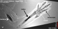
(previous page) (next page)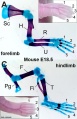 Mouse limb cartilage and bone E18.5.jpg 774 × 1,200; 152 KB
 Mouse limb gene expression 01.mov ; 1.12 MB
 Mouse limb gene expression 01.mp4 ; 1.34 MB
- Mouse limb gene expression 02.mp4 ; 1.34 MB
 Mouse limb gene expression icon.jpg 446 × 550; 48 KB
 Mouse limb skeleton cartoon.jpg 1,000 × 487; 64 KB
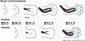
 Mouse limb tissue development.jpg 1,280 × 767; 161 KB
 Mouse Lmx1b gene expression.jpg 1,624 × 550; 104 KB
 Mouse pax7 limb 01.jpg 1,320 × 549; 112 KB
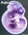 Mouse Prdm1.jpg 446 × 550; 35 KB
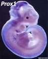 Mouse Prox1.jpg 446 × 550; 36 KB
 Mouse Ptx1.jpg 446 × 550; 21 KB
 Mouse Sim2.jpg 446 × 550; 30 KB
 Mouse SmarcD2.jpg 446 × 550; 32 KB
 Mouse Sp6.jpg 446 × 550; 34 KB
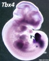 Mouse Tbx4.jpg 446 × 550; 37 KB
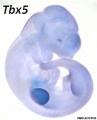 Mouse Tbx5.jpg 446 × 550; 21 KB
 Mouse- forelimb bud Shh expression and BMP pathway.jpg 905 × 308; 35 KB
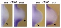 Mouse- forelimb-bud-Tbx3-Tbx2.jpg 955 × 436; 45 KB
 Mouse- Hand2 forelimb buds.jpg 458 × 600; 48 KB
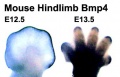 Mouse- hindlimb Bmp4 expression.jpg 374 × 241; 19 KB
 Mouse- hindlimb buds Shh expression.jpg 600 × 679; 30 KB
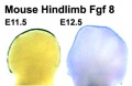 Mouse- hindlimb Fgf8 expression.jpg 374 × 243; 17 KB
 Mouse-E17.5 foot.jpg 385 × 500; 29 KB
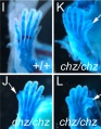 Mouse-E17.5 normal and abnormal limb.jpg 661 × 848; 105 KB
 Mouse-E17.5-foot-large.jpg 600 × 779; 53 KB
 Mouse-forelimb bud Fgf4-Hoxd13-Hoxa13.jpg 881 × 237; 30 KB
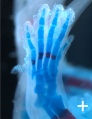
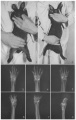
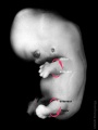
 MRI Human Embryo - upper limb 01.jpg 1,418 × 940; 106 KB
 MRI Human Embryo - upper limb 02.jpg 1,000 × 497; 74 KB
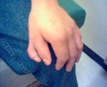
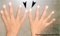
 Nail patella syndrome 02.jpg 809 × 525; 51 KB
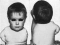
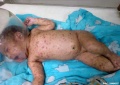 Neonatal varicella.jpg 795 × 561; 35 KB
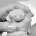
 Polydactylia.jpg 700 × 541; 37 KB
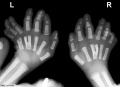 Polydactyly and y-shaped central metacarpals x-ray.jpg 800 × 579; 51 KB
 Polydactyly.jpg 230 × 188; 10 KB
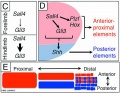
 Posterior forelimb bud identity.jpg 600 × 406; 46 KB
 Posterior forelimb bud identity.png 600 × 406; 895 KB
 Rabbitmalformation3.jpg 363 × 574; 44 KB
 Sall4 limb skeletal elements model.jpg 800 × 619; 88 KB
 Senior1919 fig10.jpg 1,280 × 679; 134 KB
 Shoulder cartoon.jpg 592 × 597; 36 KB
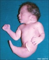 Sirenomelia 01.jpg 488 × 600; 23 KB
 Sirenomelia 02.jpg 498 × 598; 14 KB
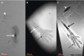
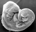
 Stage13 sem1.jpg 1,000 × 886; 80 KB
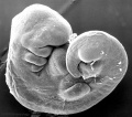 Stage13 sem1a.jpg 800 × 709; 57 KB
 Stage13 sem1b.jpg 600 × 532; 36 KB
 Stage13 sem1c.jpg 400 × 355; 18 KB
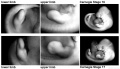 Stage16-17-limbs01.jpg 1,000 × 572; 77 KB
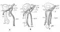
 Stage19- limb rotation.jpg 600 × 800; 27 KB
 Stage20-23 limbs a.jpg 800 × 301; 19 KB
 Stage20-23 limbs b.jpg 600 × 226; 13 KB
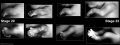 Stage20-23 limbs.jpg 1,000 × 376; 26 KB
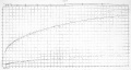 Streeter1920chart3.jpg 1,200 × 635; 79 KB
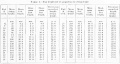 Streeter1920table4.jpg 1,120 × 593; 80 KB
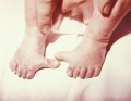
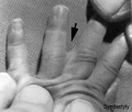 Syndactyly.jpg 443 × 377; 16 KB
 Syndactyly2.JPG 220 × 221; 7 KB
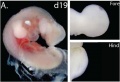 Tammar wallaby limb day 19 development.jpg 700 × 477; 39 KB
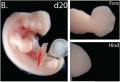 Tammar wallaby limb day 20 development.jpg 700 × 477; 36 KB
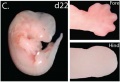 Tammar wallaby limb day 22 development.jpg 700 × 477; 36 KB
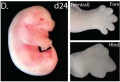 Tammar wallaby limb day 24 development.jpg 700 × 477; 39 KB
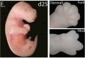 Tammar wallaby limb day 25 development.jpg 700 × 477; 37 KB
 Tammar wallaby limb development 01.jpg 1,200 × 551; 101 KB
 Thalidomide - CPS49 vascular effect.jpg 1,000 × 1,002; 197 KB
 Thalidomide - limb signaling.jpg 628 × 540; 88 KB
 Thalidomide abnormalities 01.jpg 800 × 598; 71 KB
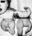 Thalidomide abnormalities 02.jpg 724 × 800; 74 KB
 Thomson1899 fig03.jpg 1,209 × 1,678; 82 KB
 Triphalangeal-thumb.jpg 450 × 273; 14 KB
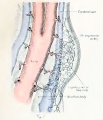
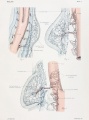 Woollard-plate01.jpg 744 × 1,000; 153 KB
 Woollard-plate02.jpg 788 × 1,000; 196 KB
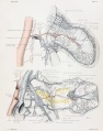
 Woollard001.jpg 739 × 858; 142 KB
 Woollard002.jpg 699 × 873; 125 KB
 Woollard003.jpg 1,107 × 848; 159 KB
 Woollard004.jpg 1,037 × 717; 148 KB
 Woollard005.jpg 1,034 × 655; 144 KB


