Category:Hippocampus: Difference between revisions
From Embryology
(Created page with "This {{Embryology}} category shows pages and media related to Hippocampus Development. Category:Neural") |
mNo edit summary |
||
| (2 intermediate revisions by the same user not shown) | |||
| Line 1: | Line 1: | ||
This {{Embryology}} category shows pages and media related to Hippocampus Development. | This {{Embryology}} category shows pages and media related to Hippocampus Development. The hippocampus forms originally from the neural tube forebrain (prosencephalon) primary vesicle and then secondary vesicle telencephalon region. | ||
:'''Links:''' [[Neural - Hippocampus Development]] | |||
{{Neural Table}} | |||
{{Neural Links}} | |||
{{Neural Links 2}} | |||
[[Category:Neural]] | [[Category:Neural]] | ||
Latest revision as of 11:18, 31 October 2015
This Embryology category shows pages and media related to Hippocampus Development. The hippocampus forms originally from the neural tube forebrain (prosencephalon) primary vesicle and then secondary vesicle telencephalon region.
| Neural Tube | Primary Vesicles | Secondary Vesicles | Adult Structures |
|---|---|---|---|
| week 3 | week 4 | week 5 | adult |
| prosencephalon (forebrain) | telencephalon | Rhinencephalon, Amygdala, hippocampus, cerebrum (cortex), hypothalamus, pituitary | Basal Ganglia, lateral ventricles | |
| diencephalon | epithalamus, thalamus, Subthalamus, pineal, posterior commissure, pretectum, third ventricle | ||
| mesencephalon (midbrain) | mesencephalon | tectum, Cerebral peduncle, cerebral aqueduct, pons | |
| rhombencephalon (hindbrain) | metencephalon | cerebellum | |
| myelencephalon | medulla oblongata, isthmus | ||
| spinal cord, pyramidal decussation, central canal | |||
Pages in category 'Hippocampus'
The following 6 pages are in this category, out of 6 total.
Media in category 'Hippocampus'
The following 11 files are in this category, out of 11 total.
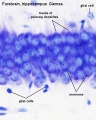 Brain histology 02.jpg 480 × 600; 51 KB
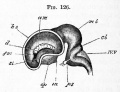 Foster126.jpg 614 × 471; 53 KB
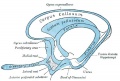 Gray0732.jpg 600 × 406; 31 KB
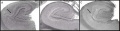 Hippocampal Formation.jpg 600 × 153; 34 KB
 Keith1921 fig115.jpg 1,200 × 627; 144 KB
 Kollmann628.jpg 823 × 543; 126 KB
 Mouse Brain E17 MRI 01.jpg 963 × 403; 88 KB
 Mouse cerebellum connections 01.jpg 470 × 600; 44 KB
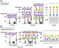 Mouse cortex and hippocampus development 01.jpg 761 × 622; 112 KB
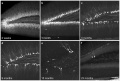 Mouse- hippocampus dentate granule cells.jpg 1,000 × 671; 130 KB
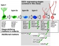 Neural - Human hippocampus marker expression.jpg 600 × 472; 62 KB


